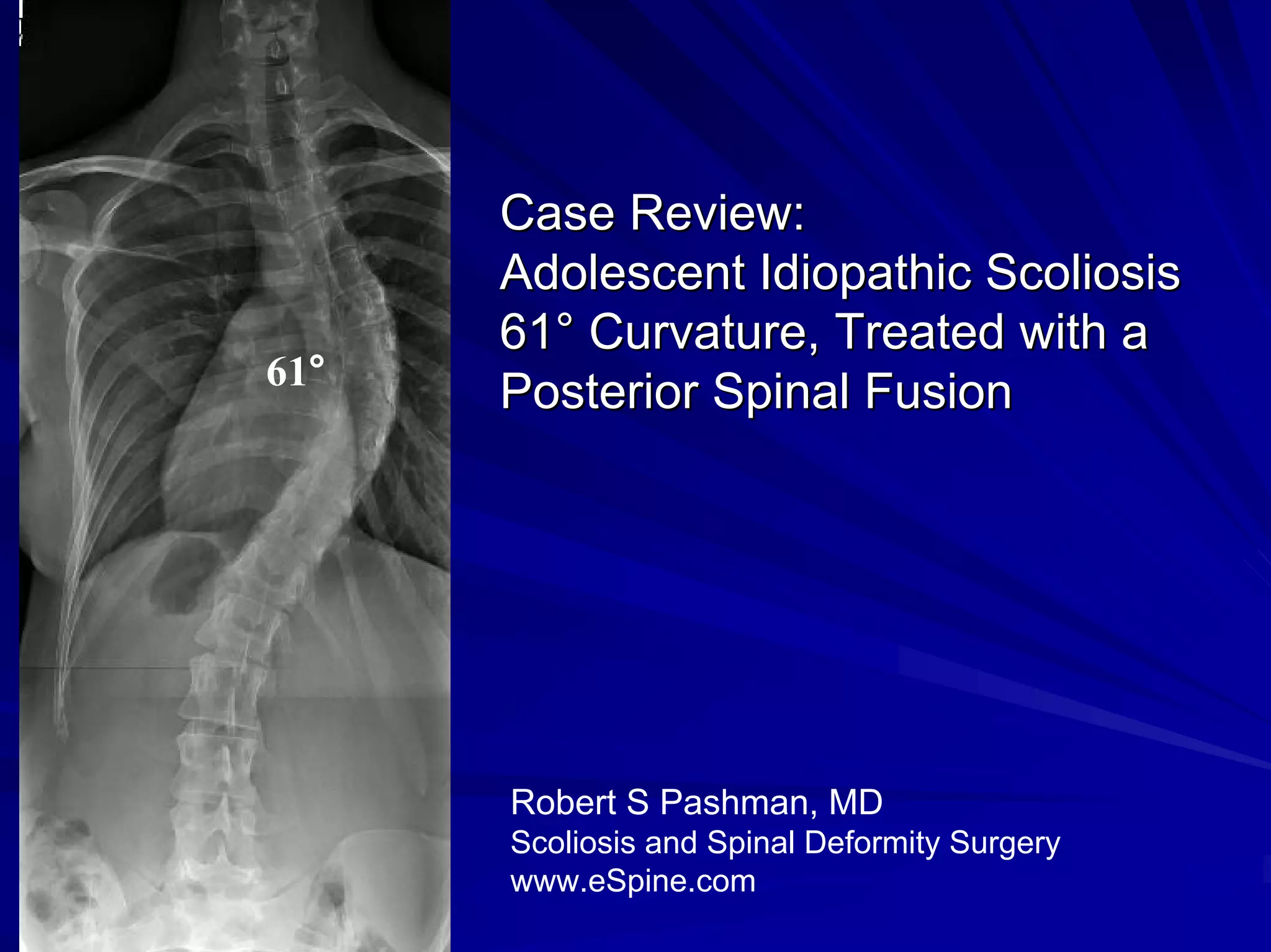
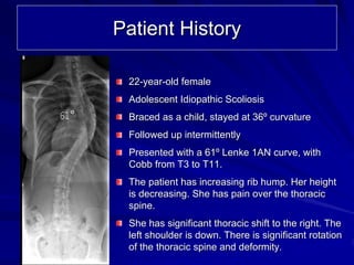
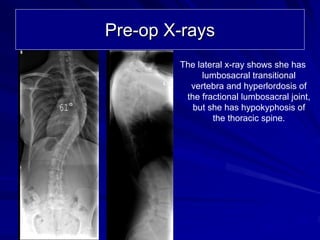
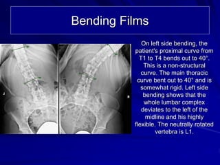
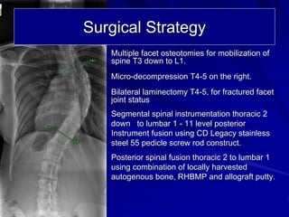
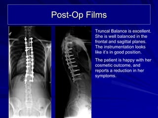
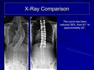
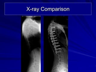
Case Review:
      Adolescent Idiopathic Scoliosis
      61° Curvature, Treated with a
61°
      Posterior Spinal Fusion


      Robert S Pashman, MD
      Scoliosis and Spinal Deformity Surgery
      www.eSpine.com
Patient History

       22-year-old female
       Adolescent Idiopathic Scoliosis
61°    Braced as a child, stayed at 36º curvature
       Followed up intermittently
       Presented with a 61º Lenke 1AN curve, with
       Cobb from T3 to T11.
       The patient has increasing rib hump. Her height
       is decreasing. She has pain over the thoracic
       spine.
       She has significant thoracic shift to the right. The
       left shoulder is down. There is significant rotation
       of the thoracic spine and deformity.
Pre-op X-rays
              The lateral x-ray shows she has
                   lumbosacral transitional
                vertebra and hyperlordosis of
               the fractional lumbosacral joint,
61°              but she has hypokyphosis of
                      the thoracic spine.
Bending Films
             On left side bending, the
          patient's proximal curve from
            T1 to T4 bends out to 40°.
              This is a non-structural
             curve. The main thoracic
           curve bent out to 40° and is
             somewhat rigid. Left side
              bending shows that the
              whole lumbar complex
             deviates to the left of the
               midline and his highly
          flexible. The neutrally rotated
                   vertebra is L1.
Surgical Strategy
   Multiple facet osteotomies for mobilization of
   spine T3 down to L1.
   Micro-decompression T4-5 on the right.
   Bilateral laminectomy T4-5, for fractured facet
   joint status
   Segmental spinal instrumentation thoracic 2
   down to lumbar 1 - 11 level posterior
   Instrument fusion using CD Legacy stainless
   steel 55 pedicle screw rod construct.
   Posterior spinal fusion thoracic 2 to lumbar 1
   using combination of locally harvested
   autogenous bone, RHBMP and allograft putty.
Post-Op Films
          Truncal Balance is excellent.
          She is well balanced in the
          frontal and sagittal planes.
          The instrumentation looks
          like it’s in good position.
          The patient is happy with her
          cosmetic outcome, and
          reports a reduction in her
          symptoms.
X-Ray Comparison

                     The curve has been
                  reduced 36%, from 61° to
                     approximately 22°.
61°       22°
X-ray Comparison

Case Review #4: Adolescent Idiopathic Scoliosis with 61 degree curvature

  • 1.
    Case Review: Adolescent Idiopathic Scoliosis 61° Curvature, Treated with a 61° Posterior Spinal Fusion Robert S Pashman, MD Scoliosis and Spinal Deformity Surgery www.eSpine.com
  • 2.
    Patient History 22-year-old female Adolescent Idiopathic Scoliosis 61° Braced as a child, stayed at 36º curvature Followed up intermittently Presented with a 61º Lenke 1AN curve, with Cobb from T3 to T11. The patient has increasing rib hump. Her height is decreasing. She has pain over the thoracic spine. She has significant thoracic shift to the right. The left shoulder is down. There is significant rotation of the thoracic spine and deformity.
  • 3.
    Pre-op X-rays The lateral x-ray shows she has lumbosacral transitional vertebra and hyperlordosis of the fractional lumbosacral joint, 61° but she has hypokyphosis of the thoracic spine.
  • 4.
    Bending Films On left side bending, the patient's proximal curve from T1 to T4 bends out to 40°. This is a non-structural curve. The main thoracic curve bent out to 40° and is somewhat rigid. Left side bending shows that the whole lumbar complex deviates to the left of the midline and his highly flexible. The neutrally rotated vertebra is L1.
  • 5.
    Surgical Strategy Multiple facet osteotomies for mobilization of spine T3 down to L1. Micro-decompression T4-5 on the right. Bilateral laminectomy T4-5, for fractured facet joint status Segmental spinal instrumentation thoracic 2 down to lumbar 1 - 11 level posterior Instrument fusion using CD Legacy stainless steel 55 pedicle screw rod construct. Posterior spinal fusion thoracic 2 to lumbar 1 using combination of locally harvested autogenous bone, RHBMP and allograft putty.
  • 6.
    Post-Op Films Truncal Balance is excellent. She is well balanced in the frontal and sagittal planes. The instrumentation looks like it’s in good position. The patient is happy with her cosmetic outcome, and reports a reduction in her symptoms.
  • 7.
    X-Ray Comparison The curve has been reduced 36%, from 61° to approximately 22°. 61° 22°
  • 8.