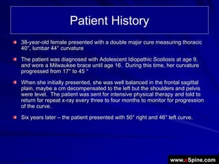
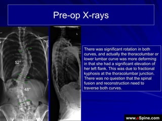
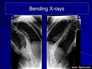
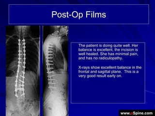
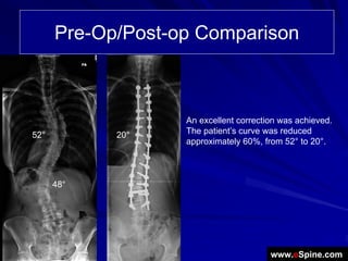
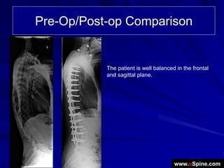
Case Review:
52°
            Adult Idiopathic Scoliosis
            treated with a posterior
            fusion from T3 to L4 fusion
      48°


            Robert S Pashman, MD
            Scoliosis and Spinal Deformity
            www.eSpine.com
Patient History
38-year-old female presented with a double major cure measuring thoracic
40°, lumbar 44° curvature

The patient was diagnosed with Adolescent Idiopathic Scoliosis at age 9,
and wore a Milwaukee brace until age 16. During this time, her curvature
progressed from 17° to 45 °

When she initially presented, she was well balanced in the frontal sagittal
plain, maybe a cm decompensated to the left but the shoulders and pelvis
were level. The patient was sent for intensive physical therapy and told to
return for repeat x-ray every three to four months to monitor for progression
of the curve.

Six years later – the patient presented with 50° right and 46° left curve.


                                                                www.eSpine.com
Pre-op X-rays


                   There was significant rotation in both
                   curves, and actually the thoracolumbar or
                   lower lumbar curve was more deforming
52°                in that she had a significant elevation of
                   her left flank. This was due to fractional
                   kyphosis at the thoracolumbar junction.
                   There was no question that the spinal
                   fusion and reconstruction need to
      48°          traverse both curves.


                                          www.eSpine.com
Bending X-rays
L                         R


                     www.eSpine.com
Indications for Surgery
Adult idiopathic scoliosis double major curve, measuring 52 and 48,
respectively, thoracolumbar spine.
Rigid kyphosis, thoracolumbar junction
Severe superimposed degenerative disk disease and facet arthropathy, with
mid lumbar degeneration causing rigid compensatory curve with
thoracolumbar kyphosis.
Low back/lower extremity symptoms, with spinal stenosis, neural foraminal
stenosis lumbar spine.
Failed conservative therapy.


                                                            www.eSpine.com
Surgical Strategy
Segmental spinal instrumentation, thoracic 3 to lumbar 4; this is a 13-level
instrumentation, with 5.5 stainless steel Legacy screw-rod construct.
Posterior spinal fusion, thoracic 3 to lumbar 4, with autogenous bone and
Rh bone morphogenic protein.
Because of the rigidity of this curve, it required significant mobilization,
which necessitated multiple-level osteotomies for correction of the curve in
a posterior only basis. Ponte osteotomy, radical mobilization of the spine,
with complete facetectomy resection, thoracic 5 to lumbar 3-4. This is a 10-
level osteotomy, with correction of coronal and sagittal plane deformity.
Subtotal laminectomy, thoracic 12 to lumbar 1, lumbar 1-2, lumbar 2- 3, and
lumbar 3-4 under loupe magnification for spinal canal decompression and
spinal stenosis.
Intraoperative motor evoked potential interpretation.
Intraoperative fluoroscopic interpretation.
Plastic closure.

                                                               www.eSpine.com
Post-Op Films


      The patient is doing quite well. Her
      balance is excellent, the incision is
      well healed. She has minimal pain,
      and has no radiculopathy.

      X-rays show excellent balance in the
      frontal and sagittal plane. This is a
      very good result early on.


                               www.eSpine.com
Pre-Op/Post-op Comparison


                   An excellent correction was achieved.
52°         20°    The patient’s curve was reduced
                   approximately 60%, from 52° to 20°.


      48°


                                        www.eSpine.com
Pre-Op/Post-op Comparison


           The patient is well balanced in the frontal
           and sagittal plane.


                                       www.eSpine.com

Case Review #8: 44 year old woman presented with a Double Major Scoliosis Curvature

  • 1.
    Case Review: 52° Adult Idiopathic Scoliosis treated with a posterior fusion from T3 to L4 fusion 48° Robert S Pashman, MD Scoliosis and Spinal Deformity www.eSpine.com
  • 2.
    Patient History 38-year-old femalepresented with a double major cure measuring thoracic 40°, lumbar 44° curvature The patient was diagnosed with Adolescent Idiopathic Scoliosis at age 9, and wore a Milwaukee brace until age 16. During this time, her curvature progressed from 17° to 45 ° When she initially presented, she was well balanced in the frontal sagittal plain, maybe a cm decompensated to the left but the shoulders and pelvis were level. The patient was sent for intensive physical therapy and told to return for repeat x-ray every three to four months to monitor for progression of the curve. Six years later – the patient presented with 50° right and 46° left curve. www.eSpine.com
  • 3.
    Pre-op X-rays There was significant rotation in both curves, and actually the thoracolumbar or lower lumbar curve was more deforming 52° in that she had a significant elevation of her left flank. This was due to fractional kyphosis at the thoracolumbar junction. There was no question that the spinal fusion and reconstruction need to 48° traverse both curves. www.eSpine.com
  • 4.
    Bending X-rays L R www.eSpine.com
  • 5.
    Indications for Surgery Adultidiopathic scoliosis double major curve, measuring 52 and 48, respectively, thoracolumbar spine. Rigid kyphosis, thoracolumbar junction Severe superimposed degenerative disk disease and facet arthropathy, with mid lumbar degeneration causing rigid compensatory curve with thoracolumbar kyphosis. Low back/lower extremity symptoms, with spinal stenosis, neural foraminal stenosis lumbar spine. Failed conservative therapy. www.eSpine.com
  • 6.
    Surgical Strategy Segmental spinalinstrumentation, thoracic 3 to lumbar 4; this is a 13-level instrumentation, with 5.5 stainless steel Legacy screw-rod construct. Posterior spinal fusion, thoracic 3 to lumbar 4, with autogenous bone and Rh bone morphogenic protein. Because of the rigidity of this curve, it required significant mobilization, which necessitated multiple-level osteotomies for correction of the curve in a posterior only basis. Ponte osteotomy, radical mobilization of the spine, with complete facetectomy resection, thoracic 5 to lumbar 3-4. This is a 10- level osteotomy, with correction of coronal and sagittal plane deformity. Subtotal laminectomy, thoracic 12 to lumbar 1, lumbar 1-2, lumbar 2- 3, and lumbar 3-4 under loupe magnification for spinal canal decompression and spinal stenosis. Intraoperative motor evoked potential interpretation. Intraoperative fluoroscopic interpretation. Plastic closure. www.eSpine.com
  • 7.
    Post-Op Films The patient is doing quite well. Her balance is excellent, the incision is well healed. She has minimal pain, and has no radiculopathy. X-rays show excellent balance in the frontal and sagittal plane. This is a very good result early on. www.eSpine.com
  • 8.
    Pre-Op/Post-op Comparison An excellent correction was achieved. 52° 20° The patient’s curve was reduced approximately 60%, from 52° to 20°. 48° www.eSpine.com
  • 9.
    Pre-Op/Post-op Comparison The patient is well balanced in the frontal and sagittal plane. www.eSpine.com