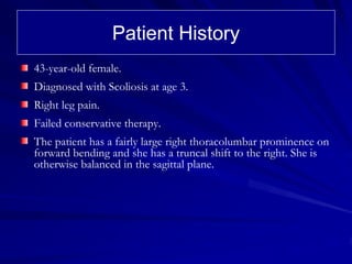
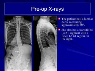
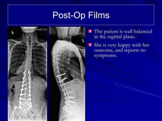
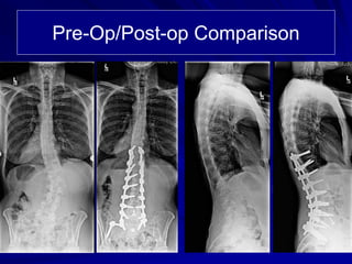
Case Review:
      43 year old female with Adult
      Scoliosis and lumbar sacral
      transitional vertebra.


40°

       Robert S Pashman, MD
       Scoliosis and Spinal Deformity Surgery
       www.eSpine.com
Patient History
43-year-old female.
Diagnosed with Scoliosis at age 3.
Right leg pain.
Failed conservative therapy.
The patient has a fairly large right thoracolumbar prominence on
forward bending and she has a truncal shift to the right. She is
otherwise balanced in the sagittal plane.
Pre-op X-rays
                  The patient has a lumbar
                  curve measuring
                  approximately 40°.
                  She also has a transitional
                  L5-S1 segment with a
                  fused L5-S1 region on
                  the right.


40°
Indications for Surgery
1. Kim/SRP type II adult idiopathic scoliosis.
2. Adult idiopathic possible congenital and degenerative scoliosis.
3. Severe low back pain due to multiple-level degenerative disk
    disease.
4. Multiple-level foraminal lateral recess stenosis with combination
    of low back and leg pain.
5. Failed conservative therapy.
6. Coronal and sagittal plane deformity.
7. Bilateral facet arthropathy due to degeneration.
8. Multiple comorbidities including narcotic tolerance.
9. Radiculopathy.
10. Lumbosacral transitional vertebrae.
Surgical Strategy
Anterior interbody fusion L4-5      Segmental spinal instrumentation using
and L5-S1 with radical              quarter-inch stainless steel screw-rod
diskectomy and epidural             construct, for Kim/SRP type II scoliosis,
decompression.                      thoracic 10 to sacral pelvis.
Abdominal retroperitoneal           Spinal osteotomy, multiple level, for
approach to the lumbosacral         correction of sagittal plane and coronal
spine.                              scoliosis deformity, L1-2, L2-3, L3-4, L4-
Interbody fusion with with          5.
autogenous bone centrally L4-5,     Posterior spinal fusion using locally-
L5-S1.                              harvested autogenous bone and rhBMP,
Anterior screw fixation L4-5, L5-   T10 to the sacral pelvis.
S1.                                 Pelvic fixation with bilateral exposure
Reduction kyphosis and              through iliac crest through separate
correction of coronal plane         incision.
deformity lumbosacral joint.        Intraoperative SSEP with motor-evoked
Intraoperative somatosensory        potentials.
evoked potentials.                  Intraoperative O-arm localization and
Intraoperative fluoroscopy.         fluoroscopy.
Post-Op Films
         The patient is well balanced
         in the sagittal plane.
         She is very happy with her
         outcome, and reports no
         symptoms.
Pre-Op/Post-op Comparison

Case Review #35: 43 year old female with Adult Scoliosis and a Transitional Vertebra

  • 1.
    Case Review: 43 year old female with Adult Scoliosis and lumbar sacral transitional vertebra. 40° Robert S Pashman, MD Scoliosis and Spinal Deformity Surgery www.eSpine.com
  • 2.
    Patient History 43-year-old female. Diagnosedwith Scoliosis at age 3. Right leg pain. Failed conservative therapy. The patient has a fairly large right thoracolumbar prominence on forward bending and she has a truncal shift to the right. She is otherwise balanced in the sagittal plane.
  • 3.
    Pre-op X-rays The patient has a lumbar curve measuring approximately 40°. She also has a transitional L5-S1 segment with a fused L5-S1 region on the right. 40°
  • 4.
    Indications for Surgery 1.Kim/SRP type II adult idiopathic scoliosis. 2. Adult idiopathic possible congenital and degenerative scoliosis. 3. Severe low back pain due to multiple-level degenerative disk disease. 4. Multiple-level foraminal lateral recess stenosis with combination of low back and leg pain. 5. Failed conservative therapy. 6. Coronal and sagittal plane deformity. 7. Bilateral facet arthropathy due to degeneration. 8. Multiple comorbidities including narcotic tolerance. 9. Radiculopathy. 10. Lumbosacral transitional vertebrae.
  • 5.
    Surgical Strategy Anterior interbodyfusion L4-5 Segmental spinal instrumentation using and L5-S1 with radical quarter-inch stainless steel screw-rod diskectomy and epidural construct, for Kim/SRP type II scoliosis, decompression. thoracic 10 to sacral pelvis. Abdominal retroperitoneal Spinal osteotomy, multiple level, for approach to the lumbosacral correction of sagittal plane and coronal spine. scoliosis deformity, L1-2, L2-3, L3-4, L4- Interbody fusion with with 5. autogenous bone centrally L4-5, Posterior spinal fusion using locally- L5-S1. harvested autogenous bone and rhBMP, Anterior screw fixation L4-5, L5- T10 to the sacral pelvis. S1. Pelvic fixation with bilateral exposure Reduction kyphosis and through iliac crest through separate correction of coronal plane incision. deformity lumbosacral joint. Intraoperative SSEP with motor-evoked Intraoperative somatosensory potentials. evoked potentials. Intraoperative O-arm localization and Intraoperative fluoroscopy. fluoroscopy.
  • 6.
    Post-Op Films The patient is well balanced in the sagittal plane. She is very happy with her outcome, and reports no symptoms.
  • 7.