Adult Scoliosis Case Review: 50° Lumbar Curve Corrected to 15
•
1 like•1,061 views

A 43 year old woman, presented with Adult Idiopathic Scoliosis, 50° lumbar curve. Dr. Pashman treated her with Posterior Spinal Fusion from T9 - L5. Curve was a KIM/SRP Classification 2.

Recommended
Recommended
More Related Content
What's hot
What's hot (20)
Viewers also liked
Viewers also liked (18)
Similar to Adult Scoliosis Case Review: 50° Lumbar Curve Corrected to 15
Similar to Adult Scoliosis Case Review: 50° Lumbar Curve Corrected to 15 (19)
More from Robert Pashman
More from Robert Pashman (11)
Recently uploaded
Recently uploaded (20)
Adult Scoliosis Case Review: 50° Lumbar Curve Corrected to 15
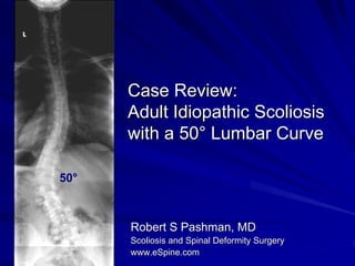
- 1. Case Review: Adult Idiopathic Scoliosis with a 50° Lumbar Curve 50° Robert S Pashman, MD Scoliosis and Spinal Deformity Surgery www.eSpine.com
- 2. Patient History 43 yo female, increasing pain over her left lumbar curve. She has tried chiropractic, physical therapy, massage, injections, anti-inflammatories. The pain is severe. On physical examination, she has typical right flank elevation. She has decompensation to the left. Sagittal plane is good. She has a slight right rib hump. 36x14 x-rays show a 45° left lumbar curve which increased from 40° in 2001. She has rotation or lateral listhesis of L3 on 4, and is decompensated approximately 3-4 cm to the left. She has good sagittal plane.
- 4. 50° May 2006
- 5. L R Bending Films On bending films, she does not correct her L4 over 5, but these are not forced bending films, as far as I can say. She also has apical degeneration through the apex of the curve which is significant. L R This was not present on 2001 films. May 2006
- 6. Indications for Surgery 50° thoracolumbar adult idiopathic scoliosis, progressive. Severe low back pain and radiculopathy. Degenerative disc disease, lumbar spine. Failed conservative therapy. Severe rotation sagittal plane deformity causing the above diagnoses.
- 7. Surgical Strategy Segmental spinal instrumentation using pedicle screw, rod construct, Legacy 5.5 stainless steel T9 to L5, eight levels. Posterior spinal fusion, T9 to L5, eight levels, using a combination of autogenous and BMP bone. Interlaminar decompression, lateral mesial facetectomy, lateral recess release, neural foraminotomy for spinal stenosis, L1-2, L2-3, L3-4, and L4-5 bilaterally. Spinal osteotomy for mobilization of stiff rigid posterior adult idiopathic spine, L1-2, L2-3, L3-4, and L4-5. Autogenous bone graft harvesting. Intraoperative somatosensory-evoked potentials. Intraoperative fluoroscopy.
- 8. Post-Operative X-Rays The patient is doing well. Hardware looks good. Balance is excellent. Two months post-op, she has minimal pain and has increased her activities. 15° Jan. 2007
- 9. Post-Operative X-Rays 50° 15° May 2006 Jan. 2007
- 10. Post-Operative X-Rays