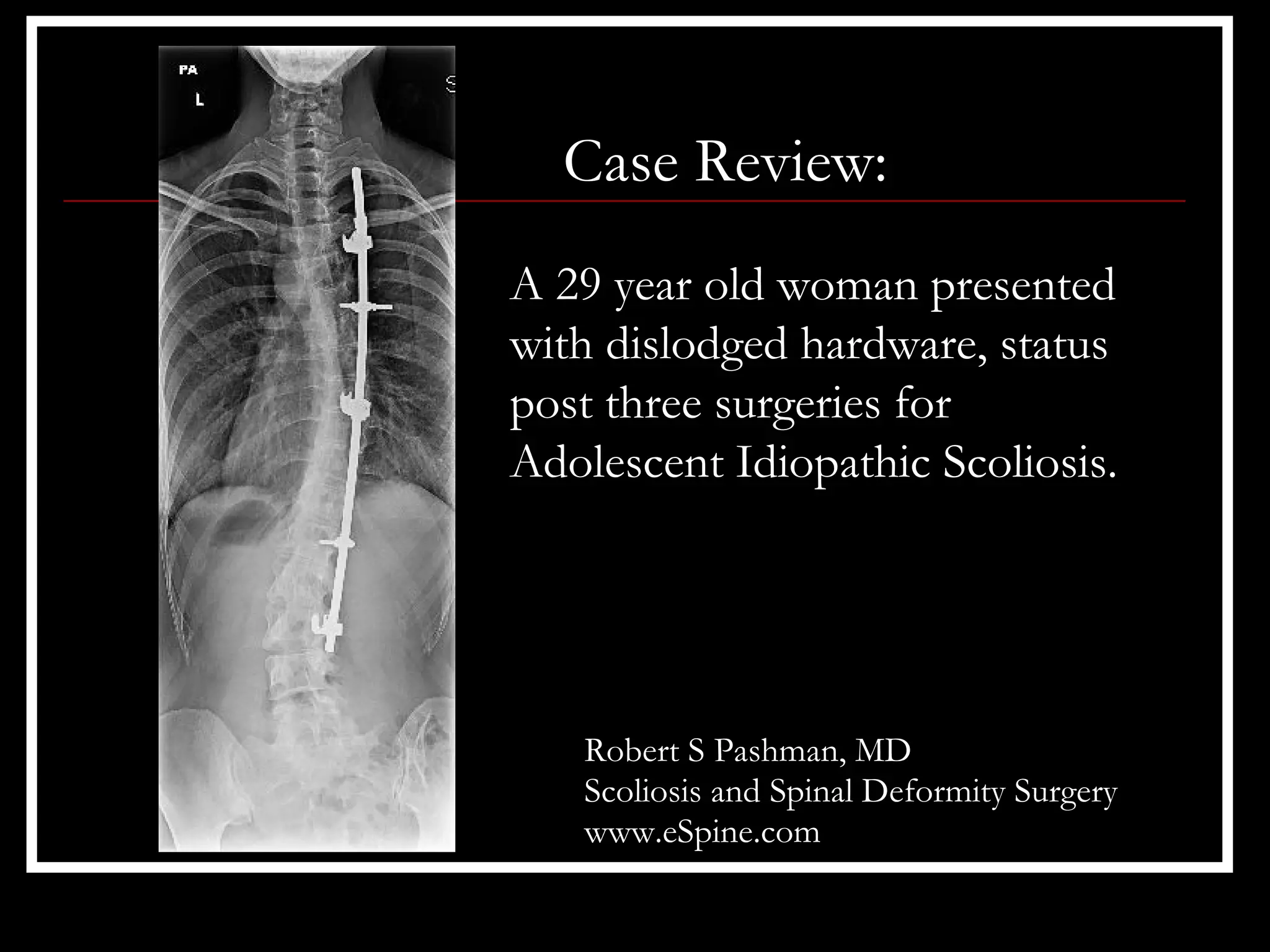
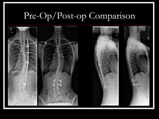
Case Review:
A 29 year old woman presented
with dislodged hardware, status
post three surgeries for
Adolescent Idiopathic Scoliosis.


   Robert S Pashman, MD
   Scoliosis and Spinal Deformity Surgery
   www.eSpine.com
Patient History
• 29 year old female
• The patient is status post 3 operations from the age of 3 to 15.
• This was done in Estonia and then ultimately in Finland.
• She had a posterior spinal fusion for adolescent idiopathic
  scoliosis that ultimately got infected, was redone, reinstrumented
  and half the instrumentation was ultimately removed.
• The patient has noted increase of pain and deformity with rib
  prominence as rotation has increased. The painful apical
  segments over the ribs are associated also with some low back
  pain.
Pre-op X-rays
   The retained hardware has disassociated
   from her spinal column is loose
   underneath her. I told the patient that
   this loose instrumentation is dangerous
   to her. That is, the pedicle hooks
   proximately are not connected to the
   spine and may be very close to the rib
   cage and her pleura, and it would be not
   be implausible that any sort of trauma to
   her can cause dislodging of the
   instrumentation and become a serious
   medical problem.
Indications for Surgery
1. Status post posterior instrumented fusion for adolescent
   idiopathic scoliosis x3.
2. Removal of hardware with infected wound.
3. Removal of half instrumentation, status post multiple operations
   for adolescent idiopathic scoliosis.
4. Now with retained hardware which is loose.
5. Rule out pseudoarthritis, thoracolumbar spine.
6. Rib prominence with pain over hemithorax, right hand side,
   apical thoracic curve.
7. Increasing low back pain and disability due to possible
   pseudoarthrosis, lumbar spine.
Surgical Strategy
• Removal of retained hardware, Cotrel-Dubousset instrumentation
  through osteotomy site, thoracolumbar spine.
• Multiple level spinal osteotomy for removal of hardware and
  regraft of pseudoarthrosis, T4-5, T5-6, T6-7, T12-L1, L1-L2 and
  L2-L3.
• Posterior spinal fusion regraft, L1 to L3.
• Segmental spinal instrumentation, L2-3, using pedicle screw/rod
  construct for through-and-through pseudoarthrosis, L2-3.
• Posterior spinal fusion using locally-harvested autologous bone
  graft and rib graft, L2-3.
Surgical Strategy – cont.
• Thoracoplasty with removal of 6 ribs over apical segment and
  thoracotomy, thoracic apex spine.
• Plastic closure of wound.
• O-arm neuro-navigation/interpretation with CT interpretation.
• Intraoperative somatosensory-evoked potential and motor-
  evoked potential interpretation
Post-op Films
      • The patient did well
        immediately post-operative.
      • She reported minimal pain.
      • Postoperative x-rays show
        excellent balance of the frontal
        sagittal plane with
        instrumentation intact.
Pre-Op/Post-op Comparison

Case Review #50: 29 year old woman presents with dislodged instrumentation following three scoliosis surgeries

  • 1.
    Case Review: A 29year old woman presented with dislodged hardware, status post three surgeries for Adolescent Idiopathic Scoliosis. Robert S Pashman, MD Scoliosis and Spinal Deformity Surgery www.eSpine.com
  • 2.
    Patient History • 29year old female • The patient is status post 3 operations from the age of 3 to 15. • This was done in Estonia and then ultimately in Finland. • She had a posterior spinal fusion for adolescent idiopathic scoliosis that ultimately got infected, was redone, reinstrumented and half the instrumentation was ultimately removed. • The patient has noted increase of pain and deformity with rib prominence as rotation has increased. The painful apical segments over the ribs are associated also with some low back pain.
  • 3.
    Pre-op X-rays The retained hardware has disassociated from her spinal column is loose underneath her. I told the patient that this loose instrumentation is dangerous to her. That is, the pedicle hooks proximately are not connected to the spine and may be very close to the rib cage and her pleura, and it would be not be implausible that any sort of trauma to her can cause dislodging of the instrumentation and become a serious medical problem.
  • 4.
    Indications for Surgery 1.Status post posterior instrumented fusion for adolescent idiopathic scoliosis x3. 2. Removal of hardware with infected wound. 3. Removal of half instrumentation, status post multiple operations for adolescent idiopathic scoliosis. 4. Now with retained hardware which is loose. 5. Rule out pseudoarthritis, thoracolumbar spine. 6. Rib prominence with pain over hemithorax, right hand side, apical thoracic curve. 7. Increasing low back pain and disability due to possible pseudoarthrosis, lumbar spine.
  • 5.
    Surgical Strategy • Removalof retained hardware, Cotrel-Dubousset instrumentation through osteotomy site, thoracolumbar spine. • Multiple level spinal osteotomy for removal of hardware and regraft of pseudoarthrosis, T4-5, T5-6, T6-7, T12-L1, L1-L2 and L2-L3. • Posterior spinal fusion regraft, L1 to L3. • Segmental spinal instrumentation, L2-3, using pedicle screw/rod construct for through-and-through pseudoarthrosis, L2-3. • Posterior spinal fusion using locally-harvested autologous bone graft and rib graft, L2-3.
  • 6.
    Surgical Strategy –cont. • Thoracoplasty with removal of 6 ribs over apical segment and thoracotomy, thoracic apex spine. • Plastic closure of wound. • O-arm neuro-navigation/interpretation with CT interpretation. • Intraoperative somatosensory-evoked potential and motor- evoked potential interpretation
  • 7.
    Post-op Films • The patient did well immediately post-operative. • She reported minimal pain. • Postoperative x-rays show excellent balance of the frontal sagittal plane with instrumentation intact.
  • 8.