A 14 year old female presented with Adolescent Idiopathic Scoliosis. The patient was non-compliant with bracing. The Scoliosis curvature and Kyphosis curvature progressed, and she required surgery.
Case Review #10: 12 year old girl with 70 degree Scoliosis Robert Pashman
A 12 year old girl presented with a 70 degree adolescent idiopathic scoliosis discovered during a routine school screening. Dr. Pashman performed a posterior spinal fusion on the patient.
Case Presentation #8: 14 year old female presented with Adolescent Idiopathic...Robert Pashman
A 14 year old girl presented with 38 degree Adolescent Idiopathic Scoliosis. The curve progressed within a few months, and she required scoliosis surgery. Dr. Pashman performed a posterior spinal fusion on her.
Case Review #15: 13 year old female with Profressive Adolescent Idiopathic Sc...Robert Pashman
A 13 year old female presented with thoracic and lumbar pain from Adolescent Idiopathic Scoliosis. The patient had a significant rib hump due to the spinal curvature. She was treated with a posterior spinal fusion.
Case Review #11: Progressive Adolescent Idiopathic ScoliosisRobert Pashman
A 17 year old female with Progressive Adolescent Idiopathic Scoliosis presented to Dr. Pashman for a surgical opinion. The patient presented with a 36° thoracic curve which progressed to 48°. Dr. Pashman treated the patient with a posterior fusion T3-L1.
Case Review #22: 21 year old with Progressive Adolescent Idiopathic ScoliosisRobert Pashman
A 21 year old female presented to Dr. Pashman with Progressive Adolescent Idiopathic Scoliosis. The patient had been followed for scoliosis, and was compliant wearing her brace. Her spinal curvature progressed despite physical therapy and bracing. Dr. Pashman treated her with a Posterior Spinal Fusion T3-L1.
Case Review #42: 39 year old female with Adult Congenital ScoliosisRobert Pashman
A 39 year old female with Congenital Scoliosis presented to Dr. Pashman. The patient had a fusion at age six, and her spinal curve continued to progress. Dr. Pashman treated the patient with a posterior spinal fusion from T3-Pelvis. KIM/SRP Classification 3.
Case Review #7: Progressive Adult Idiopathic Scoliosis with a 75 degree curva...Robert Pashman
The patient, a 19-year-old female, presented with a progressive 75° thoracic scoliosis that had increased 21° over four years and was causing her significant pain. Pre-operative images showed the curvature was rigid. The surgical plan was to perform segmental spinal instrumentation from T3 to L1, posterior spinal fusion from T3 to L2, and spinal osteotomies from T4 to T9 to correct the rigid curvature. Post-operatively, a 43° correction was achieved, reducing the curvature to 27°, and the patient was able to resume her normal activities with no restrictions after one year.
Case Review #6: 53 year old woman with Adult ScoliosisRobert Pashman
A 53 year old woman, with an 85° thoracic curve, and a 75° lumbar curve. Dr. Pashman treated her with an Anterior fusion followed by a Posterior Spinal Fusion from T1 to the Pelvis. Curve was a KIM/SRP Classification 3.
Case Review #23: 15 year old male with Adolescent Idiopathic ScoliosisScoliosisRobert Pashman
A very active 15 year old male presented with progressive Adolescent Idiopathic Scoliosis. His curve progressed after a recent growth spurt. The patient had scoliosis surgery, and returned to ROTC.
Case Presentation#56: Adult Idiopathic ScoliosisRobert Pashman
A 28 year old female with progressive Adult Idiopathic Scoliosis postponed surgery from age 17 to 28. The patient failed conservative therapy and decided to have surgery due to pain and curve progression.
Case Review #50: 29 year old woman presents with dislodged instrumentation fo...Robert Pashman
A 29-year old woman presented with dislodged hardware from three previous surgeries for adolescent idiopathic scoliosis. She had retained loose instrumentation from her past operations that posed a medical risk. The surgical strategy was to remove the retained hardware through osteotomies, perform spinal fusions from T4 to L3, and place new segmental instrumentation from L2 to L3.
Case Review #2: 41 year old female presented with Adult Scoliosis and Spodylo...Robert Pashman
A 41 year old female with a 50° thoracolumbar curve and Spondylolisthesis. Dr. Pashman treated the patient with an Posterior Spinal Fusion from T10-Pelvis. Her curve was a KIM/SRP Classification 2.
Case Review #13: 13 year old female softball player with Adolescent Idiopathi...Robert Pashman
A 13 year old female softball player presented with Adolescent Idiopathic Scoliosis. The degree of her scoliosis curve progressed to 48 degrees and she required a spinal fusion.
Case Review #24: A 15 Year Old Female with Adolescent Idiopathic ScoliosisRobert Pashman
Dr. Pashman followed the patient for three years. After a growth spurt, her scoliosis curvature progressed significantly, and she required scoliosis surgery.
Case Review #14: 16 year old female with progressive adolescent scoliosisRobert Pashman
A 16-year-old female presented with progressive adolescent idiopathic scoliosis. Her curve had increased from 40 degrees to 46 degrees over two years while wearing a brace. Pre-op x-rays showed a 46 degree thoracic curve. The indications for surgery were a progressive curve, pain, and deformity. The surgical strategy involved segmental spinal instrumentation from T2 to L1 using pedicle screws, multiple osteotomies from T5 to T10 including Smith-Peterson osteotomies, and posterior spinal fusion from T3 to L1. Post-op films showed the patient was well-balanced in the coronal and sagittal planes, and her symptoms resolved following surgery.
Case Review #27: 59 Year Old Female with Progressive Adult ScoliosisRobert Pashman
59 year old female presented with Progressive Adult Idiopathic Scoliosis, Spondylolisthesis, Flatback Deformity, and Stenosis. The patient was treated with a spinal fusion,
Case Review #9: Adult Idiopathic Scoliosis with a Double CurvatureRobert Pashman
A 54 year old female presented with Adult Idiopathic Scoliosis. In addition to lower back pain, she noticed that her height was decreasing. Her spine was significantly rotated and she required a spinal fusion.
Case Review #3: 65 year old woman with 55 degree Thoracolumbar ScoliosisRobert Pashman
A 65 year old female with a 55° thoracolumbar curve, spondylolisthesis, and flatback syndrome. Treated with an Anterior/Posterior Spinal Fusion. KIM/SRP Classification 3.
Case Review #26: 73 year old female with KyphoscoliosisRobert Pashman
73 year old female presented with Kyphoscoliosis. Dr, Pashman, treated the patient with a posterior spinal fusion from T2-Pelvis. KIM/SRP Classification 3.
Case Review #16: 59 year old woman with 25 year history of Adult Idiopathic S...Robert Pashman
59 year old female presented with 25 year history of scoliosis progression. 68° thoracic curve, 42° lumbar curve. Dr. Pashman treated her with a fusion T3-L4. KIM/SRP Classification 1.
Case Review #6: 45 year old woman with flatback syndrome after several surger...Robert Pashman
A 45-year old female with a flat back due to multiple spine surgeries for scoliosis underwent surgery to correct her sagittal imbalance. The surgical strategy included a T10 to sacrum fusion with pedicle subtraction osteotomy at L3 to induce lordosis. Post-operatively, sagittal balance was restored but a slight coronal imbalance remained due to prior fusion in a shifted position. Good functional outcomes are expected with less than 4cm of coronal imbalance.
Case Review #43: 43 year old female with Adult Idiopathic Scoliosi requiring...Robert Pashman
1. A 43-year old female with adult idiopathic scoliosis underwent anterior spinal surgery with instrumentation from T12 to L3 to correct curves of 60° thoracic and 57° lumbar, reducing them to 41°.
2. A 48-year old female with Kim/SRP type III scoliosis underwent revision surgery with anterior lumbar interbody fusion from L4-S1 and posterior spinal fusion from T3 to S1 to correct increasing proximal thoracic curvature and subadjacent degeneration.
3. Post-operatively, the patient was well-balanced in sagittal and coronal planes with thoracic and lumbar curves reduced to 26° and 27° respectively.
Case Review #9: 19 year old female with Scheuermann's DiseaseRobert Pashman
A 19 year old female from Las Vegas traveled to Los Angeles for treatment. She presented with progressive Scheurmann's Kyphosis. The patient is an equestrian rider and model. Dr. Pashman treated the patient with a Posterior Spinal Fusion from T3 to L1. She returned to modeling a few months post-op, and returned to horseback riding at 6 months post-op.
Case Review #7: 51 year old female with severe flatback after multiple surge...Robert Pashman
A 51 year old female status post multiple spine surgeries presented to Dr. Pashman with severe Flatback Syndrome and psueoarthrosis. Dr. Pashman treated the patient with a Posterior Spinal Fusion T4-Pelvis.
Case Review #D: 16 year old female with Adolescent Idiopathic ScoliosisRobert Pashman
A 16 year old female with Adolescent Idiopathic Scoliosis. Her curve progressed to 50° despite bracing. Dr. Pashman treated her with an Anterior Interbody fusion from T11 to L3.
Case Review #2: 66 year old female with severe Flatback SyndromeRobert Pashman
A 66 year old female presented with severe Flat back Syndrome, Kyphosis, and critical stenosis. Dr. Pashman treated the patient with a posterior spinal fusion T2 to the pelvis. The patient was able to stand up straight following surgery.
Case Review #5: 67 year old woman with flatback syndrome following 5 spinal s...Robert Pashman
A 67 year old female presented to Dr. Pashman with severe Flat back Syndrome after 5 previous spine surgeries for Adult Idiopathic Scoliosis. Dr. Pashman treated her with a Posterior Spinal Fusion from T8 to S1.
This 13-year-old schoolgirl is complaining that her friends say her right shoulder is higher and she has occasional low back pain during physical activity at school. A physical examination should check for deformities like a rib hump or asymmetrical hips, a tilted waistline, or prominent scapula. Differential diagnoses could include idiopathic or congenital scoliosis, hyperkyphosis, or leg length discrepancy. The progression of the condition depends on the curve magnitude and the patient's skeletal maturity - smaller curves that develop later are less likely to worsen. Management options include observation, bracing, or surgery depending on the severity and progression of any scoliotic curve.
This document discusses anterior cervical instrumentation for cervical fusion procedures. It covers indications for instrumentation including corpectomy, trauma, deformity correction, and high-risk patients. The operative technique is described including exposure, discectomy/corpectomy, grafting, plate fixation and closure. Post-operative management including bracing and follow-up are also outlined. Potential complications are listed. Various cervical plating systems and technologies to aid surgery are also briefly mentioned.
The document presents on a competitive position matrix (CPM) analysis of three IT companies in Nepal: Business Intelligence Academy PVT. LTD (BIA), Upveda Technology PVT. LTD, and Picovico PVT. LTD. It provides an overview of the companies and their products/services. A CPM is used to analyze the companies' strengths and weaknesses against strategic factors. The analysis finds that Picovico is performing better in the market than the other two companies due to factors like product quality, global expansion, and management. It recommends that BIA focus on price competitiveness, global expansion, and advertisement.
The document describes a case study of a 33-year old professional baseball player who sustained a grade 1 ankle sprain. He was treated with a class IV therapeutic laser, Polymem bandage, open basket weave tape, and ice. This new treatment approach reduced pain and swelling, allowing the player to return to play in just 2 days, much faster than the typical recovery time of 1 week. While these new modalities seemed effective in this case, more research is needed to validate their use for ankle sprains.
Clinical research is quite vital in the field of physiotherapy. Physiotherapists depends on information from researches to enhance the knowledge they have already gained through their university education and with continuous education courses.
This document discusses methods for assessing cardiorespiratory endurance through measurement of maximal oxygen uptake (VO2 max). It describes both submaximal and maximal exercise testing protocols using equipment like bikes, treadmills, and step tests to determine VO2 max either directly through gas analysis or indirectly through heart rate monitoring. Key indicators for assessing cardiorespiratory fitness include VO2 max, respiratory exchange ratio, workload, and heart rate response to exercise.
The document discusses various surgical techniques for cervical spine stabilization at C1-C2. It describes the anatomy of the atlantoaxial joint and its range of motion. Various fixation techniques are covered, including Magerl transarticular screws, C1 lateral mass screws, C2 pars screws, C2 pedicle screws, and C2 laminar screws. Entry points and trajectories for each technique are detailed. Potential risks and variations are also noted. The importance of preoperative imaging to evaluate anatomical variations is emphasized.
This powerpoint was prepared to be presented at University of Health Sciences Cambodia for the Neurosurgery , Medicine and Psychiatry Residents, by shaweta khosa
Cervical spondylosis is a disorder caused by the degeneration of discs between vertebrae, causing bones to move out of position and pressure nerves. Exercises include slow motion exercises turning and tilting the head in different directions, as well as isometric exercises pressing against parts of the head without moving. A cervical collar or neck traction may help restrict movement and reduce pain. Surgery can decompress the spinal cord if symptoms are severe.
The effectiveness of remedial massage therapy in dealing with muscular issues...Nicholaslaverty
This document summarizes a study on the effectiveness of remedial massage therapy for dealing with muscular issues in active adults. The study surveyed 47 active individuals who received remedial massage at least twice previously. Results showed remedial massage was used predominantly when needed rather than as part of regular training, and was rated most effective at increasing range of motion. Massage was often used in combination with physiotherapy and aids like stretching. Further research is still needed to verify all benefits and applications of massage therapy.
The document discusses posterior spine fixation using pedicle screw instrumentation. It provides details on the anatomy of the posterior spine elements and pedicles. Pedicle screw fixation provides a rigid construct that can increase fusion rates and reduce pain. Precise screw placement is important to avoid neurological or vascular injury. Proper preoperative planning and technique are essential to achieve a stable construct and reduce complications.
1. Physiotherapy aims to maintain and restore maximum movement and functional ability throughout a person's lifespan by providing services to address movement issues arising from aging, injury, disease or other factors.
2. Physiotherapists work in a variety of settings and have many specialties. They assess patients, determine treatment plans, and reassess progress using manual therapy techniques, therapeutic exercises, electrotherapy modalities and more.
3. Physiotherapy education programs provide students with training in anatomy, physiology, movement science and other basic sciences as well as hands-on skills and clinical experience treating patients in different areas of specialization.
The document discusses the anatomy and surgical treatment of the spine. It describes the typical divisions of the spinal column, common curves seen, vertebral anatomy including facets and ligaments, and pedicle anatomy. Surgical techniques like pedicle screw fixation are covered including entry points, trajectories, and complications. Degenerative spinal conditions and their treatment with options like bracing, decompression surgery, and stabilization are also summarized.
The document discusses pedicle screw fixation of the thoracolumbar spine. It begins with learning objectives about spinal anatomy, pedicle screws, and techniques for fixation. It then describes the anatomy of thoracic and lumbar vertebrae, with specifics on pedicle size and orientation. Entry points and techniques for screw placement are outlined for the thoracic and lumbar spine, including the intersection technique. Considerations for upper thoracic fixation and avoiding complications are also covered. The document concludes with pearls for achieving good fixation.
The document provides an overview of spinal anatomy including:
- The 7 cervical, 12 thoracic, 5 lumbar vertebrae and sacrum/coccyx bones that make up the spine.
- Key structures like the anterior/posterior columns that provide compression/tension resistance.
- Facet joints that resist rotation and displacement.
- Important anatomical features of each region like the cervical facet orientation and thoracic transverse processes.
- Neural structures like the spinal cord, nerves and nerve roots.
- Key concepts like clinical instability and relationships between structures.
A 70-year-old female presented with bilateral knee pain diagnosed as osteoarthritis. She received four weeks of physical therapy involving exercises to increase strength and flexibility in the knees and hips. The therapy resulted in decreased pain, improved range of motion and strength. While progress was made, the patient required additional therapy to further improve functional strength.
Thoraco lumbar injuries can be categorized based on which spinal columns are affected. Injuries involving the middle column and at least one other column are considered unstable. Burst fractures involve failure of the anterior and middle columns and may require early stabilization, especially if they involve over 50% canal compromise, over 20 degrees of kyphosis, or over 45-50% canal compromise. Flexion distraction injuries can be categorized into types A through D depending on whether they involve bone or ligaments at one or two spinal levels.
This document provides an overview of the anatomy of the spinal cord including its structure, blood supply, sensory and motor systems, and approach to spinal cord diseases. Key points include:
- The spinal cord has grey matter containing nuclei and white matter containing tracts that transmit sensory and motor signals.
- It receives blood supply from the anterior and posterior spinal arteries as well as segmental radicular arteries like the artery of Adamkiewicz.
- Sensory systems assess proprioception, pain, temperature and touch. Motor systems involve upper and lower motor neurons controlling voluntary movement.
- Clinical examination assesses sensory levels and motor function to localize spinal cord lesions, while investigations include CSF analysis, imaging
The document discusses spinal and spinal cord injuries, including incidence, morbidity and mortality, anatomy, assessment, types of spinal cord injuries, management, and non-traumatic conditions. It provides details on the anatomy of the spine and spinal cord, mechanisms of spinal cord injury, approaches to assessing and managing spinal injuries, and specific spinal cord syndromes. Prevention, immobilization, and avoiding further injury to the spinal cord are the primary goals in managing spinal and spinal cord trauma."
The document describes the anatomy and structure of the spinal cord. It details the external structure including its cylindrical shape and location within the vertebral canal surrounded by meninges. Internally, it describes the gray and white matter, including the ascending and descending tracts that transmit sensory and motor signals. It also discusses the blood supply, clinical correlations, and applications related to injuries and diseases of the spinal cord.
Case Review #8: 44 year old woman presented with a Double Major Scoliosis Cur...Robert Pashman
A 44 year old female presented with Adult Idiopathic Scoliosis with a double major curve. Dr. Pashman treated the patient with a Posterior Spinal Fusion T3-L4. KIM/SRP Classification 1.
Case Review #25: 39 year old female with Progressive Adult ScoliosisRobert Pashman
39 year old female presented with Progressive Adult Idiopathic Scoliosis. Dr. Pashman treated the patient with a posterior spinal fusion from T3-L4. KIM/SRP Classification 1
Case Review #22: 50 year old female presented with Scoliosis and spondylolis...Robert Pashman
50 year old female presented with Adult Idiopathic Scoliosis and Spondylolisthesis. She suffered from low back pain and leg pain. Dr. Pashman performed a spinal fusion on the patient.
Case Review #21: Triple Curvature Adult Idiopathic ScoliosisRobert Pashman
A 23 year old male presented with a triple adult idiopathic scoliosis curvature. The curvature progressed despite bracing. Dr. Pashman performed a spinal fusion from T4-L3.
Case Review #4: Adolescent Idiopathic Scoliosis with 61 degree curvatureRobert Pashman
A 22 year old female presented with Adolescent Idiopathic Scoliosis. She was braced as a child, and the curve continued to progress until she required surgical intervention.
Case Review #1: 39 year old female with Congenital Scoliosis Robert Pashman
A 39-year-old female with progressive congenital scoliosis underwent spinal fusion from T3 to the pelvis. She had a previous fusion at age 6 but her curvature had worsened. The surgery included multiple osteotomies and instrumentation to correct her frontal and sagittal plane deformities. Post-operatively, her curvature was reduced from 31 to 18 degrees and she had improved balance and pain relief.
Case Review #C: Adolescent Idiopathic ScoliosisRobert Pashman
A 14 year old female presented with Adolescent Idiopathic Scoliosis with a Thoracolumbar Curve. She was treated with an anterior throaco-abdominal approach.
Case Review #35: 43 year old female with Adult Scoliosis and a Transitional V...Robert Pashman
1. This case review summarizes the treatment of a 43-year-old female patient with adult scoliosis and a lumbar sacral transitional vertebra.
2. She had a 40 degree lumbar curve and a fused L5-S1 region on the right side.
3. Her treatment plan involved an anterior interbody fusion at L4-5 and L5-S1 followed by posterior segmental spinal instrumentation, spinal osteotomies, and posterior spinal fusion to correct her deformities and relieve her pain.
Case Review #3: 17 year old male with Scheurmann's KyphosisRobert Pashman
A 17 year old male presented with progressive Schuermann's Kyphosis. The patient had a 75° spinal curvature. Dr. Pashman treated the patient with a posterior fusion from T3 to L1.
Case Review 15: Adult Scoliosis treated with Spinal Fusion and OteotomiesRobert Pashman
A 50 year old female presented with progressive Adult Idiopathic Scoliosis. The patient had severe low back pain and leg pain. She was treated with a posterior spinal fusion with spinal osteotomies.
This case report describes a 61-year-old woman who presented with neck pain, headaches, and a hunched posture known as Dowager's hump. Chiropractic treatment over 9 months, including spinal manipulation, traction, and massage, improved her symptoms and corrected her spinal alignment as seen on x-rays. Her neck pain resolved, headaches stopped, and the hump was reduced in size. Chiropractic care successfully treated this patient's hyperkyphosis and cervicogenic headache through non-invasive techniques that realigned the spine.
Case Review #46: 56 year old female with Adult Idiopathic Scoliosis and Burst...Robert Pashman
56 year old female with Idiopathic Scoliosis, status post burst fracture, presented with junctional kyphosis. Dr. Pashman treated the patient with a posterior spinal fusion from T2-pelvis. Spinal curvature was a KIM/SRP Classification 3.
Case Review #17: 63 year old female with Denovo ScoliosisRobert Pashman
63 year old female with Adult Idiopathic Scoliosis, Spondylolisthesis, and facet screws at L4-L5. Dr. Pashman treated the patient with a posterior spinal fusion from T10 to Pelvis. KIM/SRP Classification 2
Case Review #11: Adult Idiopathic ScoliosisRobert Pashman
A 60 year old male presented with a 50+ degree curvature. He was status post lumbar fusion from L4-S1, and continued to have significant low back pain.
Case Review #18: 79 year old female with Degenerative scoliosisRobert Pashman
1) A 79-year-old female with adult degenerative scoliosis, herniated discs, and spondylolisthesis underwent spinal fusion surgery.
2) The surgery involved segmental spinal instrumentation from L1 to the pelvis, a pedicle subtraction osteotomy at L3 to create lordosis, and posterior spinal fusion from L1 to the pelvis.
3) Post-operatively, the patient's sagittal balance was normalized, with 5 degrees of lordosis achieved compared to 20 degrees of kyphosis pre-operatively, improving her ability to walk with less pain.
Similar to Case Review #12: 14 Year Old Female with Adolescent Idiopathic Scoliosis (20)
Case Review #34: 44 Year Old Woman with Adult Idiopathic ScoliosisRobert Pashman
A 44 year old woman presented with Adult Idiopathic Scoliosis. She wore a brace as a child, and the curvature progressed to the point she required surgery.
Case Review #B: Spondylolisthesis Surgery Robert Pashman
A 16-year-old female presented with lower back and left leg pain following a motor vehicle accident. Imaging showed spondylolisthesis at L5 from fractures of the pars interarticularis. She underwent microscopic bilateral L5 laminotomy, foraminotomy, and repair of the L5 pars fractures with bone grafting and instrumentation. Post-operatively, her symptoms resolved and she was able to return to normal activities.
Case Review #2: Isthmic Spondylolisthesis Grade IVRobert Pashman
Case review: A 58 year old female with Isthmic Spondylolisthesis status post surgery. She continued to experience back pain and leg pain, and was treated with spinal surgery.
Case Review #A: Major League Baseball Player has a Spinal FusionRobert Pashman
25 year old Major League Baseball Player presented with low back pain. Upon review of CT scans, he was discovered to have a pars fracture and require surgery,
Case Review #31: 60 Year Old Female with Adult Idiopathic ScoliosisRobert Pashman
60 year old female presented with Adult Idiopathic Scoliosis and Grade 2 Isthmic Spondylolisthesis. She was treated with an anterior and posterior spinal fusion.
Case Review #19: 40 year old Male with Adult Idiopathic Scoliosis with Flatba...Robert Pashman
A 40 year old male presented after scoliosis surgery at age 14. He presented with Flatback Syndrome and increasing low back pain and required revision surgery.
Case Review #28: Patient with Lumbar Scoliosis status post surgery with Harr...Robert Pashman
A 37 year old female presented status post posterior spinal fusion for Adolescent Idiopathic Scoliosis. She had Harrington Rods and the lumbar curve continued to progress, requiring surgical intervention.
Case Review #G: 15 year old male with Adolescent Scoliosis Robert Pashman
A 15 year old male with Adolescent Idiopathic Scoliosis and aspirations to become a professional golfer presented for treatment. After careful consideration, a surgical plan was devised to maximize his ability to pursue golf professionally.
Local Advanced Lung Cancer: Artificial Intelligence, Synergetics, Complex Sys...Oleg Kshivets
Overall life span (LS) was 1671.7±1721.6 days and cumulative 5YS reached 62.4%, 10 years – 50.4%, 20 years – 44.6%. 94 LCP lived more than 5 years without cancer (LS=2958.6±1723.6 days), 22 – more than 10 years (LS=5571±1841.8 days). 67 LCP died because of LC (LS=471.9±344 days). AT significantly improved 5YS (68% vs. 53.7%) (P=0.028 by log-rank test). Cox modeling displayed that 5YS of LCP significantly depended on: N0-N12, T3-4, blood cell circuit, cell ratio factors (ratio between cancer cells-CC and blood cells subpopulations), LC cell dynamics, recalcification time, heparin tolerance, prothrombin index, protein, AT, procedure type (P=0.000-0.031). Neural networks, genetic algorithm selection and bootstrap simulation revealed relationships between 5YS and N0-12 (rank=1), thrombocytes/CC (rank=2), segmented neutrophils/CC (3), eosinophils/CC (4), erythrocytes/CC (5), healthy cells/CC (6), lymphocytes/CC (7), stick neutrophils/CC (8), leucocytes/CC (9), monocytes/CC (10). Correct prediction of 5YS was 100% by neural networks computing (error=0.000; area under ROC curve=1.0).
Travel vaccination in Manchester offers comprehensive immunization services for individuals planning international trips. Expert healthcare providers administer vaccines tailored to your destination, ensuring you stay protected against various diseases. Conveniently located clinics and flexible appointment options make it easy to get the necessary shots before your journey. Stay healthy and travel with confidence by getting vaccinated in Manchester. Visit us: www.nxhealthcare.co.uk
Adhd Medication Shortage Uk - trinexpharmacy.comreignlana06
The UK is currently facing a Adhd Medication Shortage Uk, which has left many patients and their families grappling with uncertainty and frustration. ADHD, or Attention Deficit Hyperactivity Disorder, is a chronic condition that requires consistent medication to manage effectively. This shortage has highlighted the critical role these medications play in the daily lives of those affected by ADHD. Contact : +1 (747) 209 – 3649 E-mail : sales@trinexpharmacy.com
share - Lions, tigers, AI and health misinformation, oh my!.pptxTina Purnat
• Pitfalls and pivots needed to use AI effectively in public health
• Evidence-based strategies to address health misinformation effectively
• Building trust with communities online and offline
• Equipping health professionals to address questions, concerns and health misinformation
• Assessing risk and mitigating harm from adverse health narratives in communities, health workforce and health system
Cell Therapy Expansion and Challenges in Autoimmune DiseaseHealth Advances
There is increasing confidence that cell therapies will soon play a role in the treatment of autoimmune disorders, but the extent of this impact remains to be seen. Early readouts on autologous CAR-Ts in lupus are encouraging, but manufacturing and cost limitations are likely to restrict access to highly refractory patients. Allogeneic CAR-Ts have the potential to broaden access to earlier lines of treatment due to their inherent cost benefits, however they will need to demonstrate comparable or improved efficacy to established modalities.
In addition to infrastructure and capacity constraints, CAR-Ts face a very different risk-benefit dynamic in autoimmune compared to oncology, highlighting the need for tolerable therapies with low adverse event risk. CAR-NK and Treg-based therapies are also being developed in certain autoimmune disorders and may demonstrate favorable safety profiles. Several novel non-cell therapies such as bispecific antibodies, nanobodies, and RNAi drugs, may also offer future alternative competitive solutions with variable value propositions.
Widespread adoption of cell therapies will not only require strong efficacy and safety data, but also adapted pricing and access strategies. At oncology-based price points, CAR-Ts are unlikely to achieve broad market access in autoimmune disorders, with eligible patient populations that are potentially orders of magnitude greater than the number of currently addressable cancer patients. Developers have made strides towards reducing cell therapy COGS while improving manufacturing efficiency, but payors will inevitably restrict access until more sustainable pricing is achieved.
Despite these headwinds, industry leaders and investors remain confident that cell therapies are poised to address significant unmet need in patients suffering from autoimmune disorders. However, the extent of this impact on the treatment landscape remains to be seen, as the industry rapidly approaches an inflection point.
Hiranandani Hospital in Powai, Mumbai, is a premier healthcare institution that has been serving the community with exceptional medical care since its establishment. As a part of the renowned Hiranandani Group, the hospital is committed to delivering world-class healthcare services across a wide range of specialties, including kidney transplantation. With its state-of-the-art facilities, advanced medical technology, and a team of highly skilled healthcare professionals, Hiranandani Hospital has earned a reputation as a trusted name in the healthcare industry. The hospital's patient-centric approach, coupled with its focus on innovation and excellence, ensures that patients receive the highest standard of care in a compassionate and supportive environment.
Case Review #12: 14 Year Old Female with Adolescent Idiopathic Scoliosis
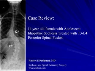
1. Case Review:
14 year old female with Adolescent
42°
Idiopathic Scoliosis Treated with T3-L4
Posterior Spinal Fusion
47°
Robert S Pashman, MD
Scoliosis and Spinal Deformity Surgery
www.eSpine.com
2. Patient History
14-year-old female
Adolescent Idiopathic Scoliosis
Type 6 curve
Patient was braced, and was noncompliant
The past medical history is contributory for renal surgery as a child for
hydronephrosis, but there are no other indications of relevant medical history as
to non-idiopathic causes for scoliosis.
Left thoracic rib hump, right lumbar fullness with some element of
hyperkyphosis in the thoracic spine.
Neurologically, she is intact.
An MRI had been obtained does not show any significant segmentation
development, defects in the cervical spine or problems and no significant non-
idiopathic causes of scoliosis, such as a spinal cord tumor compression or non-
segmentation in the thoracic spine.
3. Pre-op X-rays
On 36-x-14 x-rays, she has a structural 42° left
thoracic curve and a 47° highly rotated
thoracolumbar curve also.
On sagittal x-ray, the patient has a 57° thoracic
kyphosis, with compensatory hyperlordosis of
the lumbar spine. No spondylolisthesis is seen.
Spina bifida occulta has been noted and the
42° patient is balanced in the frontal and sagittal
plane.
She has an element of a depressed right shoulder
47° also. She is a Risser 4. The x-rays indicate to me
that this is a type 6 C positive curve, with
element of hyperkyphosis of the thoracic spine,
correlating also with a thoracolumbar structural
curve and a structural compensatory high
thoracic curve also.
4. Bending X-rays
L R
Right and left side bending
x-rays indicate mobility to
the distal lumbar curve, so
the instrumentation levels
would be set more
proximally into the curve.
5. Indications for Surgery
Progressive adolescent idiopathic scoliosis, type 6.
Rigid structural curves in excess, 57° thoracolumbar, 47° thoracic, with
significant rotation.
Cosmetic deformity due to massive left rib hump and flank fullness.
Failed conservative therapy.
Kyphoscoliosis.
6. Surgical Strategy
1. Segmental spinal instrumentation, thoracic 3 to lumbar 4, using 5.5
stainless steel screw-rod construct.
2. Multiple-level spinal osteotomy for mobilization of rigid thoracic and
lumbar curve, T5 to T11 and T12 to L3.
3. Interlaminar decompression with takedown of mesial facet in midline for
decompression of nerves and mobilization of lumbar curve, T12 to L4.
4. Posterior spinal fusion using a combination of harvested autogenous bone
and left thoracoplasty rib bone, T3 to L4.
5. Takedown of chest wall multiple-level rib osteotomies, 5 levels, and
thoracoplasty for removal of rib hump.
6. Repair of pleura incidental pleurotomy.
7. Intraoperative somatosensory evoked potentials and motor evoked
potentials.
8. Intraoperative fluoroscopy.
7. Post-Op Films
15°
15°
The patient has done quite well.
Incision is well-healed. She is very
happy with the outcome of her
surgery and the cosmetic results.
8. Pre-Op/Post-op Comparison
42° 15°
47° 15°
The patient's x- rays show an
excellent correction. The curve
was reduced 32°, her shoulders
and hips are even, and she is well
balanced in the saggital plane.