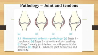
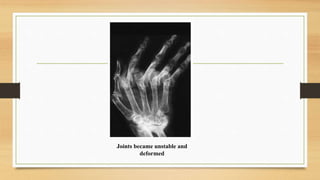
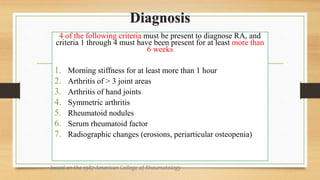
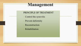
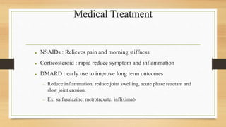
RHEUMATOID
ARTHRITIS
Definition
Chronic systemic autoimmune disease influenced by combination
of genetic, environmental and hormonal factors
 hyperproliferation and inflammation of synovium - pannus
 increased production of synovial fluid - swelling
 irritation of local nerve endings - pain
 erosion of cartilage and bone - joint deformity
Causes
1. Genetic susceptibility
• HLA-DR4
2. Abnormal Immunological reaction
• Production of anti-IgG autoantibodies which detected as rheumatoid factor
in blood
3. Inflammatory reaction in joints and tendon sheaths
• Cytokines (TNF, IL-1,IL-2)  activate inflammatory cells
Pathology – Joint and tendons
 Stage 1: pre-clinical
− Raised ESR, CRP, and RF
 Stage 2: synovitis
 Synovial membrane becomes inflamed and thickened, giving
rise to cell-rich effusion.
 Painful and swollen but tendon and joint still intact.
 Reversible condition.
 Stage 3: destruction
 Persistent inflammation cause tissues destruction.
 Articular cartilage is eroded and tendon fiber may rupture.
 Stage 4: deformity
 Combination of articular destruction, capsular stretching and
tendon rupture leads to progressive instability and deformity
of joint.
Pathology – extra-articular tissue
• Rheumatoid nodule
• Lymphadenopathy
• Vasculitis
• Muscle weakness
• Visceral disease
Clinical features
• Insidious onset
• Polysynovitis, with soft tissue swelling and stiffness
Early stage:
 c/o pain, swelling and stiffness in proximal joints of fingers
 Later, disease ‘spread’ to elbow, shoulder, knee, ankle and feet
 Generalized stiffness after periods of inactivity (early morning stiffnes) >
30 minutes
Symmetrical swelling and
tenderness of
metacarpophalangeal,
interphalangeal joints and
the wrist
Subcutaneous nodule felt
over olecranon process.
Late Stage :
• joint instability and tendon ruptures produces typical
‘rheumatoid deformities:
Ulnar deviation of the
fingers
Valgus knees
Clawed toes and
painful callosities
under metatarsal heads
• Joint movements are restricted and often very painful
• Muscle wasting
• 1/3 RA : painful and stiffness in cervical spine
• Long standing case : may be vasculitis and peripheral
neuropathy
Investigation
• Blood investigation
• Normocytic, hypocromic anaemia
• ESR & CRP raised
• Serology test of Rheumatoid factor positive
• Anti-cyclic citrullinated peptide antibodies (anti-CCP)
Imaging (radiology)
Soft tissue swelling and
peri-articular osteoporosis Articular erosion
Joints became unstable and
deformed
Diagnosis
4 of the following criteria must be present to diagnose RA, and
criteria 1 through 4 must have been present for at least more than
6 weeks
1. Morning stiffness for at least more than 1 hour
2. Arthritis of > 3 joint areas
3. Arthritis of hand joints
4. Symmetric arthritis
5. Rheumatoid nodules
6. Serum rheumatoid factor
7. Radiographic changes (erosions, periarticular osteopenia)
based on the 1987 American College of Rheumatology
Management
PRINCIPLE OF TREATMENT
 Control the synovitis
 Prevent deformity
 Reconstruction
 Rehabilitation
Medical Treatment
 NSAIDs : Relieves pain and morning stiffness
 Corticosteroid : rapid reduce symptom and inflammation
 DMARD : early use to improve long term outcomes
 Reduce inflammation, reduce joint swelling, acute phase reactant and
slow joint erosion.
 Ex: salfasalazine, metrotrexate, infliximab
PHYSIOTHERAPY AND OCCUPATIONAL THERAPY
• preventive splinting and orthotic device may delay
Surgical treatment
• soft-tissue procedures: synovectomy, tendon repair or
replacement and joint stabilization
• late rheumatoid disease (5-20 years):
• severe joint destruction fixed deformity and loss of function
indication for recontructive surgery
• arthrodesis, osteotomy, arthroplasty
Complication
 Infection
 Long standing corticosteroid therapy more susceptible to infection
 Sudden deterioration or increase pain in single joint -->suspected
septic arthritis
 Tendon Rupture
 Due to nodular infiltration
 Joint rupture
 Joint lining rupture and synovial content spill into soft tissues
 Secondary OA
 Articular cartilage erosion lead to joint damage.
DEFINITION
• Chronic joint disorder
• Progressive softening and disintegration of articular cartilage
• Accompanied by new growth of cartilage and bone at the joint margins
(osteophytes) and capsular fibrosis
• PRIMARY:
no obvious causes
 aging  lead to progressive
disintegration of articular
cartilage (wear and tear)
Generally more common in ♀,
but if age< 45y/o common in
♂ while if age>45 y/o common
in ♀
• SECONDARY :
 d/t underlying dx or problem
 Causes:
• Increased mechanical stress:
>Injury / trauma
>Morbid obesity
• Weakening of articular cartilage :
> Defect in type II collagen
>Other inflammatory dx
(eg: Rheumatoid Arthritis)
• Inactivity
• Genetics ( eg: defect in type II
collagen)
• Occupation
CAUSES/ RISK
FACTORS OF OA
Spine (cervical and lumbar)
Hip
Knee
Fingers
Toes
Subarticular bone react, cyst form in greatest stress area, trabeculae
become thickened and sclerotic
Cartilage break down, exposed underlying bone (friction cause patient felt
pain)
Imbalance btw stress applied and ability of cartilage to withstand the
stress
Cartilage in peripheral, unstressed area proliferates, ossifies
producing bony spur (osteophytes)
• Pain
• Weakness
• Stiffness
• Neurological deficit
• Loss of mobility
(ROBBINS Basic Pathology, 2007)
LOOK:
• Antalgic gait
• Muscle wasting
• Swelling of the joint
(eg: hand: Heberdens & bouchard
nodes, knee: baker’s cyst)
• Joint deformity ( eg: genu varus @ valgus)
FEEL:
• > Crepitus
• >Tenderness over joint line
MOVE (active and passive)
• > Decrease Range of Motion (ROM)
• > Stiffness of the joint
(Heberdens & bouchard nodes)
(genu varus deformity)
Variants Description
Monoarticular &
pauciarticular OA
- Pain and dysfunction in 1 or 2 large weight bearing
joint
- May have obvious underlying abnormality
Polyarticular OA
- Middle age women with pain, swelling & stiffness of
finger joints
- Produce knobby appearance, pain may disappear but
stiffness & deform persist
- Heberden’s (DIJ) & Bouchard’s nodes (PIJ)
OA in unusual sites
- Shoulder, elbow, wrist, ankle
- Suspect of congenital or traumatic
• History taking
• Physical examination
• Investigation: Radiology (X- ray)
• joint space narrowing
• osteophytes
• Eburnation /
destruction of bone
• subchondral sclerosis
• subchondral cysts
(http://www.orthobullets.com/recon/5005/osteoarthritis)
(kellgren lawrence classification knee osteoarthritis, 2011)
CONSERVATIVE
Non-
pharmacological
Pharmacological
OPERATIVE
Joint
debridement
Osteotomy
Arthroplasty
Arthrodesis
Non- pharmacological Pharmacological treatment
 Education  Pain killer
 Life- style changes  Glucocorticoid / steroid
Physiotherapy  NSAIDS: eg. Celebrex, Mobic
 Load reduction-  Glucosamine supplement
 Nutrient supplements
• Intra- articular injection:
 Corticosteroid injection :
- injecting an anti-inflammatory medication
- faster effect, useful in decreasing pain and swelling.
 Visco- supplement:
- injection of artificial synovial fluid
(hyaluronan)
- increase viscosity and elasticity
of fluid
- eg: Synvisc-one
 Advantage : directly act on
joints, does not go into
systemic circulation
• Indication:
 Pain persist despite conservative tx
Frequent locking or joint stiffness episode
Hemarthroses d/t loose bodies or osteochondral fracture
Deformity ( eg: genu varum)
Limitation in motion or mobility
• Joint debridement:
• Removal osteophytes
cartilage tags and loose bodies
• Younger patient
• Osteotomy:
- considered for both
knee and hip joint to correct
the alignment
- must be done while
joint is still stable
• Arthroplasty
- total joint replacement
- indication:
> unrelieved pain
> progressive disability
> age >60
- Advantages over osteotomy:
1) shorter recovery time
2) immediate weight bearing
3) preservation of bone stalk
• Arthrodesis
- artificial induction of joint ossification between
two bones via surgery.
-implant such as plates, screws, wires, or rods
inserted to position the bones together until they fuse.
- Bone graft are sometimes needed if there is significant bone loss
A seronegative spondyloarthropathy ( RF negative)
A chronic inflammatory disease of skin( psoriasis) and joint
(arthritis).
Classified with HLA-B27 associated spondyloarthropathies
• Psoriasis: a rash characterized by patches of raised red skin
covered by white scales ( is a inflammatory and hyperplastic
disease of skin)
• Psoriatic arthritis occurs in people with psoriasis,
but not everyone with psoriasis develops the arthritis.
Epidemiology
• Affect up to 5-20% of patient with psoriasis
• Men = Women
• Risk factor: positive family history and genetically a/w HLA-B27
What cause psoriatic arthritis?
- Is currently unknown
- A combination of genetic factor, immune factor and environmental factor.
Sign and Symptoms
• Swollen, painful, hot red joint – frequent in knee, ankle and feet
• Swollen finger or toes that appear sausage like / dactylitis
• Joint stiffness – worse in morning
• Lower back pain
Extra-articular finding
• Pitted nail
• Inflammation of tendon
• Ocular inflammation: uveitis, iritis
5 pattern of psoriatic arthritis
• DIP joint pattern
• Oligoarticular (<5 joints) pattern, usually asymmetric
• Polyarticular, symmetric in half
• Arthritis mutilans
• spondylitis
diagnosed by a combination of clinical findings.
1-personal medical history and family history of psoriasis or psoriatic arthritis
2- performs a physical examination of your joints.
3- X-rays may be done to detect changes in cartilage or bone injury.
4- Blood tests may include sedimentation rate to detect inflammation
** Rheumatoid factor to exclude rheumatoid arthritis
** HLA-B27, found in more than 50% of PSA.
** Arthrocentesis (draining fluid from a joint)
Diagnostic
Imaging
• Erosive changes , joint narrowing and
new bone formation in distal joints
• Lysis of terminal phalanges
• Fluffy periostitis and new bone formation at sites of enthesitis
• ‘pencil in cup’ appearance
 sacroilitis
Syndesmophytes
Paravertebral ossification
Destructive discovertebral lesion
Principle of management
• Non operative
- NSAID, local steroid injection, DMARD (MTX, sulfasalazine) , TNF
inhibitor ( intuximab)
• Operative
- digit fusion VS resection arthroplasty
GOUT
Disorder of purine metabolism characterised by
• hyperuricemia,
• deposition of monosodium urate monohydrate crystals in joints and peri articular tissues
• recurrent attack of acute synovitis
Late changes include
• cartilage degeneration
• Renal dysfunction
• Uric acid urolithiasis
PATHOGENESIS
Monosodium urate
appear 70% from
endogenous purine
metabolism & 30
% from diet
Urate crystal
deposited in
connective tissue
including articular
cartilage
Local trauma cause
needle like crystal
dispersed into joint
and excite acute
inflammation
Classification
Primary gout
• 95 % occur in absence any
obvious cause may be due to
overproduction urate
Secondary gout
• Prolonged hyperuricaemia due to
acquired disorder
 Myeloproliferative disease
 Renal failure
 Administration diuretics (increase
tubular reabsorption uric acid)
Clinical features
Acute attack
• Severe joint pain
• Swelling
• Joint feels hot
Chronic gout
• Chronic pain
• Stiffness
• Deformity
• Tophi
Diagnosis
• Joint aspiration
• Send synovial fluid for gram stain, culture
examine for crystals under polarized light
microscopy.
Negatively birefringent needle
shaped crystals.
Radiology
• Soft tissue swelling
• Joint space narrowing
• Tophi appear as characteristic punched out “cysts”
• Deep erosion in para-articular bone ends
Calcified tophus
Overhanging margins
Exclude septic arthritis & suppress pain and inflammation
Rest the joint, elevate the joint, use ice packs
NSAID (including coxibs) ± PPI
or
Colchicine
or
Corticosteroids (I/A, oral, IM, IV)
Review at 4-6 weeks
Assess lifestyle factors, BP & perform
serum urate, renal function & glucose in
ALL patients
Resolution
Further attacks (or risk factors+++)
Treat acute attack, when resolved add
Allopurinol* + prophylactic cover with low dose
NSAID ± PPI or colchicine
(risk of precipitating acute attacks for approximately 12
months)
*Titrate allopurinol dose dependent on SUA,
may require doses up to 900 mg/day
DO NOT STOP ALLOPURINOL
DURING ACUTE ATTACKS
All patients
• Optimise weight
• Increase exercise
• Modify diet
• Reduce alcohol intake
• Maintain fluid intake
• Treat underlying CV risk factors
Continuing acute attacks
Treat acute attacks and when resolved go to
No renal impairment
Change to Sulphinpyrazone
Or
Benzbromarone
Or
Probenecid
Consider combination therapy
Renal impairment
Change to Benzbromarone
Consider combination therapy with
low dose allopurinol
Pseudogout Gout
Ca pyrophosphate dihydrate Crystals Urate
Large joints Location Smaller joints
Moderate Pain Intense
Swollen, less inflamed Joint condition Inflamed
Positive birefringent needle
like crystals
Joint aspiration result Negative birefringent
rhomboid shaped crystals
• Soft tissue swelling
• Chondrocalcinosis
• Joint destruction (joint
space narrowing with
minimal subchondral
sclerosis and large
osteophytes)
Radiological findings • Soft tissue swelling
• Calcified tophi
• Bony erosion with
sclerotic margin and
overhanging edges
• Joint destruction (late
finding)

DEGENERATIVE JOINT DISEASE.pptx

  • 2.
  • 3.
    Definition Chronic systemic autoimmunedisease influenced by combination of genetic, environmental and hormonal factors  hyperproliferation and inflammation of synovium - pannus  increased production of synovial fluid - swelling  irritation of local nerve endings - pain  erosion of cartilage and bone - joint deformity
  • 4.
    Causes 1. Genetic susceptibility •HLA-DR4 2. Abnormal Immunological reaction • Production of anti-IgG autoantibodies which detected as rheumatoid factor in blood 3. Inflammatory reaction in joints and tendon sheaths • Cytokines (TNF, IL-1,IL-2)  activate inflammatory cells
  • 5.
  • 6.
     Stage 1:pre-clinical − Raised ESR, CRP, and RF  Stage 2: synovitis  Synovial membrane becomes inflamed and thickened, giving rise to cell-rich effusion.  Painful and swollen but tendon and joint still intact.  Reversible condition.  Stage 3: destruction  Persistent inflammation cause tissues destruction.  Articular cartilage is eroded and tendon fiber may rupture.  Stage 4: deformity  Combination of articular destruction, capsular stretching and tendon rupture leads to progressive instability and deformity of joint.
  • 7.
    Pathology – extra-articulartissue • Rheumatoid nodule • Lymphadenopathy • Vasculitis • Muscle weakness • Visceral disease
  • 8.
    Clinical features • Insidiousonset • Polysynovitis, with soft tissue swelling and stiffness Early stage:  c/o pain, swelling and stiffness in proximal joints of fingers  Later, disease ‘spread’ to elbow, shoulder, knee, ankle and feet  Generalized stiffness after periods of inactivity (early morning stiffnes) > 30 minutes
  • 9.
    Symmetrical swelling and tendernessof metacarpophalangeal, interphalangeal joints and the wrist Subcutaneous nodule felt over olecranon process.
  • 10.
    Late Stage : •joint instability and tendon ruptures produces typical ‘rheumatoid deformities: Ulnar deviation of the fingers Valgus knees Clawed toes and painful callosities under metatarsal heads
  • 11.
    • Joint movementsare restricted and often very painful • Muscle wasting • 1/3 RA : painful and stiffness in cervical spine • Long standing case : may be vasculitis and peripheral neuropathy
  • 12.
    Investigation • Blood investigation •Normocytic, hypocromic anaemia • ESR & CRP raised • Serology test of Rheumatoid factor positive • Anti-cyclic citrullinated peptide antibodies (anti-CCP)
  • 13.
    Imaging (radiology) Soft tissueswelling and peri-articular osteoporosis Articular erosion
  • 14.
  • 15.
    Diagnosis 4 of thefollowing criteria must be present to diagnose RA, and criteria 1 through 4 must have been present for at least more than 6 weeks 1. Morning stiffness for at least more than 1 hour 2. Arthritis of > 3 joint areas 3. Arthritis of hand joints 4. Symmetric arthritis 5. Rheumatoid nodules 6. Serum rheumatoid factor 7. Radiographic changes (erosions, periarticular osteopenia) based on the 1987 American College of Rheumatology
  • 17.
    Management PRINCIPLE OF TREATMENT Control the synovitis  Prevent deformity  Reconstruction  Rehabilitation
  • 18.
    Medical Treatment  NSAIDs: Relieves pain and morning stiffness  Corticosteroid : rapid reduce symptom and inflammation  DMARD : early use to improve long term outcomes  Reduce inflammation, reduce joint swelling, acute phase reactant and slow joint erosion.  Ex: salfasalazine, metrotrexate, infliximab
  • 19.
    PHYSIOTHERAPY AND OCCUPATIONALTHERAPY • preventive splinting and orthotic device may delay Surgical treatment • soft-tissue procedures: synovectomy, tendon repair or replacement and joint stabilization • late rheumatoid disease (5-20 years): • severe joint destruction fixed deformity and loss of function indication for recontructive surgery • arthrodesis, osteotomy, arthroplasty
  • 20.
    Complication  Infection  Longstanding corticosteroid therapy more susceptible to infection  Sudden deterioration or increase pain in single joint -->suspected septic arthritis  Tendon Rupture  Due to nodular infiltration  Joint rupture  Joint lining rupture and synovial content spill into soft tissues  Secondary OA  Articular cartilage erosion lead to joint damage.
  • 23.
    DEFINITION • Chronic jointdisorder • Progressive softening and disintegration of articular cartilage • Accompanied by new growth of cartilage and bone at the joint margins (osteophytes) and capsular fibrosis
  • 25.
    • PRIMARY: no obviouscauses  aging  lead to progressive disintegration of articular cartilage (wear and tear) Generally more common in ♀, but if age< 45y/o common in ♂ while if age>45 y/o common in ♀ • SECONDARY :  d/t underlying dx or problem  Causes: • Increased mechanical stress: >Injury / trauma >Morbid obesity • Weakening of articular cartilage : > Defect in type II collagen >Other inflammatory dx (eg: Rheumatoid Arthritis) • Inactivity • Genetics ( eg: defect in type II collagen) • Occupation CAUSES/ RISK FACTORS OF OA
  • 26.
    Spine (cervical andlumbar) Hip Knee Fingers Toes
  • 27.
    Subarticular bone react,cyst form in greatest stress area, trabeculae become thickened and sclerotic Cartilage break down, exposed underlying bone (friction cause patient felt pain) Imbalance btw stress applied and ability of cartilage to withstand the stress Cartilage in peripheral, unstressed area proliferates, ossifies producing bony spur (osteophytes)
  • 29.
    • Pain • Weakness •Stiffness • Neurological deficit • Loss of mobility (ROBBINS Basic Pathology, 2007)
  • 30.
    LOOK: • Antalgic gait •Muscle wasting • Swelling of the joint (eg: hand: Heberdens & bouchard nodes, knee: baker’s cyst) • Joint deformity ( eg: genu varus @ valgus) FEEL: • > Crepitus • >Tenderness over joint line MOVE (active and passive) • > Decrease Range of Motion (ROM) • > Stiffness of the joint (Heberdens & bouchard nodes) (genu varus deformity)
  • 31.
    Variants Description Monoarticular & pauciarticularOA - Pain and dysfunction in 1 or 2 large weight bearing joint - May have obvious underlying abnormality Polyarticular OA - Middle age women with pain, swelling & stiffness of finger joints - Produce knobby appearance, pain may disappear but stiffness & deform persist - Heberden’s (DIJ) & Bouchard’s nodes (PIJ) OA in unusual sites - Shoulder, elbow, wrist, ankle - Suspect of congenital or traumatic
  • 32.
    • History taking •Physical examination • Investigation: Radiology (X- ray)
  • 33.
    • joint spacenarrowing • osteophytes • Eburnation / destruction of bone • subchondral sclerosis • subchondral cysts (http://www.orthobullets.com/recon/5005/osteoarthritis)
  • 34.
    (kellgren lawrence classificationknee osteoarthritis, 2011)
  • 35.
  • 36.
    Non- pharmacological Pharmacologicaltreatment  Education  Pain killer  Life- style changes  Glucocorticoid / steroid Physiotherapy  NSAIDS: eg. Celebrex, Mobic  Load reduction-  Glucosamine supplement  Nutrient supplements
  • 37.
    • Intra- articularinjection:  Corticosteroid injection : - injecting an anti-inflammatory medication - faster effect, useful in decreasing pain and swelling.  Visco- supplement: - injection of artificial synovial fluid (hyaluronan) - increase viscosity and elasticity of fluid - eg: Synvisc-one  Advantage : directly act on joints, does not go into systemic circulation
  • 38.
    • Indication:  Painpersist despite conservative tx Frequent locking or joint stiffness episode Hemarthroses d/t loose bodies or osteochondral fracture Deformity ( eg: genu varum) Limitation in motion or mobility
  • 39.
    • Joint debridement: •Removal osteophytes cartilage tags and loose bodies • Younger patient • Osteotomy: - considered for both knee and hip joint to correct the alignment - must be done while joint is still stable
  • 40.
    • Arthroplasty - totaljoint replacement - indication: > unrelieved pain > progressive disability > age >60 - Advantages over osteotomy: 1) shorter recovery time 2) immediate weight bearing 3) preservation of bone stalk
  • 41.
    • Arthrodesis - artificialinduction of joint ossification between two bones via surgery. -implant such as plates, screws, wires, or rods inserted to position the bones together until they fuse. - Bone graft are sometimes needed if there is significant bone loss
  • 43.
    A seronegative spondyloarthropathy( RF negative) A chronic inflammatory disease of skin( psoriasis) and joint (arthritis). Classified with HLA-B27 associated spondyloarthropathies • Psoriasis: a rash characterized by patches of raised red skin covered by white scales ( is a inflammatory and hyperplastic disease of skin) • Psoriatic arthritis occurs in people with psoriasis, but not everyone with psoriasis develops the arthritis.
  • 44.
    Epidemiology • Affect upto 5-20% of patient with psoriasis • Men = Women • Risk factor: positive family history and genetically a/w HLA-B27 What cause psoriatic arthritis? - Is currently unknown - A combination of genetic factor, immune factor and environmental factor.
  • 45.
    Sign and Symptoms •Swollen, painful, hot red joint – frequent in knee, ankle and feet • Swollen finger or toes that appear sausage like / dactylitis • Joint stiffness – worse in morning • Lower back pain Extra-articular finding • Pitted nail • Inflammation of tendon • Ocular inflammation: uveitis, iritis
  • 46.
    5 pattern ofpsoriatic arthritis • DIP joint pattern • Oligoarticular (<5 joints) pattern, usually asymmetric • Polyarticular, symmetric in half • Arthritis mutilans • spondylitis
  • 48.
    diagnosed by acombination of clinical findings. 1-personal medical history and family history of psoriasis or psoriatic arthritis 2- performs a physical examination of your joints. 3- X-rays may be done to detect changes in cartilage or bone injury. 4- Blood tests may include sedimentation rate to detect inflammation ** Rheumatoid factor to exclude rheumatoid arthritis ** HLA-B27, found in more than 50% of PSA. ** Arthrocentesis (draining fluid from a joint) Diagnostic
  • 49.
    Imaging • Erosive changes, joint narrowing and new bone formation in distal joints • Lysis of terminal phalanges • Fluffy periostitis and new bone formation at sites of enthesitis • ‘pencil in cup’ appearance  sacroilitis Syndesmophytes Paravertebral ossification Destructive discovertebral lesion
  • 50.
    Principle of management •Non operative - NSAID, local steroid injection, DMARD (MTX, sulfasalazine) , TNF inhibitor ( intuximab) • Operative - digit fusion VS resection arthroplasty
  • 52.
    GOUT Disorder of purinemetabolism characterised by • hyperuricemia, • deposition of monosodium urate monohydrate crystals in joints and peri articular tissues • recurrent attack of acute synovitis Late changes include • cartilage degeneration • Renal dysfunction • Uric acid urolithiasis
  • 53.
    PATHOGENESIS Monosodium urate appear 70%from endogenous purine metabolism & 30 % from diet Urate crystal deposited in connective tissue including articular cartilage Local trauma cause needle like crystal dispersed into joint and excite acute inflammation
  • 54.
    Classification Primary gout • 95% occur in absence any obvious cause may be due to overproduction urate Secondary gout • Prolonged hyperuricaemia due to acquired disorder  Myeloproliferative disease  Renal failure  Administration diuretics (increase tubular reabsorption uric acid)
  • 55.
    Clinical features Acute attack •Severe joint pain • Swelling • Joint feels hot Chronic gout • Chronic pain • Stiffness • Deformity • Tophi
  • 56.
    Diagnosis • Joint aspiration •Send synovial fluid for gram stain, culture examine for crystals under polarized light microscopy. Negatively birefringent needle shaped crystals.
  • 57.
    Radiology • Soft tissueswelling • Joint space narrowing • Tophi appear as characteristic punched out “cysts” • Deep erosion in para-articular bone ends
  • 58.
  • 59.
    Exclude septic arthritis& suppress pain and inflammation Rest the joint, elevate the joint, use ice packs NSAID (including coxibs) ± PPI or Colchicine or Corticosteroids (I/A, oral, IM, IV) Review at 4-6 weeks Assess lifestyle factors, BP & perform serum urate, renal function & glucose in ALL patients Resolution Further attacks (or risk factors+++) Treat acute attack, when resolved add Allopurinol* + prophylactic cover with low dose NSAID ± PPI or colchicine (risk of precipitating acute attacks for approximately 12 months) *Titrate allopurinol dose dependent on SUA, may require doses up to 900 mg/day DO NOT STOP ALLOPURINOL DURING ACUTE ATTACKS All patients • Optimise weight • Increase exercise • Modify diet • Reduce alcohol intake • Maintain fluid intake • Treat underlying CV risk factors
  • 60.
    Continuing acute attacks Treatacute attacks and when resolved go to No renal impairment Change to Sulphinpyrazone Or Benzbromarone Or Probenecid Consider combination therapy Renal impairment Change to Benzbromarone Consider combination therapy with low dose allopurinol
  • 61.
    Pseudogout Gout Ca pyrophosphatedihydrate Crystals Urate Large joints Location Smaller joints Moderate Pain Intense Swollen, less inflamed Joint condition Inflamed Positive birefringent needle like crystals Joint aspiration result Negative birefringent rhomboid shaped crystals • Soft tissue swelling • Chondrocalcinosis • Joint destruction (joint space narrowing with minimal subchondral sclerosis and large osteophytes) Radiological findings • Soft tissue swelling • Calcified tophi • Bony erosion with sclerotic margin and overhanging edges • Joint destruction (late finding)

Editor's Notes

  • #19 DMARD:disease-modyfying antirheumatic drugs