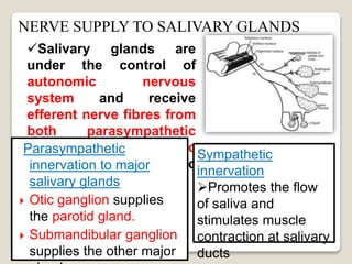
UNDER THE GUIDANCE:
Prof.Dr.C.S. Saimbi(H.O.D)
Dr.Vikash Kumar(Asst.Prof)
PRESENTED BY –
Dr.SONI BISTA
(1st year PG student)
Periodontology and Oral
Implantology
Contents:
Introduction
Salivary glands
Development and structure
Morphology
histology
innervation
Secretion
Composition
Properties
Salivary biomarker
ANOMALIES
CONCLUSION
REFERENCES….
Saliva is a clear
fluid, slightly acidic,
mucoserous secretion
which provides
chemical milieu
of the teeth and
oral soft tissue.
Complex mix of fluids from major and minor salivary glands
and from the GCF(which contains oral bacteria and food
debris)
Saliva and blood are called “brothers” in the body as they
come from same origin._Miletich I (2010)
Average daily flow : 1-1.5L
Its protein secretion:
1. Serous: contain ptylin(alpha amylase)
Enzyme for digesting starches.
2. Mucous: contain mucin for lubricating
and protective purpose.
BASED ON ANATOMIC LOCATION:
1. parotid gland
2. submandibular gland
3. sublingual gland
4. Accessory gland (labial,lingual,palatal,buccal,
glossopalatine,retromolar)
 BASED ON SIZE AND AMOUNT OF SECRETION:
1. Major salivary gland
- Parotid,submandibular,sublingual glands.
1. Minor salivary gland
- Labial,lingual,palatal,buccal,e.t.c
 BASED ON TYPE OF SECRETION (Roth G, Calmes R, 1981) :
1. Serous gland:
e.g. Parotid and lingual gland
2. Mucus gland:
e.g. lingual,buccal,palatal gland
3. Mixed gland:
e.g. submandibular,sublingual,labial gland
All the
salivary gland
show a similar
pattern of
development…
Minor salivary
gland(2nd week):
buccal epithelium.
The epithelial bud grows
into an extensively branched
system of cords of cell that
are first solid but gradually
develop a lumen and become
ducts.
The secretory portions
develop later than the duct
system and forms by
repeated branching and
budding of the finer cell
cords and ducts.
Since salivary glands are
formed from an initially
solid core of epithelial cells
–for the proper functioning
of the gland the duct needs
to undergo cavitations -to
allow free access between
the saliva producing acini
and oral cavity.- known as
Canilicular Stage.
STRUCTURE OF TERMINAL SECRETORY UNITS
oSalivary glands are made up of cells
which are arranged in small groups
around a central globular cavity called
acinus & alveolus.
oThe central cavity is continous with
the lumen of the duct.
oThe fine duct draining each acinus is
called the intercalated ducts.
oMany intercalated ducts join together
to form intralobular ducts.
oTwo or more intralobular ducts join to
form interlobular ducts , which unite
to form the main duct of the gland.
oThe gland with this type of structure &
duct system is called racemose type.
Racemose means the bunch of grapes
MORPHOLOGIC CHARACTERISTICS OF SALIVARY GLANDS
PAROTID GLAND
Largest of all the salivary
glands
Purely serous gland that
produce thin , watery amylase
rich saliva
Superficial portion lies in
front of external ear &
deeper portion lies behind the
ramus of mandible
Stensen's Duct (Parotid
Papilla) opens out adjacent to
maxillary second molar.
SUBMANDIBULAR GLAND
 Second largest salivary gland
 Mixed gland
 Located in the posterior part of floor
of mouth,adjacent to medial aspect of
mandible & wrapping around the
posterior border of mylohyoid muscle.
 Wharton's Duct opens beneath the
tongue at sub-lingual caruncle lateral to
the lingual frenum
SUBLINGUAL GLAND
Smallest salivary gland
Mixed gland but mucous
secretory cells predominate.
Located in anterior part of
floor of mouth between the
mucosa and mylohyoid muscle
Opens through series of
small ducts (ducts of rivinus)
opening along the sub-lingual
fold & often through a larger
duct (bartholin’s duct)
MINOR SALIVARY GLAND
The minor salivary glands are
located beneath the
epithelium in almost all parts
of the oral cavity.
These glands usually consist
of several small groups of
secretory units opening via
short ducts directly into
mouth.
There are 600 to 1000 minor
salivary glands lying in the oral
cavity and the oropharynx.
Predominantly mucous glands,
except for Von Ebners
glands(purely serous)
PAROTID GLAND
Arterial: Ext.Carotid Artery and its branches
Venous: Ext.Jugular Vein
Lymphatic: Parotid Nodes Upper deep
cervical nodes
SUBMANDIBULAR GLAND
Arterial: Facial Artery , Lingual Artery
Venous: Common Facial Vein /Lingual Vein
Lymphatic: Submandibular Lymph nodes
SUBLINGUAL GLAND
Arterial: Lingual and Submental Arteries
Venous: Lingual Vein
VASCULAR
SUPPLY
NERVE SUPPLY TO SALIVARY GLANDS
Salivary glands are
under the control of
autonomic nervous
system and receive
efferent nerve fibres from
both parasympathetic
and sympathetic
divisions of autonomic
nervous system.
Parasympathetic
innervation to major
salivary glands
 Otic ganglion supplies
the parotid gland.
 Submandibular ganglion
supplies the other major
Sympathetic
innervation
Promotes the flow
of saliva and
stimulates muscle
contraction at salivary
ducts
Afferent signals from sensory receptors in mouth
(Trigeminal,facial,glossopharyngeal nerves)
Salivary nuclei in the medulla oblongata of brain
Parasympathetic nerve bundle, sympathetic nerve
bundle
salivary glands
REGULATION OF SALIVARY SECRET
• This type of gland is made up of serous cells
predominantly.
• These glands secrete thin & watery saliva .
• Parotid glands and lingual glands are serous
glands.
SEROUS GLANDS
• This type of glands are made up of mucous cells
mainly .
• These glands secrete thick & viscous saliva with
more mucin .
• Lingual mucous, buccal glands & palatal glands
belongs to this type.
MUCOUS GLANDS
• Mixed glands are made up of both serous
and mucous cells .
• Submandibular , sublingual & lacrimal glands
are mixed glands
MIXED GLANDS
SECRETORY CELLS:
1.SEROUS CELLS:
a) These are spherical, consisting of 8-12 cells
surrounding a central lumen.
b) Cells are pyramidal with a broad base & narrow apex
c) The lumen usually has finger like extensions located
between adjacent cells called inter cellular canaliculi.
d) Spherical nuclei are located basally, occasionally
binucleated cells are seen.
e) Secretory granules are present in the apical
cytoplasm.
2.MUCOUS ACINI:
a) These have a tubular configuration.
b) In cross section, they appear as
round profiles with mucous cells
surrounding a central lumen of larger size
than that of serous end pieces
c) Mucous end pieces have serous cells associated with
them in the form of a demilune or cresent covering the
mucous cells at the end of the tubule.
d) The most prominent feature -accumulation of large amounts
of secretory product (mucus) in the apical cytoplasm, which
compresses the nucleus & endoplasmic reticulum & golgi
complex against the basal cell membrane.
e) Unlike serous cells, however, mucous cells lack intercellular
canaliculi, except for those covered by demilune cells.
MYOEPITHELIAL CELLS:
a) These are basket shaped cells
Contractile in nature.
b) Located between the basal lamina
& the secretory/duct cells &
are joined to the cells by desmosomes.
c) Similar to the smooth muscle cells but are derived from
the epithelium.
d) Help to expel the primary saliva from the endpiece into the
duct system.
e) Provide signals to the acinar secretory cells for maintaining
cell polarity & structural organization of the secretory end
piece.
f) Produce a no. of proteins that have tumour suppressor
activity, such as proteinase inhibitors ( ex : tissue inhibitor of
metalloproteinases ) & antiangiogenesis factors
g) Provide a barrier against invasive epithelial neoplasms.
Stage 1 : primary secretion:
Production of primary saliva from the
cells of secretory end pieces &
intercalated ducts, which is an
isotonic fluid
Stage 2 : secondary secretion:
The primary saliva is modified as it
passes through the striated &
excretory ducts mainly by
reabsorption & secretion of
electrolytes. The final saliva that
reaches the oral cavity is hypotonic.
( Mese et al., 2007, p. 711-713)
The secretory acinus produces the primary saliva, which is isotonic with an
ionic composition resembling that of plasma. In the duct system, the
primary saliva is then modified by selective reabsorption of Na+ and Cl-
(without water) and secretion of K+ and HCO3-.
Excess aldosterone secretion
Na,cl resorption K conc. increases
Copious saliva
Na,cl conc. increases K conc.decreases
Maximum salivation
Salivary ionic conc.
changes
Acinar secretion flows
through the ducts rapidly
Consistency : Slightly cloudy
Reaction : Usually slightly acidic
PH : 5-8
Specific gravity : 1.0024 – 1.0061
Freezing point :0.07 – 0.34 degree Celsius
Osmotic pressure : ( 700-1000m osmol/litre )
SALIVARY FLOW RATE
Salivary flowvaries in the stimulated and unstimulatedstate.
Stimulatedflow-
90% of average dailysaliva production
At a rate of between 0.2 and 7mL/min
Parotid glands contribute > 50% of total salivary flow.
Unstimulatedstate–
Normal flow > 0.1mL/min
Submandibularglands - 65%of total flow;
Parotid glands- 20%
Sublingual glands- 7%–8%.
Salivary flow rate = volume(ml) of saliva
min
FACTORS
DIURNAL
DURATION OF
STIMULUS
PATIENT’S POSITION
HORMONAL
SALIVA: Morethan
justwater
inthemouth.
SALIVA
Water -99.5% solids 0.5%
Organic substance Inorganic substance
Enzymes Other org. substance
1.amylase
2.maltase
3.lingual
lipase
4.lysozyme
5.phosphatas
e 6.carbonic
anhydrase
7.kalikrein
1.Proteins- mucin
& albumin
2.Blood group
antigen
3.Free amino acids
4.Non protein
nitrogenous
substances-
urea,uric
acid,creatinine,xan
thine hypoxanthine
5.Immunoglobins.
1.Sodium
2.Calcium
3.Potassium
4.Biocarbon
ate
5.Bromide
6.Chlorine
7.Fluoride
8.phosphate
1.Oxygen
2.Carbon
dioxide
3.Nitrogen
Mucins
Glycoprotein
Lubricate food
Protect teeth against acid
Help protect against
bacteria, viruses, fungi
Digestive
Enzymes
α-Amylase – digests starches
Lipase – digests fats
Protease – digests proteins
Lysozyme
Peroxidases
Lactoferrin
Histatins
Cystatins
Anti-bacterial agents
Secretory
Immunoglobulin A
Histatins
Cystatins
Anti-fungal, anti-viral
agents
Bicarbonate ions
Phosphate ions
Proteins
Help protect teeth
and soft tissues
against acidic
conditions (buffering
action)
Calcium ions
Phosphate ions
Proline-rich proteins
Help maintain
mineral content of
tooth enamel
Saliva exerts major influence on plaque initiation,
maturation and metabolism.
Salivary flow and composition influence calculus
formation, periodontal disease and caries.
Removal of salivary glands increase the incidence of
dental caries(Gilda JE,1947) and periodontal
disease(Gupta OH,1960) and also delays wound healing
(Shen LS,1979)
LYSOZYME:
•Impairs the cell wall
•Against gram positive and gram negative bacteria_Iacono VC et al(1983)
•Against Veillonella species, A.a. _ Jolles P et al(1963)
LACTOPEROXIDASE-THIOCYNATE :
•Bactericidal to lactobacillus and streptococcus
_Muhlemann HR,Schroeder H(1964)
LACTOFERRIN:
•Against A.a _ Kalmer JP, Arnold RP (1988)
MYELOPEROXIDASE:
•Bactericidal for actinobacillus_Miyasaki KT et al (1986)
•Preponderantly IgA(parotid saliva):inhibit attachment of oral
Strep.species to epithelial cells_Ellen RP(1972)
•Gibbons et al_antibodies secretions may impairs the ability of
bacteria to attach to mucosal or dental surfaces.
•Also IgG and IgM.
•Most important is bicarbonate-carbonic acid system: maintain the H
ion conc.(pH) at mucosal epithelial cell surface and tooth surface.
•Coagulation factors such as factor 8,9,10,PTA that hasten blood
coagulation and protects wounds from bacterial invasion_Leung
SW(1958)
• Also active fibrinolytic enzyme is present.
•Major enzyme: parotid amylase.
•Enzymes in increased conc.in periodontal disease:: hyaluronidase and
lipase, b-glucoronidase, chondroitin sulfatase, aspartate
aminotransferase, Alkaline phosphatase, amino acid decarboxylases,
catalase, collagenase, Peroxidase,etc.
•Proteolytic enzymes:initiation and progression of periodontal
disease.
•Antiproteases :inhibit cathepsins(Isemura S,1984)
•Antileukoproteases:inhibit elastase(Ohlsson M,1983)
•TIMP: inhibit activity of collagen degrading enzymes.
•Glycoproteins:inhibit sorption of bacteria to tooth surface.
•Orogranulocytes: Living PMNs in saliva
gingival inflammation
Whole saliva: Complex mixture of fluid from major and
minor salivary gland and from GCF which contains oral bacteria
and food debris_Edgar (1992)
Mandel and
Wotman
(1976)
 Non invasive, non painful techniques exist to collect whole
saliva, as well as saliva from the individual major & minor
salivary glands .
 Whole saliva is easily obtained & is in most case a good
indicator of whole mouth dryness.
 Diseases of salivary gland can often be diagnosed from the
secretions obtained directly.
 The quantification of salivary output is referred to as
sialometry.
COLLECTION OF SALIVA
 University of Southern California School of Dentistry
guidelines
i. Unstimulated whole saliva collection
always should precede stimulated whole
saliva collection.
ii. The patient is advised to refrain from
intake of any food or beverage (water
exempted) one hour before the test
session.
iii. Smoking, chewing gum and intake of
coffee also are prohibited during this
hour.
iv. The subject is advised to rinse his or
1.DRAINING/SPITTING METHOD: 2.SUCTION METHOD
3.SWAB METHOD 4.ABSORBANT
METHOD
METHODS FOR INDIVIDUAL/SPECIFIC
SALIVARY GLAND:
SUBMANDIBULAR/
SUBLINGUAL GLAND::
CUSTOM MADE
COLLECTORS
MINOR SALIVARY
GLANDS:::
MICROPIPETTE,ABS
ORBENT FILTER
PAPER OR STRIPS
PAROTID GLAND:::
MODIFIED CARLSON-
CRITTENDEN DEVICE
Saliva: an emerging biofluid for early
detection of diseases- Lee YH1, Wong
DT(2009)NIDCR:use of oral fluids as the diagnostic medium
to scrutinize the health and/or disease status of individua
Oral fluid being the 'mirror of body' is a perfect
medium to be explored for health and disease
surveillance.Biomarker:A biomarker is an objective measure that has been
evaluated and confirmed either as an indicator of physiologic
health, a pathogenic process, or a pharmacologic response to a
therapeutic intervention.
 CLASSIFICATION OF SALIVARY BIOMARKERS
Locally
produced
proteins of
host and
bacterial
origin
(enzymes,
immunoglo
bulins and
cytokines)
Genetic ⁄
genomic
biomarker
s such as
DNA and
mRNA of
host origin
Bacteria
and
bacterial
products,
ions,
steroid
hormones
and
volatile
compounds
Salivary proteomic, genomic and microbial
biomarkers for periodontal diagnosis
How serum constituents(i.e.,
drugs and hormones) reach
saliva.
Saliva is used for the
diagnosis of
1. Hereditary Diseases
2. Autoimmune Diseases
3. Malignancy
4. Infectious Diseases
5. Drug Monitoring
6. The Monitoring Of
Hormone Levels
7. Diagnosis Of Oral Disease
With Relevance For
Systemic Diseases
FDA APPROVED SALIVARY
KITS
Physiologic:
• Taste
• Surface
texture
• Dehydration
• Age
• Mastication
• Emotion
Pathologic conditions:
• GI irritants
• Ill fitting dentures
• Vitamin deficiency
• Trauma from surgery
• Senile atrophy of the
salivary glands
• Irradiation therapy
• Diseases of the brain stem
• Diabetes mellitus/
insipidus
• Diarrhoea
• Acute infectious diseases
Drugs:
• Cholinesterase inhibitors-
Prostigmine
• Adrenergic stimulating drugs-
epinephrine
• Sialogogues- pilocarpine.
• Antihistamines - Atropine
• Drugs for peptic ulcer – Omeprazole,
Ranitidine.
• Antihypertensives – Captopril.
• Antiparkinsonian drugs – Levodopa.
• Antianxiety agents- Benzodiazepines.
• Antidepressants – Olanzepine.
• Diuretics – Furesemide.
CONDITIONS AFFECTING SALIVATION
ANOMALIES
I.Developmental
Aberrant Salivary Glands
Aplasia and Hyperplasia
Atresia
II.Obstructive conditions
Sialolithiasis
Mucocele
Necrotizing Sialometaplasia
III. Inflammatory Diseases
Viral- Mumps , H.I.V. Associated
Bacterial - Sialadenitis
IV.Neoplastic Diseases
Benign
Malignant
V.Degenerative
Conditions
Sjogren’s Syndrome
Ionizing Radiation
VI.Xerostomia
 XEROSTOMIA is a condition of reduced or absent salivary
flow,leading to the dryness of the mouth.
 It is not a disease by itself, but a symptom associated with
alterations of salivary function.
Systemic diseases
1. Rheumatoid conditions Collagen/vascular, connective tissue
diseases, ex: Sjogren’s syndrome
2.Dysfunction of the
immune system
AIDS
3. Hormonal Disorders Diabetes mellitus
4. Neurological disorders Parkinson’s disease
5. Dehydration
Therapeutic irradiation External beam, whole- body,131I
Drugs / medications Anticholenergics,Antidepressants,Antihy
pertensive
Antipsychotics,& Antiparkinsonism drugs
Psychogenic Disorders Depression
Surgical removal of the
glands
ORAL SYMPTOMS CLINICAL SIGNS
1. Dry mouth ( xerostomia )
2. Often thirsty
3.Dysphagia (difficulty with
swallowing )
4. Dysphonia ( difficulty with
speaking )
5. Dysgeusia ( abnormal taste
sensation )
6. Difficulty with eating dry
foods
7. Need to frequently sip
water while eating
8. Difficulty with wearing
dentures
9. Often do things to keep
mouth moist
10.Burning, tingling,sensation on
the tongue.
11.Fissures & sores at corners
of lips.
1. Dryness of lining oral tissues
2. Loss of glistening of the oral
mucosa
3. Dryness of the oral mucous
membranes
4. Oral mucosa appears thin & pale
5. Tongue blade/mirror/a gloved
finger may adhere to the soft
tissues
6. Fissuring & lobulation of the
dorsum of the tongue & lips
7. Angular cheilitis
8. Candidiasis on tongue & palate
9. Increased incidence of dental
caries
10.Thicker, more stringy saliva
11. Swelling of glands
12.Increase in inflammatory
gingival diseases.
TREATMENT
Systemic Therapy:
Bromohexine, anethole,
triothiline & pilocarpine
Hcl all three should be
used under the care of a
specialist & following
medical examination
Local Therapy
SALIVARY SUBSTITUTES
Carboxy methyl cellulose (CMC) based
 Imparts lubrication and viscosity
 Sorbitol or xylitol are added to provide surface activity and as a
sweetner.
 Have surface tension greater than natural saliva.
Mucin based
• Animal mucins derived from procine gastric tissues / bovine
salivary glands.
• Salts are addeded to mimic the electrolyte content of natural
saliva
It is also known as sialorrhea, ptyalism.
It may lead problems in oral motor coordination, including
reduced muscle tone around the mouth & a reduced ability to
swallow.
Causes:
 After extensive surgery for oral or oropharyngeal
disorders.
 As a result of stomatitis, psychological factors, & the
use of some drugs, Ex: benzodiazepines,captopril
Treatment
i) Drugs – anticholinergics.
ii) Surgical – depending on the nature of the anomaly.
HYPERSALIVATION
•Saliva is an alternative to serum as a biological fluid that can be
analysed for diagnostic purposes.
•A number of markers show promise as sensitive measures of the
disease & the effectiveness of therapy.
• Longer - term longitudinal studies , however are required to
establish the relationship between specific markers & progression
of periodontal disease.
• Further more, analysis of saliva may offer a cost effective
approach to assessment of periodontal disease in large
Saliva is a most valuable oral
fluid that often is taken for
granted.
1. Clinical Periodontology 10th Edition; Carranza,Newmann.
2. Shafers textbook of oral pathology. 5th Edtn
3. Burkitt’s textboof of oral medicine. 11th edtn
4. Periodontology 2000 volume 34: 2004
5. Tencate’s Oral histology 6th edition
6. Textbook of medical physiology- guyton and hall 9th edition
7. J. Periodontal Research 1990,1983
8. Dentomaxillofac Radiol 2007;36:59-62. T Bar, A Zagury, D London, R
Shacham, and O Nahlieli.
9. ImagingGOOGLE.oom
Saliva
Saliva

Saliva

  • 2.
    UNDER THE GUIDANCE: Prof.Dr.C.S.Saimbi(H.O.D) Dr.Vikash Kumar(Asst.Prof) PRESENTED BY – Dr.SONI BISTA (1st year PG student) Periodontology and Oral Implantology
  • 3.
    Contents: Introduction Salivary glands Development andstructure Morphology histology innervation Secretion Composition Properties Salivary biomarker ANOMALIES CONCLUSION REFERENCES….
  • 4.
    Saliva is aclear fluid, slightly acidic, mucoserous secretion which provides chemical milieu of the teeth and oral soft tissue. Complex mix of fluids from major and minor salivary glands and from the GCF(which contains oral bacteria and food debris) Saliva and blood are called “brothers” in the body as they come from same origin._Miletich I (2010) Average daily flow : 1-1.5L Its protein secretion: 1. Serous: contain ptylin(alpha amylase) Enzyme for digesting starches. 2. Mucous: contain mucin for lubricating and protective purpose.
  • 6.
    BASED ON ANATOMICLOCATION: 1. parotid gland 2. submandibular gland 3. sublingual gland 4. Accessory gland (labial,lingual,palatal,buccal, glossopalatine,retromolar)  BASED ON SIZE AND AMOUNT OF SECRETION: 1. Major salivary gland - Parotid,submandibular,sublingual glands. 1. Minor salivary gland - Labial,lingual,palatal,buccal,e.t.c  BASED ON TYPE OF SECRETION (Roth G, Calmes R, 1981) : 1. Serous gland: e.g. Parotid and lingual gland 2. Mucus gland: e.g. lingual,buccal,palatal gland 3. Mixed gland: e.g. submandibular,sublingual,labial gland
  • 7.
    All the salivary gland showa similar pattern of development… Minor salivary gland(2nd week): buccal epithelium.
  • 8.
    The epithelial budgrows into an extensively branched system of cords of cell that are first solid but gradually develop a lumen and become ducts. The secretory portions develop later than the duct system and forms by repeated branching and budding of the finer cell cords and ducts. Since salivary glands are formed from an initially solid core of epithelial cells –for the proper functioning of the gland the duct needs to undergo cavitations -to allow free access between the saliva producing acini and oral cavity.- known as Canilicular Stage.
  • 9.
    STRUCTURE OF TERMINALSECRETORY UNITS oSalivary glands are made up of cells which are arranged in small groups around a central globular cavity called acinus & alveolus. oThe central cavity is continous with the lumen of the duct. oThe fine duct draining each acinus is called the intercalated ducts. oMany intercalated ducts join together to form intralobular ducts. oTwo or more intralobular ducts join to form interlobular ducts , which unite to form the main duct of the gland. oThe gland with this type of structure & duct system is called racemose type. Racemose means the bunch of grapes
  • 10.
    MORPHOLOGIC CHARACTERISTICS OFSALIVARY GLANDS PAROTID GLAND Largest of all the salivary glands Purely serous gland that produce thin , watery amylase rich saliva Superficial portion lies in front of external ear & deeper portion lies behind the ramus of mandible Stensen's Duct (Parotid Papilla) opens out adjacent to maxillary second molar.
  • 11.
    SUBMANDIBULAR GLAND  Secondlargest salivary gland  Mixed gland  Located in the posterior part of floor of mouth,adjacent to medial aspect of mandible & wrapping around the posterior border of mylohyoid muscle.  Wharton's Duct opens beneath the tongue at sub-lingual caruncle lateral to the lingual frenum
  • 12.
    SUBLINGUAL GLAND Smallest salivarygland Mixed gland but mucous secretory cells predominate. Located in anterior part of floor of mouth between the mucosa and mylohyoid muscle Opens through series of small ducts (ducts of rivinus) opening along the sub-lingual fold & often through a larger duct (bartholin’s duct)
  • 13.
    MINOR SALIVARY GLAND Theminor salivary glands are located beneath the epithelium in almost all parts of the oral cavity. These glands usually consist of several small groups of secretory units opening via short ducts directly into mouth. There are 600 to 1000 minor salivary glands lying in the oral cavity and the oropharynx. Predominantly mucous glands, except for Von Ebners glands(purely serous)
  • 14.
    PAROTID GLAND Arterial: Ext.CarotidArtery and its branches Venous: Ext.Jugular Vein Lymphatic: Parotid Nodes Upper deep cervical nodes SUBMANDIBULAR GLAND Arterial: Facial Artery , Lingual Artery Venous: Common Facial Vein /Lingual Vein Lymphatic: Submandibular Lymph nodes SUBLINGUAL GLAND Arterial: Lingual and Submental Arteries Venous: Lingual Vein VASCULAR SUPPLY
  • 15.
    NERVE SUPPLY TOSALIVARY GLANDS Salivary glands are under the control of autonomic nervous system and receive efferent nerve fibres from both parasympathetic and sympathetic divisions of autonomic nervous system. Parasympathetic innervation to major salivary glands  Otic ganglion supplies the parotid gland.  Submandibular ganglion supplies the other major Sympathetic innervation Promotes the flow of saliva and stimulates muscle contraction at salivary ducts
  • 17.
    Afferent signals fromsensory receptors in mouth (Trigeminal,facial,glossopharyngeal nerves) Salivary nuclei in the medulla oblongata of brain Parasympathetic nerve bundle, sympathetic nerve bundle salivary glands REGULATION OF SALIVARY SECRET
  • 19.
    • This typeof gland is made up of serous cells predominantly. • These glands secrete thin & watery saliva . • Parotid glands and lingual glands are serous glands. SEROUS GLANDS • This type of glands are made up of mucous cells mainly . • These glands secrete thick & viscous saliva with more mucin . • Lingual mucous, buccal glands & palatal glands belongs to this type. MUCOUS GLANDS • Mixed glands are made up of both serous and mucous cells . • Submandibular , sublingual & lacrimal glands are mixed glands MIXED GLANDS
  • 20.
    SECRETORY CELLS: 1.SEROUS CELLS: a)These are spherical, consisting of 8-12 cells surrounding a central lumen. b) Cells are pyramidal with a broad base & narrow apex c) The lumen usually has finger like extensions located between adjacent cells called inter cellular canaliculi. d) Spherical nuclei are located basally, occasionally binucleated cells are seen. e) Secretory granules are present in the apical cytoplasm.
  • 21.
    2.MUCOUS ACINI: a) Thesehave a tubular configuration. b) In cross section, they appear as round profiles with mucous cells surrounding a central lumen of larger size than that of serous end pieces c) Mucous end pieces have serous cells associated with them in the form of a demilune or cresent covering the mucous cells at the end of the tubule. d) The most prominent feature -accumulation of large amounts of secretory product (mucus) in the apical cytoplasm, which compresses the nucleus & endoplasmic reticulum & golgi complex against the basal cell membrane. e) Unlike serous cells, however, mucous cells lack intercellular canaliculi, except for those covered by demilune cells.
  • 22.
    MYOEPITHELIAL CELLS: a) Theseare basket shaped cells Contractile in nature. b) Located between the basal lamina & the secretory/duct cells & are joined to the cells by desmosomes. c) Similar to the smooth muscle cells but are derived from the epithelium. d) Help to expel the primary saliva from the endpiece into the duct system. e) Provide signals to the acinar secretory cells for maintaining cell polarity & structural organization of the secretory end piece. f) Produce a no. of proteins that have tumour suppressor activity, such as proteinase inhibitors ( ex : tissue inhibitor of metalloproteinases ) & antiangiogenesis factors g) Provide a barrier against invasive epithelial neoplasms.
  • 23.
    Stage 1 :primary secretion: Production of primary saliva from the cells of secretory end pieces & intercalated ducts, which is an isotonic fluid Stage 2 : secondary secretion: The primary saliva is modified as it passes through the striated & excretory ducts mainly by reabsorption & secretion of electrolytes. The final saliva that reaches the oral cavity is hypotonic.
  • 24.
    ( Mese etal., 2007, p. 711-713) The secretory acinus produces the primary saliva, which is isotonic with an ionic composition resembling that of plasma. In the duct system, the primary saliva is then modified by selective reabsorption of Na+ and Cl- (without water) and secretion of K+ and HCO3-.
  • 25.
    Excess aldosterone secretion Na,clresorption K conc. increases Copious saliva Na,cl conc. increases K conc.decreases Maximum salivation Salivary ionic conc. changes Acinar secretion flows through the ducts rapidly
  • 26.
    Consistency : Slightlycloudy Reaction : Usually slightly acidic PH : 5-8 Specific gravity : 1.0024 – 1.0061 Freezing point :0.07 – 0.34 degree Celsius Osmotic pressure : ( 700-1000m osmol/litre )
  • 27.
    SALIVARY FLOW RATE Salivaryflowvaries in the stimulated and unstimulatedstate. Stimulatedflow- 90% of average dailysaliva production At a rate of between 0.2 and 7mL/min Parotid glands contribute > 50% of total salivary flow. Unstimulatedstate– Normal flow > 0.1mL/min Submandibularglands - 65%of total flow; Parotid glands- 20% Sublingual glands- 7%–8%. Salivary flow rate = volume(ml) of saliva min
  • 28.
  • 29.
    SALIVA: Morethan justwater inthemouth. SALIVA Water -99.5%solids 0.5% Organic substance Inorganic substance Enzymes Other org. substance 1.amylase 2.maltase 3.lingual lipase 4.lysozyme 5.phosphatas e 6.carbonic anhydrase 7.kalikrein 1.Proteins- mucin & albumin 2.Blood group antigen 3.Free amino acids 4.Non protein nitrogenous substances- urea,uric acid,creatinine,xan thine hypoxanthine 5.Immunoglobins. 1.Sodium 2.Calcium 3.Potassium 4.Biocarbon ate 5.Bromide 6.Chlorine 7.Fluoride 8.phosphate 1.Oxygen 2.Carbon dioxide 3.Nitrogen
  • 30.
    Mucins Glycoprotein Lubricate food Protect teethagainst acid Help protect against bacteria, viruses, fungi Digestive Enzymes α-Amylase – digests starches Lipase – digests fats Protease – digests proteins Lysozyme Peroxidases Lactoferrin Histatins Cystatins Anti-bacterial agents
  • 31.
    Secretory Immunoglobulin A Histatins Cystatins Anti-fungal, anti-viral agents Bicarbonateions Phosphate ions Proteins Help protect teeth and soft tissues against acidic conditions (buffering action) Calcium ions Phosphate ions Proline-rich proteins Help maintain mineral content of tooth enamel
  • 32.
    Saliva exerts majorinfluence on plaque initiation, maturation and metabolism. Salivary flow and composition influence calculus formation, periodontal disease and caries. Removal of salivary glands increase the incidence of dental caries(Gilda JE,1947) and periodontal disease(Gupta OH,1960) and also delays wound healing (Shen LS,1979)
  • 33.
    LYSOZYME: •Impairs the cellwall •Against gram positive and gram negative bacteria_Iacono VC et al(1983) •Against Veillonella species, A.a. _ Jolles P et al(1963) LACTOPEROXIDASE-THIOCYNATE : •Bactericidal to lactobacillus and streptococcus _Muhlemann HR,Schroeder H(1964) LACTOFERRIN: •Against A.a _ Kalmer JP, Arnold RP (1988) MYELOPEROXIDASE: •Bactericidal for actinobacillus_Miyasaki KT et al (1986)
  • 34.
    •Preponderantly IgA(parotid saliva):inhibitattachment of oral Strep.species to epithelial cells_Ellen RP(1972) •Gibbons et al_antibodies secretions may impairs the ability of bacteria to attach to mucosal or dental surfaces. •Also IgG and IgM. •Most important is bicarbonate-carbonic acid system: maintain the H ion conc.(pH) at mucosal epithelial cell surface and tooth surface. •Coagulation factors such as factor 8,9,10,PTA that hasten blood coagulation and protects wounds from bacterial invasion_Leung SW(1958) • Also active fibrinolytic enzyme is present.
  • 35.
    •Major enzyme: parotidamylase. •Enzymes in increased conc.in periodontal disease:: hyaluronidase and lipase, b-glucoronidase, chondroitin sulfatase, aspartate aminotransferase, Alkaline phosphatase, amino acid decarboxylases, catalase, collagenase, Peroxidase,etc. •Proteolytic enzymes:initiation and progression of periodontal disease. •Antiproteases :inhibit cathepsins(Isemura S,1984) •Antileukoproteases:inhibit elastase(Ohlsson M,1983) •TIMP: inhibit activity of collagen degrading enzymes. •Glycoproteins:inhibit sorption of bacteria to tooth surface. •Orogranulocytes: Living PMNs in saliva gingival inflammation
  • 36.
    Whole saliva: Complexmixture of fluid from major and minor salivary gland and from GCF which contains oral bacteria and food debris_Edgar (1992) Mandel and Wotman (1976)
  • 37.
     Non invasive,non painful techniques exist to collect whole saliva, as well as saliva from the individual major & minor salivary glands .  Whole saliva is easily obtained & is in most case a good indicator of whole mouth dryness.  Diseases of salivary gland can often be diagnosed from the secretions obtained directly.  The quantification of salivary output is referred to as sialometry. COLLECTION OF SALIVA
  • 38.
     University ofSouthern California School of Dentistry guidelines i. Unstimulated whole saliva collection always should precede stimulated whole saliva collection. ii. The patient is advised to refrain from intake of any food or beverage (water exempted) one hour before the test session. iii. Smoking, chewing gum and intake of coffee also are prohibited during this hour. iv. The subject is advised to rinse his or
  • 39.
    1.DRAINING/SPITTING METHOD: 2.SUCTIONMETHOD 3.SWAB METHOD 4.ABSORBANT METHOD
  • 40.
    METHODS FOR INDIVIDUAL/SPECIFIC SALIVARYGLAND: SUBMANDIBULAR/ SUBLINGUAL GLAND:: CUSTOM MADE COLLECTORS MINOR SALIVARY GLANDS::: MICROPIPETTE,ABS ORBENT FILTER PAPER OR STRIPS PAROTID GLAND::: MODIFIED CARLSON- CRITTENDEN DEVICE
  • 41.
    Saliva: an emergingbiofluid for early detection of diseases- Lee YH1, Wong DT(2009)NIDCR:use of oral fluids as the diagnostic medium to scrutinize the health and/or disease status of individua Oral fluid being the 'mirror of body' is a perfect medium to be explored for health and disease surveillance.Biomarker:A biomarker is an objective measure that has been evaluated and confirmed either as an indicator of physiologic health, a pathogenic process, or a pharmacologic response to a therapeutic intervention.
  • 42.
     CLASSIFICATION OFSALIVARY BIOMARKERS Locally produced proteins of host and bacterial origin (enzymes, immunoglo bulins and cytokines) Genetic ⁄ genomic biomarker s such as DNA and mRNA of host origin Bacteria and bacterial products, ions, steroid hormones and volatile compounds
  • 43.
    Salivary proteomic, genomicand microbial biomarkers for periodontal diagnosis
  • 45.
    How serum constituents(i.e., drugsand hormones) reach saliva. Saliva is used for the diagnosis of 1. Hereditary Diseases 2. Autoimmune Diseases 3. Malignancy 4. Infectious Diseases 5. Drug Monitoring 6. The Monitoring Of Hormone Levels 7. Diagnosis Of Oral Disease With Relevance For Systemic Diseases
  • 46.
  • 47.
    Physiologic: • Taste • Surface texture •Dehydration • Age • Mastication • Emotion Pathologic conditions: • GI irritants • Ill fitting dentures • Vitamin deficiency • Trauma from surgery • Senile atrophy of the salivary glands • Irradiation therapy • Diseases of the brain stem • Diabetes mellitus/ insipidus • Diarrhoea • Acute infectious diseases Drugs: • Cholinesterase inhibitors- Prostigmine • Adrenergic stimulating drugs- epinephrine • Sialogogues- pilocarpine. • Antihistamines - Atropine • Drugs for peptic ulcer – Omeprazole, Ranitidine. • Antihypertensives – Captopril. • Antiparkinsonian drugs – Levodopa. • Antianxiety agents- Benzodiazepines. • Antidepressants – Olanzepine. • Diuretics – Furesemide. CONDITIONS AFFECTING SALIVATION
  • 48.
    ANOMALIES I.Developmental Aberrant Salivary Glands Aplasiaand Hyperplasia Atresia II.Obstructive conditions Sialolithiasis Mucocele Necrotizing Sialometaplasia III. Inflammatory Diseases Viral- Mumps , H.I.V. Associated Bacterial - Sialadenitis IV.Neoplastic Diseases Benign Malignant V.Degenerative Conditions Sjogren’s Syndrome Ionizing Radiation VI.Xerostomia
  • 50.
     XEROSTOMIA isa condition of reduced or absent salivary flow,leading to the dryness of the mouth.  It is not a disease by itself, but a symptom associated with alterations of salivary function. Systemic diseases 1. Rheumatoid conditions Collagen/vascular, connective tissue diseases, ex: Sjogren’s syndrome 2.Dysfunction of the immune system AIDS 3. Hormonal Disorders Diabetes mellitus 4. Neurological disorders Parkinson’s disease 5. Dehydration Therapeutic irradiation External beam, whole- body,131I Drugs / medications Anticholenergics,Antidepressants,Antihy pertensive Antipsychotics,& Antiparkinsonism drugs Psychogenic Disorders Depression Surgical removal of the glands
  • 51.
    ORAL SYMPTOMS CLINICALSIGNS 1. Dry mouth ( xerostomia ) 2. Often thirsty 3.Dysphagia (difficulty with swallowing ) 4. Dysphonia ( difficulty with speaking ) 5. Dysgeusia ( abnormal taste sensation ) 6. Difficulty with eating dry foods 7. Need to frequently sip water while eating 8. Difficulty with wearing dentures 9. Often do things to keep mouth moist 10.Burning, tingling,sensation on the tongue. 11.Fissures & sores at corners of lips. 1. Dryness of lining oral tissues 2. Loss of glistening of the oral mucosa 3. Dryness of the oral mucous membranes 4. Oral mucosa appears thin & pale 5. Tongue blade/mirror/a gloved finger may adhere to the soft tissues 6. Fissuring & lobulation of the dorsum of the tongue & lips 7. Angular cheilitis 8. Candidiasis on tongue & palate 9. Increased incidence of dental caries 10.Thicker, more stringy saliva 11. Swelling of glands 12.Increase in inflammatory gingival diseases.
  • 52.
    TREATMENT Systemic Therapy: Bromohexine, anethole, triothiline& pilocarpine Hcl all three should be used under the care of a specialist & following medical examination Local Therapy SALIVARY SUBSTITUTES Carboxy methyl cellulose (CMC) based  Imparts lubrication and viscosity  Sorbitol or xylitol are added to provide surface activity and as a sweetner.  Have surface tension greater than natural saliva. Mucin based • Animal mucins derived from procine gastric tissues / bovine salivary glands. • Salts are addeded to mimic the electrolyte content of natural saliva
  • 53.
    It is alsoknown as sialorrhea, ptyalism. It may lead problems in oral motor coordination, including reduced muscle tone around the mouth & a reduced ability to swallow. Causes:  After extensive surgery for oral or oropharyngeal disorders.  As a result of stomatitis, psychological factors, & the use of some drugs, Ex: benzodiazepines,captopril Treatment i) Drugs – anticholinergics. ii) Surgical – depending on the nature of the anomaly. HYPERSALIVATION
  • 54.
    •Saliva is analternative to serum as a biological fluid that can be analysed for diagnostic purposes. •A number of markers show promise as sensitive measures of the disease & the effectiveness of therapy. • Longer - term longitudinal studies , however are required to establish the relationship between specific markers & progression of periodontal disease. • Further more, analysis of saliva may offer a cost effective approach to assessment of periodontal disease in large Saliva is a most valuable oral fluid that often is taken for granted.
  • 55.
    1. Clinical Periodontology10th Edition; Carranza,Newmann. 2. Shafers textbook of oral pathology. 5th Edtn 3. Burkitt’s textboof of oral medicine. 11th edtn 4. Periodontology 2000 volume 34: 2004 5. Tencate’s Oral histology 6th edition 6. Textbook of medical physiology- guyton and hall 9th edition 7. J. Periodontal Research 1990,1983 8. Dentomaxillofac Radiol 2007;36:59-62. T Bar, A Zagury, D London, R Shacham, and O Nahlieli. 9. ImagingGOOGLE.oom