Saddle nose transcribed_lecture_2012
•
2 likes•1,662 views

This lecture discusses the management of saddle nose deformities. The speaker classifies saddle nose deformities into 5 types from mild to severe. For mild cases, the goal is to "finish the job" by minimally reducing the hump. For moderate cases, a balanced approach of reducing bone and using it as an onlay graft is recommended. More severe cases may require rib or ear cartilage grafts. The most severe cases require total reconstruction using rib grafts to build a new nasal scaffold. Sharp instruments, conservative grafting, and considering vascularization are important for successful reconstruction of saddle nose deformities.

Recommended
More Related Content
What's hot
What's hot (20)
Viewers also liked
Viewers also liked (15)
Similar to Saddle nose transcribed_lecture_2012
Similar to Saddle nose transcribed_lecture_2012 (20)
Saddle nose transcribed_lecture_2012
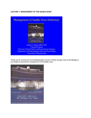
- 1. LECTURE 1: MANAGEMENT OF THE SADDLE NOSE Management of Saddle Nose Deformity Daniel G. Becker MD FACS Clinical Professor Division of Facial Plastic and Reconstructive Surgery Department of Otolaryngology-Head and Neck Surgery University of Pennsylvania Thank you for inviting me to the Multispecialty Course in Plastic Surgery here at the Bellagio in Las Vegas to discuss the management of the saddle nose.
- 2. This is my first visit to Las Vegas, and I must say, it is quite a place! I would like to thank Dr Waldman from Kentucky, Dr Paul Nassif from Beverly Hills in Los Angeles California, and the other course organizers. This is really a wonderful course both professionally and also socially. It has been nice to see friends from around the world in this festive setting. Last night we enjoyed a 50th birthday celebration, and also I have had some nice re-unions with a former resident of mine from the University of Pennsylvania, and also a college classmate from Harvard College, who is a prominent dermatologist in New York, someone I haven’t seen in 25 years. And, I’ve made a few new friends while I’ve been here. So, this meeting has been a delight. In this 20 minute lecture, my goals are to discuss the cause, the evaluation, and the treatment options for a saddle nose deformity. In the course of a lecture, we can only cover a limited amount of material. With that in mind, I would like to direct you to the website, www.RhinoplastyArchive.com for more information and further study. Dr Pietro Palma of Milano, Italy and I are the editors of this website, the world’s first free, online surgical textbook. I encourage you to go there for more detailed information. On the subject of the Saddle Nose – the subject of this lecture, this free on-line medical textbook has three individual chapters on the saddle nose deformity. There are also hundreds of videos, including video of ear cartilage harvest, rib cartilage harvest, and other subjects pertinent to treatment of the Saddle Nose. The saddle nose deformity is characterized by a distinctive scooped-out appearance of the nasal bridge that resembles a horse saddle.
- 3. The shape represents a collapse of the intrinsic cartilaginous and/or bony support structures of the nose. Characteristics include a loss of dorsal height, middle vault and dorsal depression, a loss of tip support and definition, columellar retrusion, shortened vertical length, tip-over rotation, and retrusion of the nasal and caudal spine. The causes are numerous. The most common is traumatic. Also, surgery can be a cause. There are also vascular causes such as the use of drugs like cocaine or Afrin. In this situation, damage to the septum causes a septal perforation, loss of the L-strut support, and collapse – a saddle nose. Systemic disorders such as Wegener's disease and sarcoid can also cause saddle nose. Cancers such as inverted papilloma and squamous cell cancer can lead to saddle nose deformity as well, and also there are infectious causes like syphilis, leprosy, or bacterial infection leading to a septal abscess. There are a number of classification schemes to describe saddle nose deformities. I like the one described by Daniel and Brenner in "Facial Plastic Surgery Clinics of North America," in 2006. In their classification scheme, it goes from type 0 to type 5, with type 0 being what you might call a pseudo-saddle nose, a very minor depression, type 5 being the most catastrophic, requiring a major reconstruction. And, of course, types 1 through 4 are the stages in between.
- 4. What I propose to do in this lecture is, using this classification scheme as a template, to go from the most mild to most severe and just outline what my personal treatment algorithm is. Here, you see this first patient has a very mild saddle nose deformity from a trauma, I believe it was a softball injury. You see here the before photo, and the photo after surgery by me.
- 5. My treatment for this kind of problem is what you might call, "to finish the job." This patient told me in the office that she had a little bump to begin with, she never really liked it, and the softball injury made it worse. She doesn’t like the bump and she just wants it gone. As you can see on the before picture, the saddle nose injury has created a mild bump, the cartilaginous portion of the nose has dropped making the overall bump appear worse than in her pre-injured codition. And so what I typically have done surgically in this kind of case is to do a closed, or endonasal approach, simply take down the rest of the bump. This is really what she wants, she wants me to “finish the job” that the softball started!. She wants a smaller nose and so you might say that the softball did the first half of the operation and I did the other, and so this tends to be a relatively straightforward surgery, with a closed approach, minor hump take-down, osteotomies. Here is the frontal view, before and one year postoperative. You can see in the before picture how the saddle nose creates a flattened appearance of the middle vault region, and then one year after, that flattened look is gone, and she has nice clean nasal lines after surgery. Here are the angled (oblique) view and the base view photos before and after.
- 6. Some helpful hints for hump take-down, I recommend an anatomic approach, breaking it down in your mind mentally into the various components. I like to draw out the intended reduction on the skin before injection. Also, it is really critical that you use very sharp instruments, and we will talk about that in another lecture. But, the short version – for those of you who use osteotomes to take down the bony nasal bump, they dull quickly and should be discarded after a few uses. Sharpening them with a sharpening stone or even having them sharpened by your surgical center does not seem to be sufficient. A picture is worth 1000 words: here is an electron microscopy photo of an osteotome before use, and after 9 uses.
- 7. If you were having your hump taken down, which one of these two osteotomes would you want used on you?! I hope this photo is persuasive. New osteotome……9 uses When you use the osteotome, a gentle 2-tap technique is really helpful in guiding the osteotome along the desired line, and then final refinements can be made with a rasp. I prefer a powered rasp. This next patient has a slightly greater deformity, and in cases like this a balanced approach is my preference.
- 9. In his case, what I did was to take down the bony bump, and then I used it as an onlay graft to build up the depression. What that creates is sort of a nice compromise, a nice balance. Pre- operatively, the upper third of the nose is too high and the middle third is too low, and so by balancing those we get a nice result. Indeed, that is what I did in this gentleman, with a very happy outcome as you can see. Here is a fellow, he was a motocross competitor and suffered an injury.
- 10. As you can see, he had a substantial saddle nose deformity. I repaired this with a double-layer ear cartilage graft, and because he had thin skin we wanted to create a little bit of a cushion, and so I used AlloDerm. I wrapped the double-layer graft in AlloDerm and inserted that, and in his case we used an open approach. I think you can see, as well, when you look at the side view, that when you put on an onlay graft it ends up lengthening the nose. In the before picture, you see as if the nose has been lifted, and simply by rebuilding that bridge it can tend to push the tip down and lengthen it in a very favorable way. This woman has an isolated substantial middle vault depression. In her case, I did a closed rhinoplasty with a triple-layer onlay graft. This is a 5-year postoperative result; she got a wonderful result.
- 11. And this is a woman who suffered a trauma to her nose and has a similar severe saddle nose deformity, perhaps more severe.
- 12. In her case, she had a large septal perforation and was not interested in cartilage harvest from her ears or rib, and so we discussed irradiated rib. Irradiated rib is well-described as a good option for rhinoplasty. A number of reports in the literature describe very good long-term success. And as you can see from the early postoperative picture shown here, she had a beautiful early result. Here is the intraoperative photo, the irradiated rib that I then carved to the proper size.
- 13. I made little limited marginal incisions bilaterally to make a precise pocket, and I simply inserted the graft.
- 14. However, there is a valuable lesson to be learned here. The patient came back six months after surgery; look at these photos from before and after irradiated rib graft, she is slightly better but has had significant partial resorption.
- 15. Here is the lesson to be learned. First of all, it was a very large graft in a poorly vascularized area, so I think it was asking a lot for that entire graft, a graft of that thickness, to live. if it is a poorly vascularized bed you have to be more cautious, less ambitious. I think in a case like this, if you are overly ambitious and you start the reabsorption cascade in motion, then you end up getting more reabsorption than you bargain for, so I think that if you err on the side of conservatism when you are using irradiated rib, you are better off. I think it is just like if you are working in a garden, if you have bad soil it is hard to really expect your transplanted plant to survive. You are going to lose some of the leaves, and maybe even the whole plant, if you are not careful. So you have to keep in mind the bed you are putting it in, it has to be well fertilized – or a well-vascularized bed, in the case of a patient. And so, in this patient we did a revision, and in this case we did use her ear cartilage, a double- layer graft with soft tissue. We left the postauricular soft tissue on it, which facilitated the transplanting of it. For a video of my surgical technique of ear cartilage harvest, go to www.RhinoplastyArchive.com.
- 16. This is a nice six-month postoperative result, and I think you can see that we were, in fact, able to restore a nice profile for her. Time will tell if this will be a lasting result, but I have heard from the patient (she lives at a distance) and she tells me she is doing well still, over a year since her surgery. Now, this is a gentleman with a much more severe deformity. He has a very complicated deformity.
- 17. In addition to the saddle nose that you see here, where the upper portion was really over- resected, the lower portion was under-resected. So he has a little bit of an over-projection of his tip, a pollybeak deformity, and also an over-resected bridge. What complicates this further is that his bridge is uneven. It was unevenly over-resected, so there is a depression on the right side compared to the left. In his case, we did a complex reconstruction with ear cartilage for reconstructive grafting. Surgery included takedown of the lower nasal bridge, and placement of a unilateral spreader
- 18. graft. In the prior patient earlier in this lecture, I took down the upper part of the bridge and used that tissue to build up the lower portion. in this case the resected bridge was nusable, and so we took down the lower portion and then used ear cartilage to build up the upper portion to create balance. I think you can see with this nearly three-year result, really a beautiful result in his case. This next patient represents the class 5 deformity, major reconstruction needed.
- 19. You can see from before and after she needs a total reconstruction, and in her case we used irradiated rib, shown below. This is a gentleman who has a similar sort of problem, and we did use his own rib.
- 20. See www.RhinoplastyArchive.com for more information including video, more information on rib harvest. I think that for these kinds of reconstructions, probably autogenous rib is preferable, although I think that either autogenous or irradiated rib are good options.
- 21. Here you see a picture of his rib harvest. Basically, in these sorts of situations you build an entire new architecture of the nose, a new scaffolding over what exists, because the whole nose is collapsed. You need to build a whole new structure on top of it. This is what it looks like on the inside,
- 22. and then this is the one-year postoperative result. Well, I see from the clock that I am on time, but my time is up. I hope you found this lecture useful. Thank you