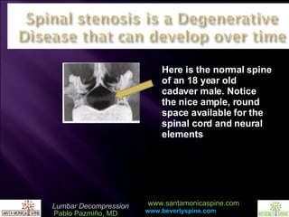
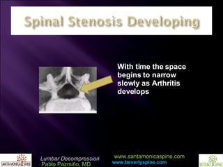
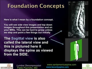
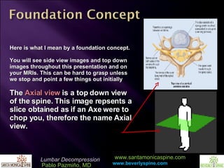
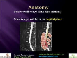
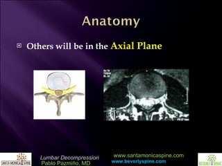
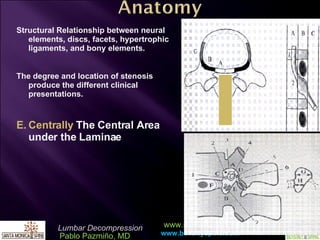
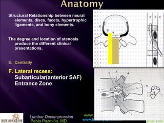
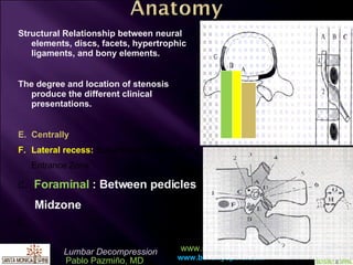
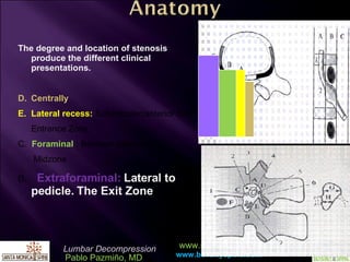
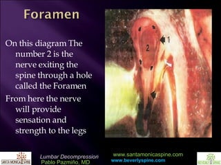
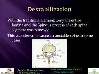
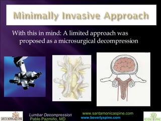
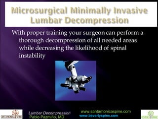
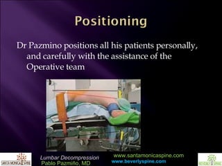
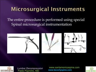
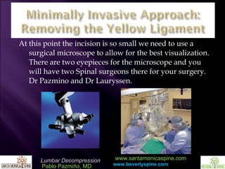
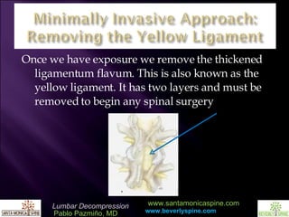
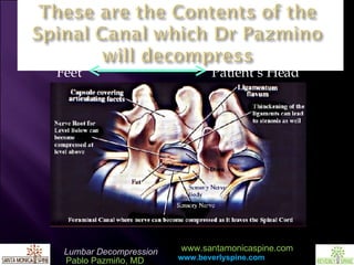
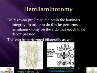
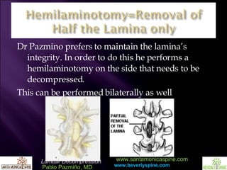
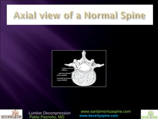
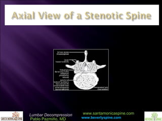
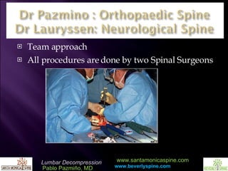
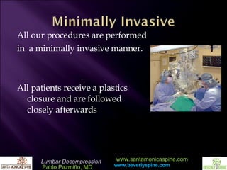
Pablo Pazmiño, MD
Orthopaedic Surgery American Academy of Orthopaedic Surgeons Clinical Faculty Olympia Medical Center Cedars Sinai Medical Center Century City Doctors Hospital Education The University of California, Los Angeles The University of Michigan, Ann Arbor
Here is the normal spine of an 18 year old cadaver male. Notice the nice ample, round space available for the spinal cord and neural elements
With time the space begins to narrow slowly as Arthritis develops
More and More Bony overgrowth occurs
With time the space begins to narrow to an almost Triangular space and can compress the neural elements within
There can be side to side differences meaning the left side may be tighter than the right and vice versa
With my background as a Third grade teacher I always make it a point to build a foundation concept before moving on to more complex ideas.  I would like to take a moment to build some foundations before continuing to discuss MRIs.
Here is what I mean by a foundation concept. You will see side view images and top down images throughout this presentation and on your MRIs. This can be hard to grasp unless we stop and point a few things out initially The  Sagittal view  is also called the lateral view and this is pictured here it displays the spine as viewed from the SIDE.
Here is what I mean by a foundation concept. You will see side view images and top down images throughout this presentation and on your MRIs. This can be hard to grasp unless we stop and point a few things out initially The  Axial view  is a top down view of the spine. This image repsents a slice obtained as if an Axe were to chop you, therefore the name Axial view.
Next we will review some basic anatomy Some images will be in the  Sagittal plane
Others will be in the  Axial Plane
Joint Facet Nucleus  Pulposus Right Nerve Root  Left Nerve Root As you can see here on MRI and CT scans the Facet joints end up looking like the buns of a hamburger. Its just the way the happen to look when they are sliced in this plane. We will show you this on some examples later Annulus Fibrosus Axial View of the Disc and Neural elements
 
 
The degree and location of stenosis can produce the different clinical presentations. Centrally Lateral recess:  Subarticular(anterior SAF) Entrance Zone C.  Foraminal  : Between pedicles   Midzone D.  Extraforaminal:  Lateral to pedicle   Exit Zone
Structural Relationship between neural elements, discs, facets, hypertrophic ligaments, and bony elements. The degree and location of stenosis produce the different clinical presentations. Centrally  The   Central Area under the Laminae
Structural Relationship between neural elements, discs, facets, hypertrophic ligaments, and bony elements. The degree and location of stenosis produce the different clinical presentations. Centrally Lateral recess:  Subarticular(anterior SAF) Entrance Zone C
Structural Relationship between neural elements, discs, facets, hypertrophic ligaments, and bony elements. The degree and location of stenosis produce the different clinical presentations. Centrally Lateral recess:  Subarticular(anterior SAF) Entrance Zone C .  Foraminal   : Between pedicles   Midzone D
The degree and location of stenosis produce the different clinical presentations. Centrally Lateral recess:  Subarticular(anterior SAF) Entrance Zone C.  Foraminal  : Between pedicles   Midzone D .  Extraforaminal:  Lateral to pedicle. The Exit Zone
On this diagram The number 2 is the nerve exiting the spine through a hole called the Foramen From here the nerve will provide sensation and strength to the legs
If the Nerve is compressed within the spine or foramen the nerve can refer pain to buttock, thigh, leg,  calf or foot. Where does your pain refer to???
 
Patients are usually an older age group >40 years of age History of recurring or persistent back pain Occasional buttocks pain and referred pain radiating down the legs Weakness Trouble walking long distances
To remove this pressure from the neural elements you may need to decompress the area highlighted here on the right
Traditional Surgery was a WIDE LAMINECTOMY.  However history has shown that this may risk destabilizing the spine
With the traditional Laminectomy the entire lamina and the Spinous process of each spinal segment was removed. This was shown to cause an unstable spine in some cases
With this in mind: A limited approach was proposed as a microsurgical decompression
With proper training your surgeon can perform a thorough decompression of all needed areas while decreasing the likelihood of spinal instability
Dr Pazmino positions all his patients personally, and carefully with the assistance of the Operative team
The entire procedure is performed using special Spinal microsurgical instrumentation
An Incision is made on your lower back The muscles on your back are not cut  instead with this approach they are simply moved out of the way. This is why patients feel pain after surgery, because we hold these muscles out of the way to do surgery and you wake up you will have a muscle sprain
At this point the incision is so small we need to use a surgical microscope to allow for the best visualization. There are two eyepieces for the microscope and you will have two Spinal surgeons there for your surgery. Dr Pazmino and Dr Lauryssen.
Once we have exposure we remove the thickened ligamentum flavum. This is also known as the yellow ligament. It has two layers and must be removed to begin any spinal surgery
Feet   Patient’s Head
Dr Pazmino prefers to maintain the lamina’s integrity. In order to do this he performs a hemilaminotomy on the side that needs to be decompressed.  This can be performed bilaterally as well
 
 
Next Dr Pazmino removes all the arthritic, extra, overgrown bone. We are trying to reapproximate the image on the left to the normal spine on the right
Dr Pazmino carefully removes all the arthritic, extra, overgrown bone, here is an intraoperative view
The wound is next thoroughly washed with an antibiotic solution and closed with Absorbable sutures Final closure is performed using Dermabond. Dr Pazmino’s brother is  a plastic surgeon.  www.miamiaesthetic.com.  This allows patients to shower the same day of surgery with NO DRESSING !!!!
Team approach All procedures are done by two Spinal Surgeons
All our procedures are performed in  a minimally invasive manner. All patients receive a plastics closure and are followed closely afterwards
Thank you for your time. If you know someone who could benefit from a consultation for a Lumbar  Minimally Invasive Decompression please refer them to our online website or call toll free to schedule an appointment 1-8SPINECAL-1 www.beverlyspine.com www.santamonicaspine.com

Lumbar Decompression

  • 1.
  • 2.
    Orthopaedic Surgery AmericanAcademy of Orthopaedic Surgeons Clinical Faculty Olympia Medical Center Cedars Sinai Medical Center Century City Doctors Hospital Education The University of California, Los Angeles The University of Michigan, Ann Arbor
  • 3.
    Here is thenormal spine of an 18 year old cadaver male. Notice the nice ample, round space available for the spinal cord and neural elements
  • 4.
    With time thespace begins to narrow slowly as Arthritis develops
  • 5.
    More and MoreBony overgrowth occurs
  • 6.
    With time thespace begins to narrow to an almost Triangular space and can compress the neural elements within
  • 7.
    There can beside to side differences meaning the left side may be tighter than the right and vice versa
  • 8.
    With my backgroundas a Third grade teacher I always make it a point to build a foundation concept before moving on to more complex ideas. I would like to take a moment to build some foundations before continuing to discuss MRIs.
  • 9.
    Here is whatI mean by a foundation concept. You will see side view images and top down images throughout this presentation and on your MRIs. This can be hard to grasp unless we stop and point a few things out initially The Sagittal view is also called the lateral view and this is pictured here it displays the spine as viewed from the SIDE.
  • 10.
    Here is whatI mean by a foundation concept. You will see side view images and top down images throughout this presentation and on your MRIs. This can be hard to grasp unless we stop and point a few things out initially The Axial view is a top down view of the spine. This image repsents a slice obtained as if an Axe were to chop you, therefore the name Axial view.
  • 11.
    Next we willreview some basic anatomy Some images will be in the Sagittal plane
  • 12.
    Others will bein the Axial Plane
  • 13.
    Joint Facet Nucleus Pulposus Right Nerve Root Left Nerve Root As you can see here on MRI and CT scans the Facet joints end up looking like the buns of a hamburger. Its just the way the happen to look when they are sliced in this plane. We will show you this on some examples later Annulus Fibrosus Axial View of the Disc and Neural elements
  • 14.
  • 15.
  • 16.
    The degree andlocation of stenosis can produce the different clinical presentations. Centrally Lateral recess: Subarticular(anterior SAF) Entrance Zone C. Foraminal : Between pedicles Midzone D. Extraforaminal: Lateral to pedicle Exit Zone
  • 17.
    Structural Relationship betweenneural elements, discs, facets, hypertrophic ligaments, and bony elements. The degree and location of stenosis produce the different clinical presentations. Centrally The Central Area under the Laminae
  • 18.
    Structural Relationship betweenneural elements, discs, facets, hypertrophic ligaments, and bony elements. The degree and location of stenosis produce the different clinical presentations. Centrally Lateral recess: Subarticular(anterior SAF) Entrance Zone C
  • 19.
    Structural Relationship betweenneural elements, discs, facets, hypertrophic ligaments, and bony elements. The degree and location of stenosis produce the different clinical presentations. Centrally Lateral recess: Subarticular(anterior SAF) Entrance Zone C . Foraminal : Between pedicles Midzone D
  • 20.
    The degree andlocation of stenosis produce the different clinical presentations. Centrally Lateral recess: Subarticular(anterior SAF) Entrance Zone C. Foraminal : Between pedicles Midzone D . Extraforaminal: Lateral to pedicle. The Exit Zone
  • 21.
    On this diagramThe number 2 is the nerve exiting the spine through a hole called the Foramen From here the nerve will provide sensation and strength to the legs
  • 22.
    If the Nerveis compressed within the spine or foramen the nerve can refer pain to buttock, thigh, leg, calf or foot. Where does your pain refer to???
  • 23.
  • 24.
    Patients are usuallyan older age group >40 years of age History of recurring or persistent back pain Occasional buttocks pain and referred pain radiating down the legs Weakness Trouble walking long distances
  • 25.
    To remove thispressure from the neural elements you may need to decompress the area highlighted here on the right
  • 26.
    Traditional Surgery wasa WIDE LAMINECTOMY. However history has shown that this may risk destabilizing the spine
  • 27.
    With the traditionalLaminectomy the entire lamina and the Spinous process of each spinal segment was removed. This was shown to cause an unstable spine in some cases
  • 28.
    With this inmind: A limited approach was proposed as a microsurgical decompression
  • 29.
    With proper trainingyour surgeon can perform a thorough decompression of all needed areas while decreasing the likelihood of spinal instability
  • 30.
    Dr Pazmino positionsall his patients personally, and carefully with the assistance of the Operative team
  • 31.
    The entire procedureis performed using special Spinal microsurgical instrumentation
  • 32.
    An Incision ismade on your lower back The muscles on your back are not cut instead with this approach they are simply moved out of the way. This is why patients feel pain after surgery, because we hold these muscles out of the way to do surgery and you wake up you will have a muscle sprain
  • 33.
    At this pointthe incision is so small we need to use a surgical microscope to allow for the best visualization. There are two eyepieces for the microscope and you will have two Spinal surgeons there for your surgery. Dr Pazmino and Dr Lauryssen.
  • 34.
    Once we haveexposure we remove the thickened ligamentum flavum. This is also known as the yellow ligament. It has two layers and must be removed to begin any spinal surgery
  • 35.
    Feet Patient’s Head
  • 36.
    Dr Pazmino prefersto maintain the lamina’s integrity. In order to do this he performs a hemilaminotomy on the side that needs to be decompressed. This can be performed bilaterally as well
  • 37.
    Dr Pazmino prefersto maintain the lamina’s integrity. In order to do this he performs a hemilaminotomy on the side that needs to be decompressed. This can be performed bilaterally as well
  • 38.
  • 39.
  • 40.
    Next Dr Pazminoremoves all the arthritic, extra, overgrown bone. We are trying to reapproximate the image on the left to the normal spine on the right
  • 41.
    Dr Pazmino carefullyremoves all the arthritic, extra, overgrown bone, here is an intraoperative view
  • 42.
    The wound isnext thoroughly washed with an antibiotic solution and closed with Absorbable sutures Final closure is performed using Dermabond. Dr Pazmino’s brother is a plastic surgeon. www.miamiaesthetic.com. This allows patients to shower the same day of surgery with NO DRESSING !!!!
  • 43.
    Team approach Allprocedures are done by two Spinal Surgeons
  • 44.
    All our proceduresare performed in a minimally invasive manner. All patients receive a plastics closure and are followed closely afterwards
  • 45.
    Thank you foryour time. If you know someone who could benefit from a consultation for a Lumbar Minimally Invasive Decompression please refer them to our online website or call toll free to schedule an appointment 1-8SPINECAL-1 www.beverlyspine.com www.santamonicaspine.com