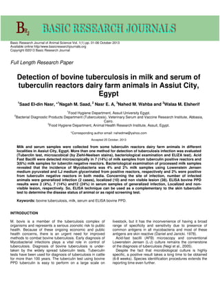
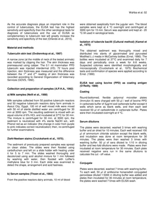
Basic Research Journal of Animal Science Vol. 1(1) pp. 01-06 October 2013
Available online http//www.basicresearchjournals.org
Copyright ©2013 Basic Research Journal
Full Length Research Paper
Detection of bovine tuberculosis in milk and serum of
tuberculin reactors dairy farm animals in Assiut City,
Egypt
1
Saad El-din Nasr, *1
Nagah M. Saad, 2
Nasr E. A, 3
Nahed M. Wahba and 3
Walaa M. Elsherif
1
Food Hygiene Department, Assuit University Egypt.
2
Bacterial Diagnostic Products Department (Tuberculosis), Veterinary Serum and Vaccine Research Institute, Abbasia,
Cairo.
3
Food Hygiene Department, Animal Health Research Institute, Assuit, Egypt.
*Corresponding author email: nahedmw@yahoo.com
Accepted 25 October, 2013
Milk and serum samples were collected from some tuberculin reactors dairy farm animals in different
localities in Assiut City, Egypt. More than one method for detection of tuberculosis infection was evaluated
(Tuberclin test, microscobical (by Ziehl-Neelsen stains), bacteriological examination and ELIZA test). Acid
Fast Bacilli were detected microscopically in 7 (14%) of milk samples from tuberculin positive reactors and
3(6%) milk samples for tuberclin negative reactors. Bacteriological examination of processed milk samples
revealed that the incidence of Mycobacteria was 4% and 2% milk samples using Lowenstein Jensen
medium pyruvated and LJ medium glycerinated from positive reactors, respectively and 2% were positive
from tuberculin negative reactors in both media. Concerning the site of infection, number of infected
animals were generalized infection (2 cases), localized (10) and non visible lesion (38). ELISA bovine PPD
results were 2 (4%), 7 (14%) and12 (24%) in serum samples of generalized infection, Localized and non-
visible lesion, respectively. So, ELISA technique can be used as a complementary to the skin tuberculin
test to determine the disease status of animal or as rapid screening test.
Keywords: bovine tuberculosis, milk, serum and ELISA bovine PPD.
INTRODUCTION
M. bovis is a member of the tuberculosis complex of
organisms and represents a serious zoonotic risk to public
health. Because of these ongoing economic and public
health concerns, there is an urgent need for improved
methods to combat bovine tuberculosis. Early diagnosis of
Mycobacterial infections plays a vital role in control of
tuberculosis. Diagnosis of bovine tuberculosis is under-
taken by the widely spread tuberculin tests. Tuberculin
tests have been used for diagnosis of tuberculosis in cattle
for more than 100 years. The tuberculin test using bovine
PPD tuberculin is easy to perform on a large scale on
livestock, but it has the inconvenience of having a broad
range of specificity and sensitivity due to presence of
common antigens in all mycobacteria and most of these
antigens are skin reactive (Daniel and Janicki. 1978).
Acid-fast bacilli (AFB) microscopy and conventional
Lowenstein Jensen (L-J) culture remains the cornerstone
of the diagnosis of tuberculosis (Negi et al., 2005).
Despite the fact that microbiological culture is highly
specific, a positive result takes a long time to be obtained
(6-8 weeks). Species identification procedures extends the
reporting time even further.
As the accurate diagnosis plays an important role in the
control of tuberculosis, the ELISA test has the highest
sensitivity and specificity than the other serological tests for
diagnosis of tuberculosis and the use of ELISA as
complementary to tuberculin test will greatly increase the
sensitivity and specificity of the test (Orlando et al., 2010).
Material and methods
Tuberculin skin test (Ovdiennkop et al., 1987)
A narrow zone (at the middle of neck of the tested animals)
was marked by clipping the hair. The skin thickness was
premeasured using caliper. The 0.1 ml mammalian PPD
tuberculin was injected interdermal (I/D). 72 hours post
injection, skin thickness was measured and the difference
between the 1
st
and 2
nd
reading of skin thickness was
recorded according to General Organization of Veterinary
Services (GOVS, 1992).
Collection and preparation of samples (A.P.H.A., 1992)
a) Milk samples (Neill et al., 1988)
Milk samples collected from 50 positive tuberculin reactors
and 50 negative tuberculin reactors dairy farm animals in
Assiut City, Egypt. 100 ml of well mixed milk were mixed
with 50 ml of sterile distilled water are centrifuged for 30
min at 3000 rpm. The resulting sediment is mixed with an
equal volume of 6% HCL and incubated at 37°C for 30 min.
The mixture is centrifuged for 30 min. at 3000 rpm, the
sediment is neutralized with 4% sterile NaOH sol., with
Phenol red as an indicator (the change in color from purple
to pink indicates correct neutralization) then, re-centrifuged
for further examinations.
Ziehl-Neelsen stains (Cruickshank et al., 1975).
The sediment of previously prepared samples was spread
on clean slides. The slides were then flooded using
carbolfuchsin and left on a slide holder with heating under
it for 5 to 7 min. The slides washed thoroughly and
decolorized using acid alcohol mixture for 1 min followed
by washing with water, then flooded with Loffler's
methylene blue for 3 min. Each slide was examined to
detect the shape, arrangement and acid fastness.
b) Serum samples (Thoen et al., 1983)
From the positive reactors dairy animals, 10 ml of blood
Wahba et al. 02
were obtained aseptically from the jugular vein. The blood
samples were kept at 4 °C overnight and centrifuged at
3000 rpm for 15 min. Serum was aspirated and kept at - 20
°C till used in serological tests.
Isolation of tubercle bacilli (Cultural method) (Kamel et
al., 1975)
The obtained sediment was thoroughly mixed and
distributed into slants of glycerinated and pyruvated
Modified L-J media in McCartney bottles (Ernst, 1990). The
bottles were incubated at 37°C and examined daily for 7
days and periodically once a week for 6-8 weeks.
Suspected colonies were identified morphologically and
microscopically (Cruickshank et al., 1975) Biochemical
tests and confirmation of species were applied according to
Ernst. (1990)
ELISA test using bovine (PPD) as coating antigen
(O’Reilly. 1989)
Coating
Round-bottomed, flexible polyvinyl microtiter plates
(Immulon II) were charged with 50 µl / well of bovine PPD
in carbonate buffer of 5µg/ml sod carbonate buffer except 4
wells which serve as blank wells, and then each well
received 50 µl of carbodiimide in carbonate buffer. Plates
were then incubated overnight at 4 °C.
Serum dilutions
The plates were decanted, washed 3 times with washing
buffer and air dried for 10 minutes. Each well received 100
µl of ammonium chloride solution except the blank wells,
and incubation was done at room temperature for 30
minutes. The plates were then washed 3 times with
washing buffer. 50 µl of sera diluted to 1/10 in diluting
buffer and two-fold dilutions were made. Plates were then
incubated at room temperature for 30 minutes. Each plate
received negative sera run in the same dilutions to
calculate the cut - off value.
Conjugate
Plates were decanted, washed 7 times with washing buffer.
To each well, 50 µl of antibovine horseradish conjugated
peroxidase diluted 1:5000 in diluting buffer was added and
plates then incubated for 30 minutes at room temperature,
the plates were washed 7 times with ELISA wash.
03. Basic Res. J. Anim. Sci.
Table 1. Incidence of Acid Fast Bacilli in milk samples examined
microscopically and culturing
Positive samples Negative
samples
No. % No. %
Milkfrom tuberculin
positive
reactors
50 7 14 43 86
tuberculin
negative
reactors
Total 100 10 10 90 90
Table 2. Incidence of Mycobacteria isolated from milk samples using conventional
culture method
Lowenstein- Jensen media
pyruvated glycerinated
+ve samples % +ve samples %
Milkfrom
tuberculin
positive
reactors
50 2 4 1 2 3
tuberculin
negative
reactors
Total 100 3 3 1 1 4
Substrate
50 µl of freshly prepared OPD Substrate buffer was added
and the plates were incubated in dark for 10 minutes.
Optical density measurement
The optical density OD was measured at 450 nm using
ELISA reader. The serum dilution was considered positive
if it yielded a mean OD of each group equal to/or greater
than the cut off value (Dimitri and Mikhail, 1996).
RESULTS
The results of AFB in milk samples examined by direct
smear using ZN stain were 7 (14%) and 3 (6%) from
tuberculin positive reactors and tuberculin negative
reactors, respectively (Table 1). Mycobacteria could be
detected by culturing in 3 (6%) and 1 (2%) milk samples
from tuberculin positive and negative reactors. The
incidence of Mycobacteria was 4% and 2% using
Lowenstein Jensen medium pyruvated and LJ medium
glycerinated from positive reactors, respectively and 2%
were positive from tuberculin negative reactors in both
media (Table 2). M. bovis was isolated in percentages of 4
and 2 % of tuberculin positive and negative reactors
however Mott was isolated from 2% of tuberculin positive
reactors only (Table 3). ELISA results indicated that
Mycobacteria could be detected among tuberculin positive
reactors in percentages of 2 (4%), 7 (14%) and 12 (24%) in
generalized TB, localized lesion and non-visible lesion,
respectively (Table 4).
DISCUSSION
Bovine tuberculosis infection in cattle is usually diagnosed
in the live animal on the basis of delayed hypersensitivity
reactions. Delayed hypersensitivity test is the standard
method for detection of bovine tuberculosis (Ereny Markos,
2011).
The I/D tuberculin test has been the widest used
diagnostic technique although it has some sensitivity and
specificity deficiencies and requires a second inspection of
the animal for its interpretation beside the three traditional
diagnostic techniques (Postmortem examination, Ziehl
Neelson stain and bacteriological culture) used for
tuberculosis diagnosis in slaughtered animals (Retmal and
Abolas, 2004). It allows detection of cattle that have been
exposed to M. bovis. However, in herds where control of
TB based on the identification and removal of reactors to
this test, some animals in advanced stages of the disease
Wahba et al. 04
Table 3. Identification of Mycobacteria isolated from milk samples
Type of milk samples Total No. of
samples
Positive culture
for AFB
Types of Mycobacteria
M. bovis MOTT
No. % No. % No. %
Milkfrom
tuberculin
positive
reactors
50 3 6 2 4 1 2
tuberculin
negative
reactors
50 1 2 1 2 0 0
Table 4. Correlation between the types of infection, cultural and serum ELISA test results
(among tuberculin positive reactors)
PM finding
No. of infected
animals
Culture exam.
ELISA
results
%
(/50)Milk %
(/50)
Generalized TB 2 1 2 2 4
Localized TB 2 4 7 14
Non visible lesion 38 0 0 12 24
Total 50 3 6 21 42
and with open lesions do not show reactivity to tuberculin
(anergic) and might remain in the herd, thus constituting a
potential source of infection in susceptible cattle (Diaz -
Otero et al., 2003).
The results of AFB in milk samples examined by direct
smear using ZN stain were higher than that recorded by
culturing among tuberculin positive reactors and tuberculin
negative reactors. These results may be due to the fact
that ZN method was used for staining of tubercle, other
acid – fast and saprophytic bacilli and can't differentiate
between them (Mackie and McCartney. 1960 and
Cruickshank et al., 1975).
Mycobacteria could be detected by culturing in 3 (6%)
and 1 (2%) milk samples from tuberculin positive and
negative reactors. These results are nearly similar to Gad
et al. (2000) (5.6%) Higher results (9.3%) were detected in
Canadian cattle by Ameni et al. (2003) (13.3%). Hamid et
al. (2003) recorded 28.07% from milk of tuberculin positive
buffaloes and from milk of 25% tuberculin positive cows.
In this study the percentage of Mycobacteria on L-J
pyruvate were 4 and 2% while on L-J glycerol were 2 and
0% for tuberculin positive and negative reactors,
respectively. These results indicated that L-J medium could
be used for primary isolation, sensitivity testing,
identification and sub-culturing of the majority of
Mycobacteria as reported by Maureen (1981).
Although the number of M. bovis positive samples was
low, the habit of pooling milk may still pose a public health
danger to milk consumers (Schelling et al., 2000).
Furthermore, M. bovis is the major cause of extra-
pulmonary tuberculosis like tuberculosis of gastrointestinal
tract and tuberculosis of cervical and mesenteric lymph
nodes, the peritoneum and the genitourinary tract (Bonsu
et al., 2001). The distribution of the tuberculosis lesion
varied greatly, being pulmonary, extra-pulmonary, mixed or
generalized. The variation of distribution of the lesion may
governed by the portal of entry and the possibility of
dissemination (Adway, 1986). The present study revealed
that the number of infected animals with generalized TB,
localized lesions and non visible lesions were 2, 10 and 38
among tuberculin positive reactors (Table 4). Freitas et al.
(2001) recorded 27.9% generalized lesions and 72.1%
localized lesions in Brazil. The rate of recovery of M. bovis
from reactor animals with visible lesions was (84%) where
105 isolates had been isolated from 125 reactor buffaloes
with V.L. that nearly agree with the recorded by Jacob
(1994). On the other hand Stefan et al. (2009) found that
out of approximately 32,800 inspected cattle 4.7% showed
suspected tuberculous lesions, Culture of suspected
lesions yielded acid-fast bacilli in 11% of cases.
Furthermore, the routine abattoir inspection was able to
detect only 117 of the total 3322 carcasses inspected
(Demelash et al., 2010). Moreover, Huitema. (1994)
attributed the cause of non specific reaction to the
assumption that those animals might be slaughtered at
early stage of the disease where the tubercles lesions were
invisible or lesions may be found in parts of the body not
routinely examined in the carcass such as bone or brain.
The ELISA is one of the main important serological tests
for diagnosis of bovine tuberculosis. Correlation between
the types of infection, cultural of milk samples and ELISA
on the sera among tuberculin positive reactors (using
bovine (PPD) as coating antigen) was investigated in Table
(4). It was found that 1 from 2 of generalized TB cases
among animals was positive for cultural of milk samples, 2
from 10 of localized TB forms had infected milk while,
05. Basic Res. J. Anim. Sci.
negative results obtained in milk of non visible lesion
cases. So, culture of milk could not be used for detection of
tuberculosis but there was danger for the presence of
tubercle bacilli in milk to the consumer. Dunn and
Hodgson. (1982) proved that milk samples with enough
viable tubercle bacilli excreted can infect the milk of 100
clean cows to be of infectious level. ELISA results
indicated that all serum of generalized TB cases was found
positive to ELISA, only 7 from 10 of localized TB and 12
from 38 of non visible lesion cases was positive. Ereny
Markos. (2011) found that out of 43 tuberculin reactor
cattle with visible lesion and Out of 19 tuberculin reactor
cattle with non visible lesion, 39 (90.6%) and 3 (15.7%)
were positive for ELISA , respectively.
False negative ELISA results obtained in the current
study (only 7 from 10 of localized TB) explained by the fact
that low titer of antibodies to Mycobacterial antigens which
may be associated with heavy infection and that antigens
may be released into the blood circulation and cause
temporary suppression of antibody formation Krambovitis.
(1986) and that agree with Thorns and . (1983) who
cleared that the level of specific antibodies in many M.
bovis infected cattle may be low or undetectable. This is
supported with Amadori et al. (1998) who pointed that
antibodies to Mycobacterial antigens were investigated
with various rates of success since the humeral immune
response to M. bovis is late and irregular during the course
of the disease.
Sensitivity of ELISA was 65.6% and specificity was
56.4%. When ELISA and caudal fold tuberculin test results
in cattle were interpreted in parallel, sensitivity was 95%,
specificity was 92.7%. The results of the ELISA and single
cervical tuberculin test were interpreted in parallel or
series. The ELISA was able to detect 47% of 68 (69%)
cattle infected with M. bovis that did not have visible
tuberculous lesions at P/M examination (Gaborick at al.,
1996). For this Sensitivity of ELISA and specificity in
comparison with prolonged time cultural consuming and
false positive results of tuberculin skin test, Nasr and
Osman. (2007) suggested that ELISA technique can be
used with the skin test to determine the disease status of
animal and reduce the frequency of misdiagnosis of
animals free of bovine tuberculosis.
CONCLUSION
It could be concluded that Bovine tuberculosis still
constitutes a public health hazard and every effort should
be made to control such disease in Egypt. The
aforementioned proved that culture of milk could not be
used for detection of tuberculosis but there was danger for
the presence of tubercle bacilli in milk to the consumer.
Moreover, the ELISA technique can be used as a
complementary to the tuberculin skin test to determine the
disease status of animal or as a rapid screening test for
herd testing program.
ACKNOWLEDGEMENTS
Authors are sincerely grateful for the great help provided
by all the staff members of Bacterial Diagnostic Products
Dep. (Tuberculosis), Vet. Serum and Vaccine Res. Inst.,
Abbasia, Cairo.
REFERENCES
A.P.H.A. (American Public Health Association) (1992). Standard Methods
for the Examination of Dairy Products. 16th
Ed., American Public Health
Association, New York.
Adway TA (1986). Studies on Tuberculosis in Slaughtered Cattle, Ph. D.
Thesis, Fac. Vet. Med., Cairo Uni.
Amadori M, Tameni S, Scaccaglia P, Cavirani AIL, Quondam RG (1998).
Antibody tests for identification of M. bovis infected bovine herds. J.
Clin. Microbiol., 36: 566-568.
Ameni G, Bonnet P, Tibbo M (2003). Across sectional study of bovine
tuberculosis in selected dairy farms in Ethiopia. Inter. J. App. Res. Vet.
Med., 1 (4. 253-258.
Bonsu OA, laing E, Akanmori BD (2001). Prevalence of tuberculosis in
cattle in the Dangme, west distinct of Ghana, public health
amplifications. Acta. Trop. J., 76 (1. 9-14.
Cruickshank R, Duguid JP, Marmion BP, Swain RHA (1975). Medical
Microbiology. Vol. 11 part I: Churchill living stone. Edinburgh, London
and New York.
Daniel TM, Janicki BW (1978). Mycobacterial antigens : A review of their
isolation, chemistry and immunological properties .Microbiol. Rev., 113
– 119.
Demelash B, Asseged B, Eystein S (2010). Diagnostic efficiency of
abattoir meat inspection service in Ethiopia to detect carcasses infected
with M. bovis: Implications for public health. BMC Public Health, 10:
462.
Dimitri RA, Mikhail DG (1996). Specific skin reactivity and ELISA for the
diagnosis of bovine tuberculosis using 30,000 Da and PPD antigens in
guinea pigs. J. Egypt. Vet. Med. Ass., 56 (4. 531-546.
Dunn BL, Hodgson DJ (1982). Atypical mycobacteria in milk. J. App.
Bacteriol., 52: 373-376.
Ereny MSL (2011). Evaluation of New Advanced Diagnostic Techniques
for Detection of Bovine Tuberculosis. Ph. D. Thesis, Fac. Vet. Med.
Beni-Suif Uni.
Ernst LB (1990). Mycobacteria species: The Agents of Animal
Tuberculosis. In Ernst L. Biberstein and Yuan Chung Zee (Ed). Review
of Vet. Microbiol. Blakwell Scientific Pub., INC. Boston, Oxford, London,
Edinburgh, Melbourne. Ch. 28, Pp. 202-212.
Freitas JA, Guerra JL, Panetta JC (2001). Characteristics of T.B. in
slaughtered water buffaloes. Brez. J. Vet. Res. Anim. Sci., 38 (4. 170-
176.
Gaborick CM, Salaman MD, Ellis RP, Triantis J (1996). Valuation of A five
antigen. ELISA for diagnosis of tuberculosis in cattle and cervidae. Am.
Vet. Med. Ass., 209 (9. 962- 966.
Gad EWA, El-Abeedy A, Mettias KN, Manal A (2000). The present state
and public health importance of tuberculosis of bovine udder. J. Egypt.
Vet. Med. Ass., 60 (6. 189- 194.
GOVS (1992). General Organization for the Veterinary Services. Ministry
of Agriculture, Egypt.
Hamid J, Puran D, Asif S (2003). Bovine tuberculosis in dairy animals at
Lahore, threat to the public health. .http://priory.com/ vet/ bovinetb.htm.
Huitema H (1994). Tuberculosis in Animal and Man. 1st
Ed., Publication of
the Royal Netherlands Tuberculosis Association (KNCV).
Jacob AK (1994). Some studies on the evaluation of BCG vaccines in
cattle. M.V. Sc. Thesis, Internal Vet. Med., Zagazig Uni.
Kamel YY, Ismail AA, Ahmed AA, Moustafa TH, Mahmoud MS (1975).
Sanitary condition of market milk in Assuit city, incidence of M. bovis
and a modified scheme for typing. Assuit Vet. Med. J., 11 (3&4.169-
180.
Krambovitis E (1986). Detection of antibodies to M. tuberculosis plasma
membrane antigen by enzyme linked immuno sorbent assay. J. Med.
Microbiol., 21: 257-264.
Mackie TJ, McCarteney JE (1960). Handbook of Bacteriology. 10th
Ed. E.
and S. Living stone limited, Edinburgh and London.
Maureen CV (1981). Mycobacteria. Printed in Great Britian by John
Wright and sons Ltd, at the stone bridge press. Bristol B545.
Nasr EA, Osman HS (2007). Evaluation of Rapid Blood Test for Detection
of M. bovis Antibody in Cattle. 9th
Sci. Cong., Egyptian Society For
Cattle Diseases, Assiut, Egypt: (64-71).
Negi SS, Khan SFB, Gupta S, Pasha ST, Khare S, Lal S (2005).
Comparison of the conventional diagnostic modalities, bactec culture
and PCR test for diagnosis of tuberculosis. Indian J. Med. Microbiol., 23
(1. 29-33.
Neill SD, Brien JJ, MacCracken RM (1988). M. bovis in the anterior
respiratory tract in the herds of tuberculin reacting cattle. Vet. Rec.,
122: 184-186.
O'Reilly LM (1989). Preliminary Evaluation of the Usefulness of An
Enzyme Linked Immunosorbent Assay for The Diagnosis of Bovine
Tuberculosis. I.U.A.T.L.D. Scientific Committee "Tuberculosis in
animal" Paris.
Wahba et al. 06
Orlando G, Cordier L, Mazza F, Casazza G, Villa AM, Codecasa L, Negri
E, Cargnel A, Ferrarese M, Rizzardini G (2010). Interferon-gamma
releasing assay versus tuberculin skin testing for latent tuberculosis
infection in targeted screening programs for high risk immigrants.
Infection, 38: 195-204.
Ovdiennkop NP, Shchur EVE, Naimanor AK, Yokuskeva OV, Sharov A,
Planikov ES (1987). Frequency of tuberculin injection in cattle. Vet -
Muscow, us sr., 8: 29-33.
Schelling E, Diguimbaye C, Daoud S (2000). Zoonoses in nomadic
populations of Chad- preliminary results obtained in humans and
livestock. 9th
conf. of the Inter. Soc. Vet. Epidemiol. and Eco.
Breckenridge, 6-11 August, abstract No. 396.
Stefan B, Rebuma F, Meseret H, Endalamaw G, Araya M, Lawrence Y,
Gobena A, Vordermeier M, Brian D, Robertson NH, Smith HE, Douglas
YR, Glyn H, Abraham A, Stephen VG (2009). The burden of
Mycobacterial disease in Ethiopian cattle: Implications for public health.
Plos. One, 4 (4): 5068-5070.
Thoen C, Hall MR, Petersburg TA, Harrington R (1983). Detection of
mycobacteria antibodies in sera of cattle experimentally exposed to M.
bovis by use of modified ELISA. 26th
Annual proc. Amer. Ass. Vet. Lab.
Diag. USA. Pp. 25- 38.
Thorns CJ, Morris JA (1983). The immune spectrum of M. bovis infections
in some mammalian species. Vet. Bull., 53 (6): 543-547.

Saad et al

  • 1.
    Basic Research Journalof Animal Science Vol. 1(1) pp. 01-06 October 2013 Available online http//www.basicresearchjournals.org Copyright ©2013 Basic Research Journal Full Length Research Paper Detection of bovine tuberculosis in milk and serum of tuberculin reactors dairy farm animals in Assiut City, Egypt 1 Saad El-din Nasr, *1 Nagah M. Saad, 2 Nasr E. A, 3 Nahed M. Wahba and 3 Walaa M. Elsherif 1 Food Hygiene Department, Assuit University Egypt. 2 Bacterial Diagnostic Products Department (Tuberculosis), Veterinary Serum and Vaccine Research Institute, Abbasia, Cairo. 3 Food Hygiene Department, Animal Health Research Institute, Assuit, Egypt. *Corresponding author email: nahedmw@yahoo.com Accepted 25 October, 2013 Milk and serum samples were collected from some tuberculin reactors dairy farm animals in different localities in Assiut City, Egypt. More than one method for detection of tuberculosis infection was evaluated (Tuberclin test, microscobical (by Ziehl-Neelsen stains), bacteriological examination and ELIZA test). Acid Fast Bacilli were detected microscopically in 7 (14%) of milk samples from tuberculin positive reactors and 3(6%) milk samples for tuberclin negative reactors. Bacteriological examination of processed milk samples revealed that the incidence of Mycobacteria was 4% and 2% milk samples using Lowenstein Jensen medium pyruvated and LJ medium glycerinated from positive reactors, respectively and 2% were positive from tuberculin negative reactors in both media. Concerning the site of infection, number of infected animals were generalized infection (2 cases), localized (10) and non visible lesion (38). ELISA bovine PPD results were 2 (4%), 7 (14%) and12 (24%) in serum samples of generalized infection, Localized and non- visible lesion, respectively. So, ELISA technique can be used as a complementary to the skin tuberculin test to determine the disease status of animal or as rapid screening test. Keywords: bovine tuberculosis, milk, serum and ELISA bovine PPD. INTRODUCTION M. bovis is a member of the tuberculosis complex of organisms and represents a serious zoonotic risk to public health. Because of these ongoing economic and public health concerns, there is an urgent need for improved methods to combat bovine tuberculosis. Early diagnosis of Mycobacterial infections plays a vital role in control of tuberculosis. Diagnosis of bovine tuberculosis is under- taken by the widely spread tuberculin tests. Tuberculin tests have been used for diagnosis of tuberculosis in cattle for more than 100 years. The tuberculin test using bovine PPD tuberculin is easy to perform on a large scale on livestock, but it has the inconvenience of having a broad range of specificity and sensitivity due to presence of common antigens in all mycobacteria and most of these antigens are skin reactive (Daniel and Janicki. 1978). Acid-fast bacilli (AFB) microscopy and conventional Lowenstein Jensen (L-J) culture remains the cornerstone of the diagnosis of tuberculosis (Negi et al., 2005). Despite the fact that microbiological culture is highly specific, a positive result takes a long time to be obtained (6-8 weeks). Species identification procedures extends the reporting time even further.
  • 2.
    As the accuratediagnosis plays an important role in the control of tuberculosis, the ELISA test has the highest sensitivity and specificity than the other serological tests for diagnosis of tuberculosis and the use of ELISA as complementary to tuberculin test will greatly increase the sensitivity and specificity of the test (Orlando et al., 2010). Material and methods Tuberculin skin test (Ovdiennkop et al., 1987) A narrow zone (at the middle of neck of the tested animals) was marked by clipping the hair. The skin thickness was premeasured using caliper. The 0.1 ml mammalian PPD tuberculin was injected interdermal (I/D). 72 hours post injection, skin thickness was measured and the difference between the 1 st and 2 nd reading of skin thickness was recorded according to General Organization of Veterinary Services (GOVS, 1992). Collection and preparation of samples (A.P.H.A., 1992) a) Milk samples (Neill et al., 1988) Milk samples collected from 50 positive tuberculin reactors and 50 negative tuberculin reactors dairy farm animals in Assiut City, Egypt. 100 ml of well mixed milk were mixed with 50 ml of sterile distilled water are centrifuged for 30 min at 3000 rpm. The resulting sediment is mixed with an equal volume of 6% HCL and incubated at 37°C for 30 min. The mixture is centrifuged for 30 min. at 3000 rpm, the sediment is neutralized with 4% sterile NaOH sol., with Phenol red as an indicator (the change in color from purple to pink indicates correct neutralization) then, re-centrifuged for further examinations. Ziehl-Neelsen stains (Cruickshank et al., 1975). The sediment of previously prepared samples was spread on clean slides. The slides were then flooded using carbolfuchsin and left on a slide holder with heating under it for 5 to 7 min. The slides washed thoroughly and decolorized using acid alcohol mixture for 1 min followed by washing with water, then flooded with Loffler's methylene blue for 3 min. Each slide was examined to detect the shape, arrangement and acid fastness. b) Serum samples (Thoen et al., 1983) From the positive reactors dairy animals, 10 ml of blood Wahba et al. 02 were obtained aseptically from the jugular vein. The blood samples were kept at 4 °C overnight and centrifuged at 3000 rpm for 15 min. Serum was aspirated and kept at - 20 °C till used in serological tests. Isolation of tubercle bacilli (Cultural method) (Kamel et al., 1975) The obtained sediment was thoroughly mixed and distributed into slants of glycerinated and pyruvated Modified L-J media in McCartney bottles (Ernst, 1990). The bottles were incubated at 37°C and examined daily for 7 days and periodically once a week for 6-8 weeks. Suspected colonies were identified morphologically and microscopically (Cruickshank et al., 1975) Biochemical tests and confirmation of species were applied according to Ernst. (1990) ELISA test using bovine (PPD) as coating antigen (O’Reilly. 1989) Coating Round-bottomed, flexible polyvinyl microtiter plates (Immulon II) were charged with 50 µl / well of bovine PPD in carbonate buffer of 5µg/ml sod carbonate buffer except 4 wells which serve as blank wells, and then each well received 50 µl of carbodiimide in carbonate buffer. Plates were then incubated overnight at 4 °C. Serum dilutions The plates were decanted, washed 3 times with washing buffer and air dried for 10 minutes. Each well received 100 µl of ammonium chloride solution except the blank wells, and incubation was done at room temperature for 30 minutes. The plates were then washed 3 times with washing buffer. 50 µl of sera diluted to 1/10 in diluting buffer and two-fold dilutions were made. Plates were then incubated at room temperature for 30 minutes. Each plate received negative sera run in the same dilutions to calculate the cut - off value. Conjugate Plates were decanted, washed 7 times with washing buffer. To each well, 50 µl of antibovine horseradish conjugated peroxidase diluted 1:5000 in diluting buffer was added and plates then incubated for 30 minutes at room temperature, the plates were washed 7 times with ELISA wash.
  • 3.
    03. Basic Res.J. Anim. Sci. Table 1. Incidence of Acid Fast Bacilli in milk samples examined microscopically and culturing Positive samples Negative samples No. % No. % Milkfrom tuberculin positive reactors 50 7 14 43 86 tuberculin negative reactors Total 100 10 10 90 90 Table 2. Incidence of Mycobacteria isolated from milk samples using conventional culture method Lowenstein- Jensen media pyruvated glycerinated +ve samples % +ve samples % Milkfrom tuberculin positive reactors 50 2 4 1 2 3 tuberculin negative reactors Total 100 3 3 1 1 4 Substrate 50 µl of freshly prepared OPD Substrate buffer was added and the plates were incubated in dark for 10 minutes. Optical density measurement The optical density OD was measured at 450 nm using ELISA reader. The serum dilution was considered positive if it yielded a mean OD of each group equal to/or greater than the cut off value (Dimitri and Mikhail, 1996). RESULTS The results of AFB in milk samples examined by direct smear using ZN stain were 7 (14%) and 3 (6%) from tuberculin positive reactors and tuberculin negative reactors, respectively (Table 1). Mycobacteria could be detected by culturing in 3 (6%) and 1 (2%) milk samples from tuberculin positive and negative reactors. The incidence of Mycobacteria was 4% and 2% using Lowenstein Jensen medium pyruvated and LJ medium glycerinated from positive reactors, respectively and 2% were positive from tuberculin negative reactors in both media (Table 2). M. bovis was isolated in percentages of 4 and 2 % of tuberculin positive and negative reactors however Mott was isolated from 2% of tuberculin positive reactors only (Table 3). ELISA results indicated that Mycobacteria could be detected among tuberculin positive reactors in percentages of 2 (4%), 7 (14%) and 12 (24%) in generalized TB, localized lesion and non-visible lesion, respectively (Table 4). DISCUSSION Bovine tuberculosis infection in cattle is usually diagnosed in the live animal on the basis of delayed hypersensitivity reactions. Delayed hypersensitivity test is the standard method for detection of bovine tuberculosis (Ereny Markos, 2011). The I/D tuberculin test has been the widest used diagnostic technique although it has some sensitivity and specificity deficiencies and requires a second inspection of the animal for its interpretation beside the three traditional diagnostic techniques (Postmortem examination, Ziehl Neelson stain and bacteriological culture) used for tuberculosis diagnosis in slaughtered animals (Retmal and Abolas, 2004). It allows detection of cattle that have been exposed to M. bovis. However, in herds where control of TB based on the identification and removal of reactors to this test, some animals in advanced stages of the disease
  • 4.
    Wahba et al.04 Table 3. Identification of Mycobacteria isolated from milk samples Type of milk samples Total No. of samples Positive culture for AFB Types of Mycobacteria M. bovis MOTT No. % No. % No. % Milkfrom tuberculin positive reactors 50 3 6 2 4 1 2 tuberculin negative reactors 50 1 2 1 2 0 0 Table 4. Correlation between the types of infection, cultural and serum ELISA test results (among tuberculin positive reactors) PM finding No. of infected animals Culture exam. ELISA results % (/50)Milk % (/50) Generalized TB 2 1 2 2 4 Localized TB 2 4 7 14 Non visible lesion 38 0 0 12 24 Total 50 3 6 21 42 and with open lesions do not show reactivity to tuberculin (anergic) and might remain in the herd, thus constituting a potential source of infection in susceptible cattle (Diaz - Otero et al., 2003). The results of AFB in milk samples examined by direct smear using ZN stain were higher than that recorded by culturing among tuberculin positive reactors and tuberculin negative reactors. These results may be due to the fact that ZN method was used for staining of tubercle, other acid – fast and saprophytic bacilli and can't differentiate between them (Mackie and McCartney. 1960 and Cruickshank et al., 1975). Mycobacteria could be detected by culturing in 3 (6%) and 1 (2%) milk samples from tuberculin positive and negative reactors. These results are nearly similar to Gad et al. (2000) (5.6%) Higher results (9.3%) were detected in Canadian cattle by Ameni et al. (2003) (13.3%). Hamid et al. (2003) recorded 28.07% from milk of tuberculin positive buffaloes and from milk of 25% tuberculin positive cows. In this study the percentage of Mycobacteria on L-J pyruvate were 4 and 2% while on L-J glycerol were 2 and 0% for tuberculin positive and negative reactors, respectively. These results indicated that L-J medium could be used for primary isolation, sensitivity testing, identification and sub-culturing of the majority of Mycobacteria as reported by Maureen (1981). Although the number of M. bovis positive samples was low, the habit of pooling milk may still pose a public health danger to milk consumers (Schelling et al., 2000). Furthermore, M. bovis is the major cause of extra- pulmonary tuberculosis like tuberculosis of gastrointestinal tract and tuberculosis of cervical and mesenteric lymph nodes, the peritoneum and the genitourinary tract (Bonsu et al., 2001). The distribution of the tuberculosis lesion varied greatly, being pulmonary, extra-pulmonary, mixed or generalized. The variation of distribution of the lesion may governed by the portal of entry and the possibility of dissemination (Adway, 1986). The present study revealed that the number of infected animals with generalized TB, localized lesions and non visible lesions were 2, 10 and 38 among tuberculin positive reactors (Table 4). Freitas et al. (2001) recorded 27.9% generalized lesions and 72.1% localized lesions in Brazil. The rate of recovery of M. bovis from reactor animals with visible lesions was (84%) where 105 isolates had been isolated from 125 reactor buffaloes with V.L. that nearly agree with the recorded by Jacob (1994). On the other hand Stefan et al. (2009) found that out of approximately 32,800 inspected cattle 4.7% showed suspected tuberculous lesions, Culture of suspected lesions yielded acid-fast bacilli in 11% of cases. Furthermore, the routine abattoir inspection was able to detect only 117 of the total 3322 carcasses inspected (Demelash et al., 2010). Moreover, Huitema. (1994) attributed the cause of non specific reaction to the assumption that those animals might be slaughtered at early stage of the disease where the tubercles lesions were invisible or lesions may be found in parts of the body not routinely examined in the carcass such as bone or brain. The ELISA is one of the main important serological tests for diagnosis of bovine tuberculosis. Correlation between the types of infection, cultural of milk samples and ELISA on the sera among tuberculin positive reactors (using bovine (PPD) as coating antigen) was investigated in Table (4). It was found that 1 from 2 of generalized TB cases among animals was positive for cultural of milk samples, 2 from 10 of localized TB forms had infected milk while,
  • 5.
    05. Basic Res.J. Anim. Sci. negative results obtained in milk of non visible lesion cases. So, culture of milk could not be used for detection of tuberculosis but there was danger for the presence of tubercle bacilli in milk to the consumer. Dunn and Hodgson. (1982) proved that milk samples with enough viable tubercle bacilli excreted can infect the milk of 100 clean cows to be of infectious level. ELISA results indicated that all serum of generalized TB cases was found positive to ELISA, only 7 from 10 of localized TB and 12 from 38 of non visible lesion cases was positive. Ereny Markos. (2011) found that out of 43 tuberculin reactor cattle with visible lesion and Out of 19 tuberculin reactor cattle with non visible lesion, 39 (90.6%) and 3 (15.7%) were positive for ELISA , respectively. False negative ELISA results obtained in the current study (only 7 from 10 of localized TB) explained by the fact that low titer of antibodies to Mycobacterial antigens which may be associated with heavy infection and that antigens may be released into the blood circulation and cause temporary suppression of antibody formation Krambovitis. (1986) and that agree with Thorns and . (1983) who cleared that the level of specific antibodies in many M. bovis infected cattle may be low or undetectable. This is supported with Amadori et al. (1998) who pointed that antibodies to Mycobacterial antigens were investigated with various rates of success since the humeral immune response to M. bovis is late and irregular during the course of the disease. Sensitivity of ELISA was 65.6% and specificity was 56.4%. When ELISA and caudal fold tuberculin test results in cattle were interpreted in parallel, sensitivity was 95%, specificity was 92.7%. The results of the ELISA and single cervical tuberculin test were interpreted in parallel or series. The ELISA was able to detect 47% of 68 (69%) cattle infected with M. bovis that did not have visible tuberculous lesions at P/M examination (Gaborick at al., 1996). For this Sensitivity of ELISA and specificity in comparison with prolonged time cultural consuming and false positive results of tuberculin skin test, Nasr and Osman. (2007) suggested that ELISA technique can be used with the skin test to determine the disease status of animal and reduce the frequency of misdiagnosis of animals free of bovine tuberculosis. CONCLUSION It could be concluded that Bovine tuberculosis still constitutes a public health hazard and every effort should be made to control such disease in Egypt. The aforementioned proved that culture of milk could not be used for detection of tuberculosis but there was danger for the presence of tubercle bacilli in milk to the consumer. Moreover, the ELISA technique can be used as a complementary to the tuberculin skin test to determine the disease status of animal or as a rapid screening test for herd testing program. ACKNOWLEDGEMENTS Authors are sincerely grateful for the great help provided by all the staff members of Bacterial Diagnostic Products Dep. (Tuberculosis), Vet. Serum and Vaccine Res. Inst., Abbasia, Cairo. REFERENCES A.P.H.A. (American Public Health Association) (1992). Standard Methods for the Examination of Dairy Products. 16th Ed., American Public Health Association, New York. Adway TA (1986). Studies on Tuberculosis in Slaughtered Cattle, Ph. D. Thesis, Fac. Vet. Med., Cairo Uni. Amadori M, Tameni S, Scaccaglia P, Cavirani AIL, Quondam RG (1998). Antibody tests for identification of M. bovis infected bovine herds. J. Clin. Microbiol., 36: 566-568. Ameni G, Bonnet P, Tibbo M (2003). Across sectional study of bovine tuberculosis in selected dairy farms in Ethiopia. Inter. J. App. Res. Vet. Med., 1 (4. 253-258. Bonsu OA, laing E, Akanmori BD (2001). Prevalence of tuberculosis in cattle in the Dangme, west distinct of Ghana, public health amplifications. Acta. Trop. J., 76 (1. 9-14. Cruickshank R, Duguid JP, Marmion BP, Swain RHA (1975). Medical Microbiology. Vol. 11 part I: Churchill living stone. Edinburgh, London and New York. Daniel TM, Janicki BW (1978). Mycobacterial antigens : A review of their isolation, chemistry and immunological properties .Microbiol. Rev., 113 – 119. Demelash B, Asseged B, Eystein S (2010). Diagnostic efficiency of abattoir meat inspection service in Ethiopia to detect carcasses infected with M. bovis: Implications for public health. BMC Public Health, 10: 462. Dimitri RA, Mikhail DG (1996). Specific skin reactivity and ELISA for the diagnosis of bovine tuberculosis using 30,000 Da and PPD antigens in guinea pigs. J. Egypt. Vet. Med. Ass., 56 (4. 531-546. Dunn BL, Hodgson DJ (1982). Atypical mycobacteria in milk. J. App. Bacteriol., 52: 373-376. Ereny MSL (2011). Evaluation of New Advanced Diagnostic Techniques for Detection of Bovine Tuberculosis. Ph. D. Thesis, Fac. Vet. Med. Beni-Suif Uni. Ernst LB (1990). Mycobacteria species: The Agents of Animal Tuberculosis. In Ernst L. Biberstein and Yuan Chung Zee (Ed). Review of Vet. Microbiol. Blakwell Scientific Pub., INC. Boston, Oxford, London, Edinburgh, Melbourne. Ch. 28, Pp. 202-212. Freitas JA, Guerra JL, Panetta JC (2001). Characteristics of T.B. in slaughtered water buffaloes. Brez. J. Vet. Res. Anim. Sci., 38 (4. 170- 176. Gaborick CM, Salaman MD, Ellis RP, Triantis J (1996). Valuation of A five antigen. ELISA for diagnosis of tuberculosis in cattle and cervidae. Am. Vet. Med. Ass., 209 (9. 962- 966. Gad EWA, El-Abeedy A, Mettias KN, Manal A (2000). The present state and public health importance of tuberculosis of bovine udder. J. Egypt. Vet. Med. Ass., 60 (6. 189- 194. GOVS (1992). General Organization for the Veterinary Services. Ministry of Agriculture, Egypt. Hamid J, Puran D, Asif S (2003). Bovine tuberculosis in dairy animals at Lahore, threat to the public health. .http://priory.com/ vet/ bovinetb.htm. Huitema H (1994). Tuberculosis in Animal and Man. 1st Ed., Publication of the Royal Netherlands Tuberculosis Association (KNCV). Jacob AK (1994). Some studies on the evaluation of BCG vaccines in cattle. M.V. Sc. Thesis, Internal Vet. Med., Zagazig Uni. Kamel YY, Ismail AA, Ahmed AA, Moustafa TH, Mahmoud MS (1975). Sanitary condition of market milk in Assuit city, incidence of M. bovis
  • 6.
    and a modifiedscheme for typing. Assuit Vet. Med. J., 11 (3&4.169- 180. Krambovitis E (1986). Detection of antibodies to M. tuberculosis plasma membrane antigen by enzyme linked immuno sorbent assay. J. Med. Microbiol., 21: 257-264. Mackie TJ, McCarteney JE (1960). Handbook of Bacteriology. 10th Ed. E. and S. Living stone limited, Edinburgh and London. Maureen CV (1981). Mycobacteria. Printed in Great Britian by John Wright and sons Ltd, at the stone bridge press. Bristol B545. Nasr EA, Osman HS (2007). Evaluation of Rapid Blood Test for Detection of M. bovis Antibody in Cattle. 9th Sci. Cong., Egyptian Society For Cattle Diseases, Assiut, Egypt: (64-71). Negi SS, Khan SFB, Gupta S, Pasha ST, Khare S, Lal S (2005). Comparison of the conventional diagnostic modalities, bactec culture and PCR test for diagnosis of tuberculosis. Indian J. Med. Microbiol., 23 (1. 29-33. Neill SD, Brien JJ, MacCracken RM (1988). M. bovis in the anterior respiratory tract in the herds of tuberculin reacting cattle. Vet. Rec., 122: 184-186. O'Reilly LM (1989). Preliminary Evaluation of the Usefulness of An Enzyme Linked Immunosorbent Assay for The Diagnosis of Bovine Tuberculosis. I.U.A.T.L.D. Scientific Committee "Tuberculosis in animal" Paris. Wahba et al. 06 Orlando G, Cordier L, Mazza F, Casazza G, Villa AM, Codecasa L, Negri E, Cargnel A, Ferrarese M, Rizzardini G (2010). Interferon-gamma releasing assay versus tuberculin skin testing for latent tuberculosis infection in targeted screening programs for high risk immigrants. Infection, 38: 195-204. Ovdiennkop NP, Shchur EVE, Naimanor AK, Yokuskeva OV, Sharov A, Planikov ES (1987). Frequency of tuberculin injection in cattle. Vet - Muscow, us sr., 8: 29-33. Schelling E, Diguimbaye C, Daoud S (2000). Zoonoses in nomadic populations of Chad- preliminary results obtained in humans and livestock. 9th conf. of the Inter. Soc. Vet. Epidemiol. and Eco. Breckenridge, 6-11 August, abstract No. 396. Stefan B, Rebuma F, Meseret H, Endalamaw G, Araya M, Lawrence Y, Gobena A, Vordermeier M, Brian D, Robertson NH, Smith HE, Douglas YR, Glyn H, Abraham A, Stephen VG (2009). The burden of Mycobacterial disease in Ethiopian cattle: Implications for public health. Plos. One, 4 (4): 5068-5070. Thoen C, Hall MR, Petersburg TA, Harrington R (1983). Detection of mycobacteria antibodies in sera of cattle experimentally exposed to M. bovis by use of modified ELISA. 26th Annual proc. Amer. Ass. Vet. Lab. Diag. USA. Pp. 25- 38. Thorns CJ, Morris JA (1983). The immune spectrum of M. bovis infections in some mammalian species. Vet. Bull., 53 (6): 543-547.