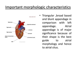
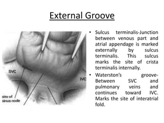
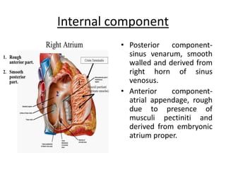
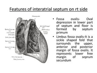
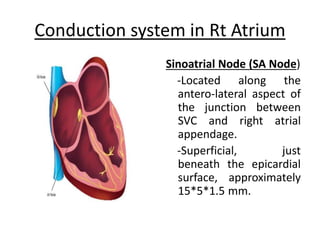
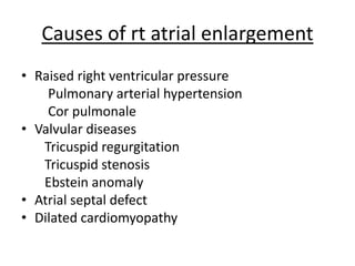
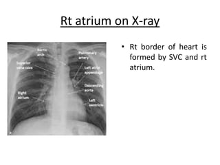
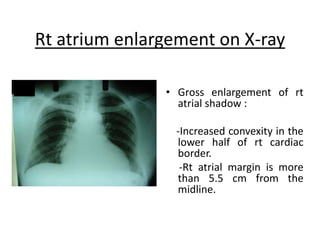
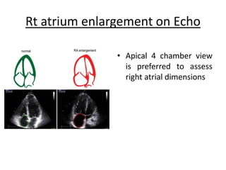
Clinical Presentation
Dr Anup Kumar Sarkar
MS (CVTS) Final part trainee
NHFH&RI
Right Atrium
• The right atrial chamber forms right border of
cardiac silhouette and most superficial
chamber.
• Recieves systemic venous drainage from SVC
& IVC
• Also receives coronary venous drainage from
coronary sinus.
Important morphologic characteristics
• Triangular ,broad based
and blunt appendage in
comparison with left
appendage. Atrial
appendage is of major
significance because of
their shape is the best
guide to atrial
morphology and hence
to atrial situs.
Important morphologic characteristics
• Eustachian valve at the orifice of IVC and
thebesian valve at the orifice of coronary
sinus.
• Crista terminalis- separates trabeculated from
non-trabeculated portion.
• Limbus of fossa ovalis.
External Groove
• Sulcus terminalis-Junction
between venous part and
atrial appendage is marked
externally by sulcus
terminalis. This sulcus
marks the site of crista
terminalis internally.
• Waterston’s groove-
Between SVC and
pulmonary veins and
continues toward IVC.
Marks the site of interatrial
fold.
Internal component
• Posterior component-
sinus venarum, smooth
walled and derived from
right horn of sinus
venosus.
• Anterior component-
atrial appendage, rough
due to presence of
musculi pectiniti and
derived from embryonic
atrium proper.
Features of interatrial septum on rt side
• Fossa ovalis- Oval
depression in lower part
of septum and floor is
formed by septum
primum
• Limbus fossa ovalis-It is a
sickle shaped fold that
surrounds the upper,
anterior and posterior
margin of fossa ovalis. It
represents lower free
margin of seprum
secundum
Conduction system in Rt Atrium
Sinoatrial Node (SA Node)
-Located along the
antero-lateral aspect of
the junction between
SVC and right atrial
appendage.
-Superficial, just
beneath the epicardial
surface, approximately
15*5*1.5 mm.
Conduction system in Rt Atrium
AV Node
• In Triangle of Koch
• Bounded by opening
of coronary sinuses,
septal tricuspid leaflet
and Tendon of Torado.
Interatrial Septum development and anomalies
Causes of rt atrial enlargement
• Raised right ventricular pressure
Pulmonary arterial hypertension
Cor pulmonale
• Valvular diseases
Tricuspid regurgitation
Tricuspid stenosis
Ebstein anomaly
• Atrial septal defect
• Dilated cardiomyopathy
Rt atrium on X-ray
• Rt border of heart is
formed by SVC and rt
atrium.
Rt atrium enlargement on X-ray
• Gross enlargement of rt
atrial shadow :
-Increased convexity in the
lower half of rt cardiac
border.
-Rt atrial margin is more
than 5.5 cm from the
midline.
Rt atrium enlargement on ECG
• Amplitude of P wave
in lead II >2.5 mm,
often demonstrate
peaked morphology
• Initial positive
deflection of P wave
in lead V1 >1.5 mm
Rt atrium enlargement on Echo
• Apical 4 chamber view
is preferred to assess
right atrial dimensions
Right  ATRIUM.pptx

Right ATRIUM.pptx

  • 1.
    Clinical Presentation Dr AnupKumar Sarkar MS (CVTS) Final part trainee NHFH&RI
  • 2.
    Right Atrium • Theright atrial chamber forms right border of cardiac silhouette and most superficial chamber. • Recieves systemic venous drainage from SVC & IVC • Also receives coronary venous drainage from coronary sinus.
  • 3.
    Important morphologic characteristics •Triangular ,broad based and blunt appendage in comparison with left appendage. Atrial appendage is of major significance because of their shape is the best guide to atrial morphology and hence to atrial situs.
  • 4.
    Important morphologic characteristics •Eustachian valve at the orifice of IVC and thebesian valve at the orifice of coronary sinus. • Crista terminalis- separates trabeculated from non-trabeculated portion. • Limbus of fossa ovalis.
  • 5.
    External Groove • Sulcusterminalis-Junction between venous part and atrial appendage is marked externally by sulcus terminalis. This sulcus marks the site of crista terminalis internally. • Waterston’s groove- Between SVC and pulmonary veins and continues toward IVC. Marks the site of interatrial fold.
  • 6.
    Internal component • Posteriorcomponent- sinus venarum, smooth walled and derived from right horn of sinus venosus. • Anterior component- atrial appendage, rough due to presence of musculi pectiniti and derived from embryonic atrium proper.
  • 7.
    Features of interatrialseptum on rt side • Fossa ovalis- Oval depression in lower part of septum and floor is formed by septum primum • Limbus fossa ovalis-It is a sickle shaped fold that surrounds the upper, anterior and posterior margin of fossa ovalis. It represents lower free margin of seprum secundum
  • 8.
    Conduction system inRt Atrium Sinoatrial Node (SA Node) -Located along the antero-lateral aspect of the junction between SVC and right atrial appendage. -Superficial, just beneath the epicardial surface, approximately 15*5*1.5 mm.
  • 9.
    Conduction system inRt Atrium AV Node • In Triangle of Koch • Bounded by opening of coronary sinuses, septal tricuspid leaflet and Tendon of Torado.
  • 10.
  • 11.
    Causes of rtatrial enlargement • Raised right ventricular pressure Pulmonary arterial hypertension Cor pulmonale • Valvular diseases Tricuspid regurgitation Tricuspid stenosis Ebstein anomaly • Atrial septal defect • Dilated cardiomyopathy
  • 12.
    Rt atrium onX-ray • Rt border of heart is formed by SVC and rt atrium.
  • 13.
    Rt atrium enlargementon X-ray • Gross enlargement of rt atrial shadow : -Increased convexity in the lower half of rt cardiac border. -Rt atrial margin is more than 5.5 cm from the midline.
  • 14.
    Rt atrium enlargementon ECG • Amplitude of P wave in lead II >2.5 mm, often demonstrate peaked morphology • Initial positive deflection of P wave in lead V1 >1.5 mm
  • 15.
    Rt atrium enlargementon Echo • Apical 4 chamber view is preferred to assess right atrial dimensions