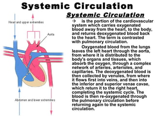
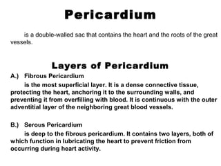
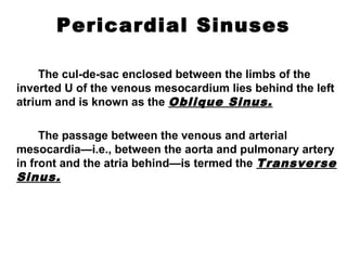
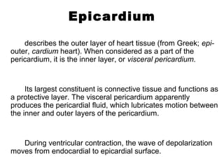
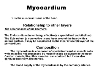
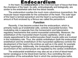
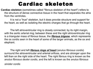
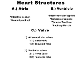
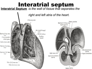
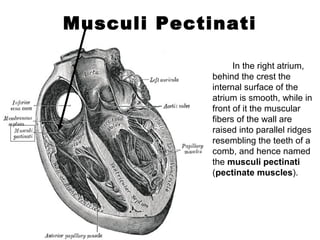
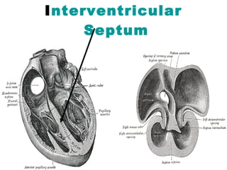
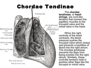
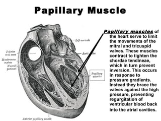
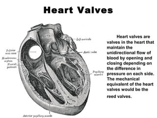
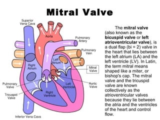
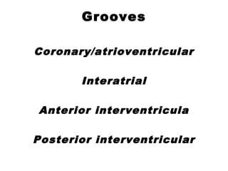
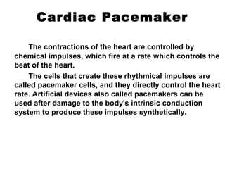
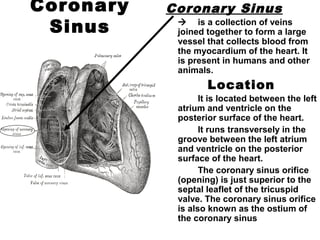
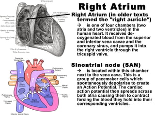
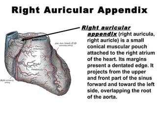
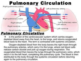
The human heart is a muscular organ that pumps blood through the body. It is divided into four chambers - two atria that receive blood and two ventricles that pump blood out. The right side receives deoxygenated blood from the body and pumps it to the lungs. The left side receives oxygenated blood from the lungs and pumps it out to the body through the aorta. Valves control the direction of blood flow between the chambers and vessels. The heart is a vital organ that circulates blood continuously through two circuits - pulmonary circulation to the lungs and systemic circulation to the body.


![Aorta
The aorta (generally
pronounced [e tə] orɪˈɔː
"ay-orta") is the largest
artery in the human body,
originating from the left
ventricle of the heart and
bringing oxygenated blood
to all parts of the body in
the systemic circulation.
The course of the
Aorta
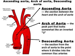
The aorta is usually divided
into five
segments/sections:
• Ascending aorta
• Arch of aorta
• Descending aorta
• Thoracic aorta
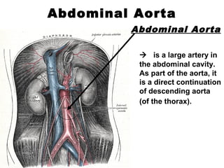
• Abdominal aorta](https://image.slidesharecdn.com/268099-human-heart-131002030353-phpapp01/85/268099-human-heart-32-320.jpg)