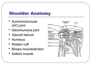
Rotator Cuff Injuries
Rotator cuff serves as a stabilizer for the
shoulder
Cuff is comprised of the supraspinatus,
infraspinatus, subscapularis and teres
minor muscles
Common rotator cuff injuries occur to the
underside of the supraspinatus tendon
Increase in risk of tear at age 40
5.
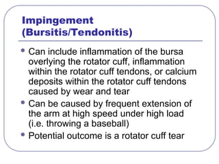
Impingement
(Bursitis/Tendonitis)
Can includeinflammation of the bursa
overlying the rotator cuff, inflammation
within the rotator cuff tendons, or calcium
deposits within the rotator cuff tendons
caused by wear and tear
Can be caused by frequent extension of
the arm at high speed under high load
(i.e. throwing a baseball)
Potential outcome is a rotator cuff tear
6.
Instability
Shoulder laxityneeds to be differentiated from
frank instability
Laxity is common in the swimmer and
throwing athlete, as the shoulder must be
loose enough to allow excessive external
rotation
Instability is unwanted translation of the
humeral head on the glenoid, and
compromises the comfort and function of the
shoulder
7.
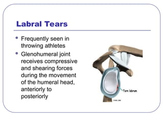
Labral Tears
Frequentlyseen in
throwing athletes
Glenohumeral joint
receives compressive
and shearing forces
during the movement
of the humeral head,
anteriorly to
posteriorly
8.
Bicipital Tendonitis
Inflammationof the biceps tendon
Diagnosis made principally by palpation
of the tendon during clinical examination
Occurs frequently in the throwing athlete:
• Modest biceps activity during cocking and
acceleration phase
• High level of biceps activity during follow-
through phase
9.
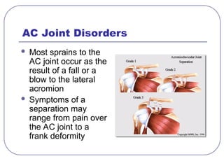
AC Joint Disorders
Most sprains to the
AC joint occur as the
result of a fall or a
blow to the lateral
acromion
Symptoms of a
separation may
range from pain over
the AC joint to a
frank deformity
10.
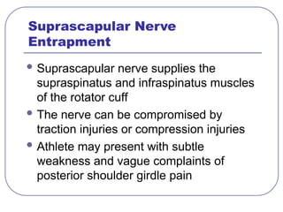
Suprascapular Nerve
Entrapment
Suprascapularnerve supplies the
supraspinatus and infraspinatus muscles
of the rotator cuff
The nerve can be compromised by
traction injuries or compression injuries
Athlete may present with subtle
weakness and vague complaints of
posterior shoulder girdle pain
The Clinical Examination
Inspection
Examination of the cervical spine
Palpation
Range of motion assessment
Strength assessment
Glenohumeral stability assessment
Neurovascular examination
Special tests
13.
Inspection
Should beperformed from different
perspectives (front, side, back, top)
Should assess for symmetry, atrophy,
hypertrophy, deformities, bruising and
swelling
Note scars as evidence of prior surgical
procedures
14.
Examination of theCervical
Spine
Have the patient look up at the ceiling,
touch his chin to his chest, look over
each shoulder
Any numbness, tingling or pain referred
to the affected shoulder points to the
cervical spine as the etiology of the
shoulder pain
Range of Motion
Includes testing of both active and
passive range of motion
For example, in the setting of a rotator
cuff tear, passive range of motion will be
normal but active range of motion will be
diminished due to the tear in the muscle
17.
Range of Motion(norms)
External rotation in a 0° plane (90°)
External rotation in a 90° plane (90°)
Abduction (150°)
Internal rotation (90°)
Forward flexion (180°)
ALWAYS compare both shoulders!
18.
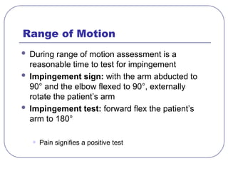
Range of Motion
During range of motion assessment is a
reasonable time to test for impingement
Impingement sign: with the arm abducted to
90° and the elbow flexed to 90°, externally
rotate the patient’s arm
Impingement test: forward flex the patient’s
arm to 180°
• Pain signifies a positive test
19.
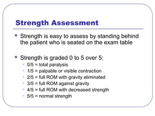
Strength Assessment
Strengthis easy to assess by standing behind
the patient who is seated on the exam table
Strength is graded 0 to 5 over 5:
• 0/5 = total paralysis
• 1/5 = palpable or visible contraction
• 2/5 = full ROM with gravity eliminated
• 3/5 = full ROM against gravity
• 4/5 = full ROM with decreased strength
• 5/5 = normal strength
20.
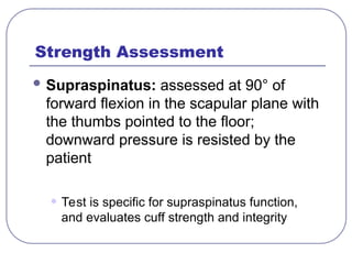
Strength Assessment
Supraspinatus:assessed at 90° of
forward flexion in the scapular plane with
the thumbs pointed to the floor;
downward pressure is resisted by the
patient
• Test is specific for supraspinatus function,
and evaluates cuff strength and integrity
21.
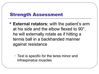
Strength Assessment
Externalrotators: with the patient’s arm
at his side and the elbow flexed to 90°,
he will externally rotate as if hitting a
tennis ball in a backhanded manner
against resistance
• Test is specific for the teres minor and
infraspinatus muscles
22.
Strength Assessment
Abduction:assessed in the coronal
plane against resistance
• May be suggestive of either deltoid or cuff
deficiency
Subscapularis: with the dorsum of the
patient’s hand on his ipsalateral back
pocket, instruct him to push backward
against resistance
23.
Glenohumeral Stability Assessment
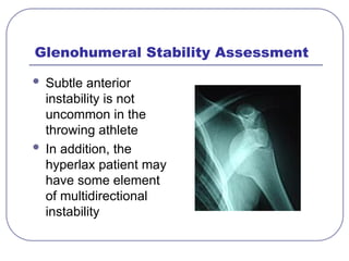
Subtle anterior
instability is not
uncommon in the
throwing athlete
In addition, the
hyperlax patient may
have some element
of multidirectional
instability
24.
Glenohumeral Stability Assessment
Sulcus sign: distraction force is placed
on the elbow and the space created
between the undersurface of the
acromion and the apex of the humeral
head is noted
• This distance is recorded in centimeters, and
indicates laxity in the joint
25.
Glenohumeral Stability Assessment
“Load and shift” test: with the humeral head
reduced (“loaded”) into the glenoid fossa, the
examiner steadies the limb girdle with one hand
and translates the humeral head both anteriorly
and posteriorly with the opposite hand
• The amount of translation is graded as 1+, 2+, or 3+
• This test is also repeated in the supine position
• Glenohumeral translation depends upon the skill of the
examiner as well as the patient’s ability to relax
26.
Glenohumeral Stability Assessment
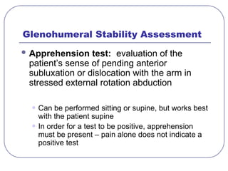
Apprehension test: evaluation of the
patient’s sense of pending anterior
subluxation or dislocation with the arm in
stressed external rotation abduction
• Can be performed sitting or supine, but works best
with the patient supine
• In order for a test to be positive, apprehension
must be present – pain alone does not indicate a
positive test
27.
Glenohumeral Stability Assessment
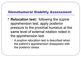
Relocation test: following the supine
apprehension test, apply posterior
pressure to the proximal humerus at the
same level of external rotation noted in
the apprehension test
• A positive relocation test is described when
the patient’s apprehension disappears with
the posterior stress
28.
Neurovascular Examination
Dermatomalsensory examination
Deep tendon reflexes at the wrist and
elbow
Cervical root testing – wrist extension,
finger abduction and adduction, thumb
abduction, elbow flexion
Palpation of the brachial and radial pulses
29.
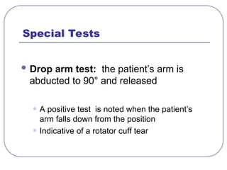
Special Tests
Droparm test: the patient’s arm is
abducted to 90° and released
• A positive test is noted when the patient’s
arm falls down from the position
• Indicative of a rotator cuff tear
30.
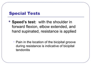
Special Tests
Speed’stest: with the shoulder in
forward flexion, elbow extended, and
hand supinated, resistance is applied
• Pain in the location of the bicipital groove
during resistance is indicative of bicipital
tendonitis
31.
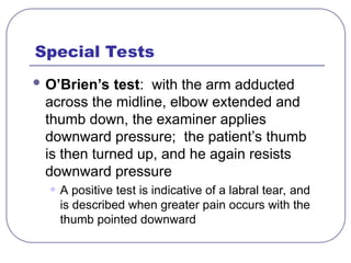
Special Tests
O’Brien’stest: with the arm adducted
across the midline, elbow extended and
thumb down, the examiner applies
downward pressure; the patient’s thumb
is then turned up, and he again resists
downward pressure
• A positive test is indicative of a labral tear, and
is described when greater pain occurs with the
thumb pointed downward
32.
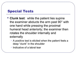
Special Tests
Clunktest: while the patient lies supine
the examiner abducts the arm past 90° with
one hand while pressing the proximal
humeral head anteriorly; the examiner then
rotates the shoulder internally and
externally
• A positive test is elicited when the patient feels a
deep “clunk” in the shoulder
• Indicative of a labral tear
33.
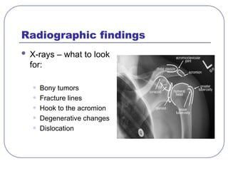
Radiographic findings
X-rays– what to look
for:
• Bony tumors
• Fracture lines
• Hook to the acromion
• Degenerative changes
• Dislocation
34.
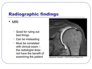
Radiographic findings
MRI
•Good for ruling out
bad things
• Can be misleading
• Must be correlated
with clinical exam –
the radiologist does
not have the benefit of
examining the patient
35.
Conservative treatment
Physicaltherapy
• Excellent form of strengthening and
rehabilitating weak or injured muscles
• Formal physical therapy will reassure you that
the exercises are actually being done
• The most successful conservative form of
therapy for the musculoskeletal system
36.
Conservative treatment
Oralanti-inflammatories
• Sometimes just a short course of anti-
inflammatories can provide permanent relief
• Non-selective COX inhibitors still work great if
the patient can tolerate them
• COX-2 inhibitors:
• Celebrex 200 mg daily
• Vioxx 25 mg daily
• Bextra 20 mg daily
37.
Conservative treatment
Cortisoneinjection (short-acting + local)
• Can be a permanent cure, but is frequently a
short-term fix
• Relief from the injection gives an excellent
prognosis for surgical success
• Should only be given every 3 months
38.
If the abovefail…
Refer to orthopedic surgeon
Surgery is a measure of last resort!
“There is no pain so terrible that surgery can’t
make worse.”


![Shoulder_joint_and_applied_aspects[1].pptx](https://cdn.slidesharecdn.com/ss_thumbnails/shoulderjointandappliedaspects1-240425164911-e75cbd49-thumbnail.jpg?width=640&height=640&fit=bounds)


![[DSC Europe 25] Dubravko Culibrk - Deep Learning for Mammography.pptx](https://cdn.slidesharecdn.com/ss_thumbnails/yiscimuktacgqoiu4dkp-deep-learning-for-mammography-260119121559-aad59182-thumbnail.jpg?width=640&height=640&fit=bounds)
![[DSC Europe 25] Paula Garcia Esteban -Building the Future: The Role of Data S...](https://cdn.slidesharecdn.com/ss_thumbnails/9ld1r1bsqpwve8qfvphy-paula-garcia-esteban-building-the-future-260122103838-4171f5cb-thumbnail.jpg?width=640&height=640&fit=bounds)

![[DSC Europe 25] Bojan Banjac - AI is always right when it comes to the matter...](https://cdn.slidesharecdn.com/ss_thumbnails/syoxtqierpydwxm5srcb-4-bojan-banjac-ai-is-always-right-when-it-comes-to-the-matters-of-taste-260119101519-694ee7d7-thumbnail.jpg?width=640&height=640&fit=bounds)


![[DSC Europe 25] Jovan Sumarac - Real-World Applications of Computer Vision in...](https://cdn.slidesharecdn.com/ss_thumbnails/fiksms22smcpopvvld03-jovan-sumarac-real-life-applications-of-computer-vision-in-automotive-systems-260120105855-de622abb-thumbnail.jpg?width=640&height=640&fit=bounds)
![[DSC Europe 25] Mikhail Rozhkov - AI Product Canvas: From Business Goals to T...](https://cdn.slidesharecdn.com/ss_thumbnails/d53doddtpgfqivmzqel6-mikhail-rozhkov-ai-product-canvas-v1-260121115910-9dd517a7-thumbnail.jpg?width=640&height=640&fit=bounds)
![[DSC Europe 25] Gordana Milutinovic Dumbelovic - From Insight to Oversight: A...](https://cdn.slidesharecdn.com/ss_thumbnails/t7dkjsfxqwwzceropjv4-gordana-milutinovicdumbelovic-from-insight-to-oversight-ai-driven-power-bi-moni-260119121559-9e0bf11b-thumbnail.jpg?width=640&height=640&fit=bounds)


![[DSC Europe 25] Marcos Heidemann - Beyond the Hype: Making AI Coding Assistan...](https://cdn.slidesharecdn.com/ss_thumbnails/eexkhvldrjsopspdjbur-marcos-heidemann-beyond-the-hype-getting-real-value-out-of-ai-assisted-coding-260121115910-7e9d41ec-thumbnail.jpg?width=640&height=640&fit=bounds)