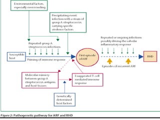
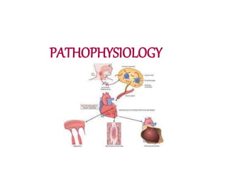
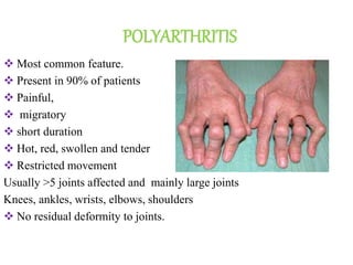
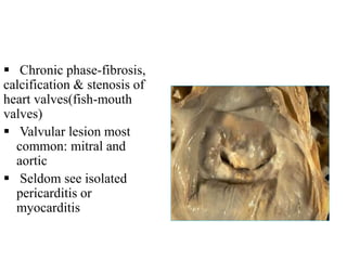
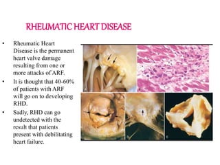
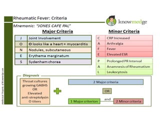
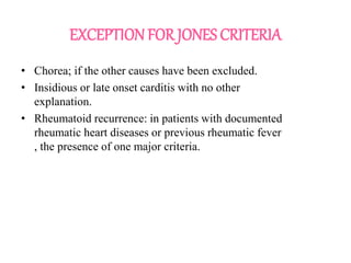
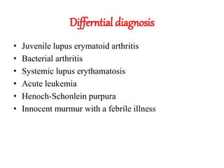
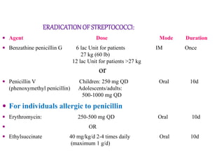
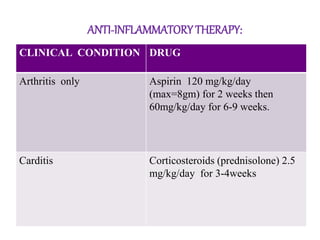
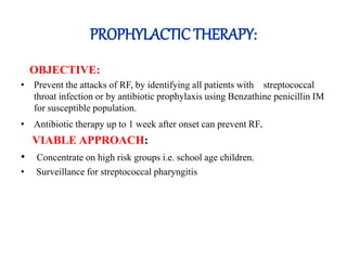
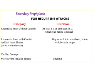
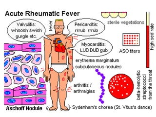
BY TOOBA IQBAL
RHEUMATIC FEVER
Rheumatic fever is an
immunologically mediated
inflammatory disorder, which
occurs as a sequel to group A
streptococcal pharyngeal
infection.
It is an Multisystem disease
affecting connective tissue
particularly of the heart, joints,
brain, cutaneous and
subcutaneous tissues.
:
Group A Streptococcal
infections.
Recently virus (coxsackie
B-4) has been suggested
as causative agent with
streptococcus acting as
conditioning agent.
epidemiology
PREDISPOSING FACTORS:
 Age
5-15 years
Rare before 4 years
 Family History
 Season
Winter and early spring
 Recurrent streptococcal infections
Recurrence rate is about 50% during first year
10% after several years
• Environmental factors:
Poverty
Over crowding
Poor housing poor hygiene
Inadequate health services
• Immuno-compromised.
• 1-5% of throat infections leads to Rheumatic Fever.
PATHOPHYSIOLOGY
PATHOGENESIS:
Acute rheumatic fever is a hypersensitivity reaction classically attributed
to antibodies directed against group A streptococcal molecules that also are
cross-reactive with host antigens.
Antibodies against M proteins of certain streptococci strains binds to
protein in the myocardium and cardiac valves and cause injury through the
activation of complement and Fc-receptor bearing cells.
CD4+ T, cells that recognize streptococcal peptides also can cross-react
with host antigens and elicit cytokine-mediated inflammatory responses.
MORPHOLOGY:
Acute rheumatic fever is
characterized by discrete
inflammatory foci within a
variety of tissues, these cardial
inflammation known as
Aschoff bodies , composed of
swollen eosinophilic collagen
surrounded by lymphocytes
and macrophages can be seen
on light microscopy. The
larger macrophages may
become Aschoff giant cells.
CLINICAL FEATURES
Following upper airway infection with GAS
Silent period of 1-5 weeks
Sudden onset of fever, pallor, malaise, joint pain.
Commonly GAS streptococcal infection is
subclinical; such cases confirmed using streptococcal
antibody testing .
FEVER
• Present at onset
of acute illness.
• High grade
fever >39ºC.
• Lasts for about
12 weeks ,tends
to recur.
Joint pain Loss of appetite
Epistaxis Abdominal pain
MAJOR MANIFESTATIONS
Polyarthritis
Carditis
Sydenham’s chorea
Erythema marginatum
Subcutaneous nodules
Minor Manifestations:
Fever
Arthralgia
Prolonged
PR
interval
ECG
Elevated
ESR, CRP
WBCs
POLYARTHRITIS
 Most common feature.
 Present in 90% of patients
 Painful,
 migratory
 short duration
 Hot, red, swollen and tender
 Restricted movement
Usually >5 joints affected and mainly large joints
Knees, ankles, wrists, elbows, shoulders
 No residual deformity to joints.
 Excellent response of salicylates and NSAIDS
 Pain and swelling come on quickly and subsides within 5-7
days
 In children below 5 yrs arthritis usually mild but carditis more
prominent
 Arthritis do not progress to chronic disease.
CARDITIS:
 Early and most serious
manifestation.
 Manifest as pancarditis.
 Occur in 60-70% of cases.
 Carditis is the only
manifestation of rheumatic
fever that leaves a squeal &
permanent damage to the
organ.
 Valvular damage is the
hallmark of RF.
 Chronic phase-fibrosis,
calcification & stenosis of
heart valves(fish-mouth
valves)
 Valvular lesion most
common: mitral and
aortic
 Seldom see isolated
pericarditis or
myocarditis
RHEUMATIC HEART DISEASE
• Rheumatic Heart
Disease is the permanent
heart valve damage
resulting from one or
more attacks of ARF.
• It is thought that 40-60%
of patients with ARF
will go on to developing
RHD.
• Sadly, RHD can go
undetected with the
result that patients
present with debilitating
heart failure.
High pulse
rate
Murmur
mitral or aortic regurgitation-endocardium
involved
Cardiomegaly myocardium involvement
Pericardial
friction rub
Pericarditis
Prolonged PR
interval
Myocardial inflammation affecting
electrical conduction
Cardiac
failure
SYNDENHAM’ S CHOREA
• Occur in 5-10% of cases
• Mainly in girls of 1-15 years age
• Late manifestation of RF.
• May occur up to 6 months after infection
• Spasmodic, unintentional, jerky choreiform movements,
• Speech affected, fidgety
• Choreiform movements particularly affect the head(darting
movement of tongue)and upper limb.
First sign: difficulty
walking, talking,
writing
Occurs in 30% of
patients with ARF
Usually benign and
resolves in 2 - 3
months
Disappears leaving
no residual damage.
ERYTHEMA MARGINATUM
Occur in <7%.
Unique, transient, serpiginous-
looking lesions of 1-2 inches in
size.
Pink macules - Clear centrally ,
serpiginous spreading edge .
More on trunks & limbs & non-
itchy.
Almost never on face.
Worsens with application of heat.
Often associated with chronic
carditis.
SUBCUTANEOUS NODULES
• Subcutaneous nodules usually indicate
severe carditis.
• Small, painless, mobile hard lumps
beneath skin.
• Most common along extensor surfaces
of joint (Knees, elbows, wrists)
• Also on bony prominences, tendons,
dorsi of feet, occiput or cervical spine,
mastoid process and on scapula.
• Appears 4 weeks after onset of RF.
• Delayed manifestation, disappears
leaves no residual damage.
• Occur in 9 - 20% of cases.
INVESTIGATIONS:
• High ESR
• Anemia, leucocytosis
• Elevated C-reactive protein
• Elevated ASO or other streptococcal antibody titer
• Throat swab
• ECG
• X-ray Chest
• Echocardiography
Antistreptolysin O Titre:
• Raised in 85% of the cases
• Its value 500 units indicates recent
streptococcal infections.
• A value of 333 units, it is
recommended additional antibody
• Always >200 Todd unit/ml remains
elevated for weeks or months .
• ASO and Anti-DNAse used for
diagnosis
• Anti-Hyaluronidase is the third
choice.
ECG:
• Prolongation of PR interval to greater than 0.18 sec.
• Prolongation of QT interval.
• ST wave or T wave changes of pericarditis or myocarditis.
• Complete block or second degree shows in inflammation of
the conduction system.
X-rayChest:
Cardiac
enlargement due
to congestive
cardiac failure
ECHOCARDIOGRAHY:
Changes can detect
valvular and
myocardial
involvement or
pericardial effusion.
It is helpful in
monitoring valve
problems.
EXCEPTION FOR JONES CRITERIA
• Chorea; if the other causes have been excluded.
• Insidious or late onset carditis with no other
explanation.
• Rheumatoid recurrence: in patients with documented
rheumatic heart diseases or previous rheumatic fever
, the presence of one major criteria.
Differntial diagnosis
• Juvenile lupus erymatoid arthritis
• Bacterial arthritis
• Systemic lupus erythamatosis
• Acute leukemia
• Henoch-Schonlein purpura
• Innocent murmur with a febrile illness
CURATIVE THERAPY
PROPHYLACTIC THERAPY
CURATIVE THERAPY:
BED REST:
• In case of Arthritis alone for 1 -2week.
• Minimal Carditis for 2-4 weeks.
• Severe Carditis for several months.
ERADICATIONOF STREPTOCOCCI:
 Agent Dose Mode Duration
 Benzathine penicillin G 6 lac Unit for patients IM Once
27 kg (60 lb)
12 lac Unit for patients >27 kg
or
 Penicillin V Children: 250 mg QD Oral 10d
(phenoxymethyl penicillin) Adolescents/adults:
500-1000 mg QD
 For individuals allergic to penicillin
 Erythromycin: 250-500 mg QD Oral 10d
 OR
 Ethylsuccinate 40 mg/kg/d 2-4 times daily Oral 10d
(maximum 1 g/d)
ANTI-INFLAMMATORY THERAPY:
CLINICAL CONDITION DRUG
Arthritis only Aspirin 120 mg/kg/day
(max=8gm) for 2 weeks then
60mg/kg/day for 6-9 weeks.
Carditis Corticosteroids (prednisolone) 2.5
mg/kg/day for 3-4weeks
PROPHYLACTIC THERAPY:
OBJECTIVE:
• Prevent the attacks of RF, by identifying all patients with streptococcal
throat infection or by antibiotic prophylaxis using Benzathine penicillin IM
for susceptible population.
• Antibiotic therapy up to 1 week after onset can prevent RF.
VIABLE APPROACH:
• Concentrate on high risk groups i.e. school age children.
• Surveillance for streptococcal pharyngitis
Secondary Prophylaxis:
FOR RECURRENT ATTACKS
Category Duration
Rheumatic fever without Carditis At least 5 y or until age 21 y,
whichever period is longer
Rheumatic fever with Carditis 10 y or well into adulthood, but no
residual heart disease whichever is longer
(no valvular disease)
Cardiac Damage
More severe valvular disease Lifelong
complications
• Congestive heart failure
• Rheumatic heart disease
• Arrhythmias
• Pericarditis
• Pericardial effusion
• Rheumatic pneumonitis
Rheumatic fever
Rheumatic fever
Rheumatic fever

Rheumatic fever

  • 1.
  • 2.
    RHEUMATIC FEVER Rheumatic feveris an immunologically mediated inflammatory disorder, which occurs as a sequel to group A streptococcal pharyngeal infection. It is an Multisystem disease affecting connective tissue particularly of the heart, joints, brain, cutaneous and subcutaneous tissues.
  • 3.
    : Group A Streptococcal infections. Recentlyvirus (coxsackie B-4) has been suggested as causative agent with streptococcus acting as conditioning agent.
  • 4.
  • 5.
    PREDISPOSING FACTORS:  Age 5-15years Rare before 4 years  Family History  Season Winter and early spring  Recurrent streptococcal infections Recurrence rate is about 50% during first year 10% after several years
  • 6.
    • Environmental factors: Poverty Overcrowding Poor housing poor hygiene Inadequate health services • Immuno-compromised. • 1-5% of throat infections leads to Rheumatic Fever.
  • 9.
  • 10.
    PATHOGENESIS: Acute rheumatic feveris a hypersensitivity reaction classically attributed to antibodies directed against group A streptococcal molecules that also are cross-reactive with host antigens. Antibodies against M proteins of certain streptococci strains binds to protein in the myocardium and cardiac valves and cause injury through the activation of complement and Fc-receptor bearing cells. CD4+ T, cells that recognize streptococcal peptides also can cross-react with host antigens and elicit cytokine-mediated inflammatory responses.
  • 13.
    MORPHOLOGY: Acute rheumatic feveris characterized by discrete inflammatory foci within a variety of tissues, these cardial inflammation known as Aschoff bodies , composed of swollen eosinophilic collagen surrounded by lymphocytes and macrophages can be seen on light microscopy. The larger macrophages may become Aschoff giant cells.
  • 14.
  • 15.
    Following upper airwayinfection with GAS Silent period of 1-5 weeks Sudden onset of fever, pallor, malaise, joint pain. Commonly GAS streptococcal infection is subclinical; such cases confirmed using streptococcal antibody testing .
  • 16.
    FEVER • Present atonset of acute illness. • High grade fever >39ºC. • Lasts for about 12 weeks ,tends to recur.
  • 17.
    Joint pain Lossof appetite
  • 18.
  • 20.
  • 21.
  • 22.
    POLYARTHRITIS  Most commonfeature.  Present in 90% of patients  Painful,  migratory  short duration  Hot, red, swollen and tender  Restricted movement Usually >5 joints affected and mainly large joints Knees, ankles, wrists, elbows, shoulders  No residual deformity to joints.
  • 23.
     Excellent responseof salicylates and NSAIDS  Pain and swelling come on quickly and subsides within 5-7 days  In children below 5 yrs arthritis usually mild but carditis more prominent  Arthritis do not progress to chronic disease.
  • 24.
    CARDITIS:  Early andmost serious manifestation.  Manifest as pancarditis.  Occur in 60-70% of cases.  Carditis is the only manifestation of rheumatic fever that leaves a squeal & permanent damage to the organ.  Valvular damage is the hallmark of RF.
  • 25.
     Chronic phase-fibrosis, calcification& stenosis of heart valves(fish-mouth valves)  Valvular lesion most common: mitral and aortic  Seldom see isolated pericarditis or myocarditis
  • 26.
    RHEUMATIC HEART DISEASE •Rheumatic Heart Disease is the permanent heart valve damage resulting from one or more attacks of ARF. • It is thought that 40-60% of patients with ARF will go on to developing RHD. • Sadly, RHD can go undetected with the result that patients present with debilitating heart failure.
  • 27.
    High pulse rate Murmur mitral oraortic regurgitation-endocardium involved Cardiomegaly myocardium involvement Pericardial friction rub Pericarditis Prolonged PR interval Myocardial inflammation affecting electrical conduction Cardiac failure
  • 28.
    SYNDENHAM’ S CHOREA •Occur in 5-10% of cases • Mainly in girls of 1-15 years age • Late manifestation of RF. • May occur up to 6 months after infection • Spasmodic, unintentional, jerky choreiform movements, • Speech affected, fidgety • Choreiform movements particularly affect the head(darting movement of tongue)and upper limb.
  • 29.
    First sign: difficulty walking,talking, writing Occurs in 30% of patients with ARF Usually benign and resolves in 2 - 3 months Disappears leaving no residual damage.
  • 30.
    ERYTHEMA MARGINATUM Occur in<7%. Unique, transient, serpiginous- looking lesions of 1-2 inches in size. Pink macules - Clear centrally , serpiginous spreading edge . More on trunks & limbs & non- itchy. Almost never on face. Worsens with application of heat. Often associated with chronic carditis.
  • 31.
    SUBCUTANEOUS NODULES • Subcutaneousnodules usually indicate severe carditis. • Small, painless, mobile hard lumps beneath skin. • Most common along extensor surfaces of joint (Knees, elbows, wrists) • Also on bony prominences, tendons, dorsi of feet, occiput or cervical spine, mastoid process and on scapula. • Appears 4 weeks after onset of RF. • Delayed manifestation, disappears leaves no residual damage. • Occur in 9 - 20% of cases.
  • 33.
    INVESTIGATIONS: • High ESR •Anemia, leucocytosis • Elevated C-reactive protein • Elevated ASO or other streptococcal antibody titer • Throat swab • ECG • X-ray Chest • Echocardiography
  • 34.
    Antistreptolysin O Titre: •Raised in 85% of the cases • Its value 500 units indicates recent streptococcal infections. • A value of 333 units, it is recommended additional antibody • Always >200 Todd unit/ml remains elevated for weeks or months . • ASO and Anti-DNAse used for diagnosis • Anti-Hyaluronidase is the third choice.
  • 35.
    ECG: • Prolongation ofPR interval to greater than 0.18 sec. • Prolongation of QT interval. • ST wave or T wave changes of pericarditis or myocarditis. • Complete block or second degree shows in inflammation of the conduction system.
  • 36.
  • 37.
    ECHOCARDIOGRAHY: Changes can detect valvularand myocardial involvement or pericardial effusion. It is helpful in monitoring valve problems.
  • 40.
    EXCEPTION FOR JONESCRITERIA • Chorea; if the other causes have been excluded. • Insidious or late onset carditis with no other explanation. • Rheumatoid recurrence: in patients with documented rheumatic heart diseases or previous rheumatic fever , the presence of one major criteria.
  • 41.
    Differntial diagnosis • Juvenilelupus erymatoid arthritis • Bacterial arthritis • Systemic lupus erythamatosis • Acute leukemia • Henoch-Schonlein purpura • Innocent murmur with a febrile illness
  • 43.
  • 44.
    CURATIVE THERAPY: BED REST: •In case of Arthritis alone for 1 -2week. • Minimal Carditis for 2-4 weeks. • Severe Carditis for several months.
  • 45.
    ERADICATIONOF STREPTOCOCCI:  AgentDose Mode Duration  Benzathine penicillin G 6 lac Unit for patients IM Once 27 kg (60 lb) 12 lac Unit for patients >27 kg or  Penicillin V Children: 250 mg QD Oral 10d (phenoxymethyl penicillin) Adolescents/adults: 500-1000 mg QD  For individuals allergic to penicillin  Erythromycin: 250-500 mg QD Oral 10d  OR  Ethylsuccinate 40 mg/kg/d 2-4 times daily Oral 10d (maximum 1 g/d)
  • 46.
    ANTI-INFLAMMATORY THERAPY: CLINICAL CONDITIONDRUG Arthritis only Aspirin 120 mg/kg/day (max=8gm) for 2 weeks then 60mg/kg/day for 6-9 weeks. Carditis Corticosteroids (prednisolone) 2.5 mg/kg/day for 3-4weeks
  • 47.
    PROPHYLACTIC THERAPY: OBJECTIVE: • Preventthe attacks of RF, by identifying all patients with streptococcal throat infection or by antibiotic prophylaxis using Benzathine penicillin IM for susceptible population. • Antibiotic therapy up to 1 week after onset can prevent RF. VIABLE APPROACH: • Concentrate on high risk groups i.e. school age children. • Surveillance for streptococcal pharyngitis
  • 48.
    Secondary Prophylaxis: FOR RECURRENTATTACKS Category Duration Rheumatic fever without Carditis At least 5 y or until age 21 y, whichever period is longer Rheumatic fever with Carditis 10 y or well into adulthood, but no residual heart disease whichever is longer (no valvular disease) Cardiac Damage More severe valvular disease Lifelong
  • 49.
    complications • Congestive heartfailure • Rheumatic heart disease • Arrhythmias • Pericarditis • Pericardial effusion • Rheumatic pneumonitis