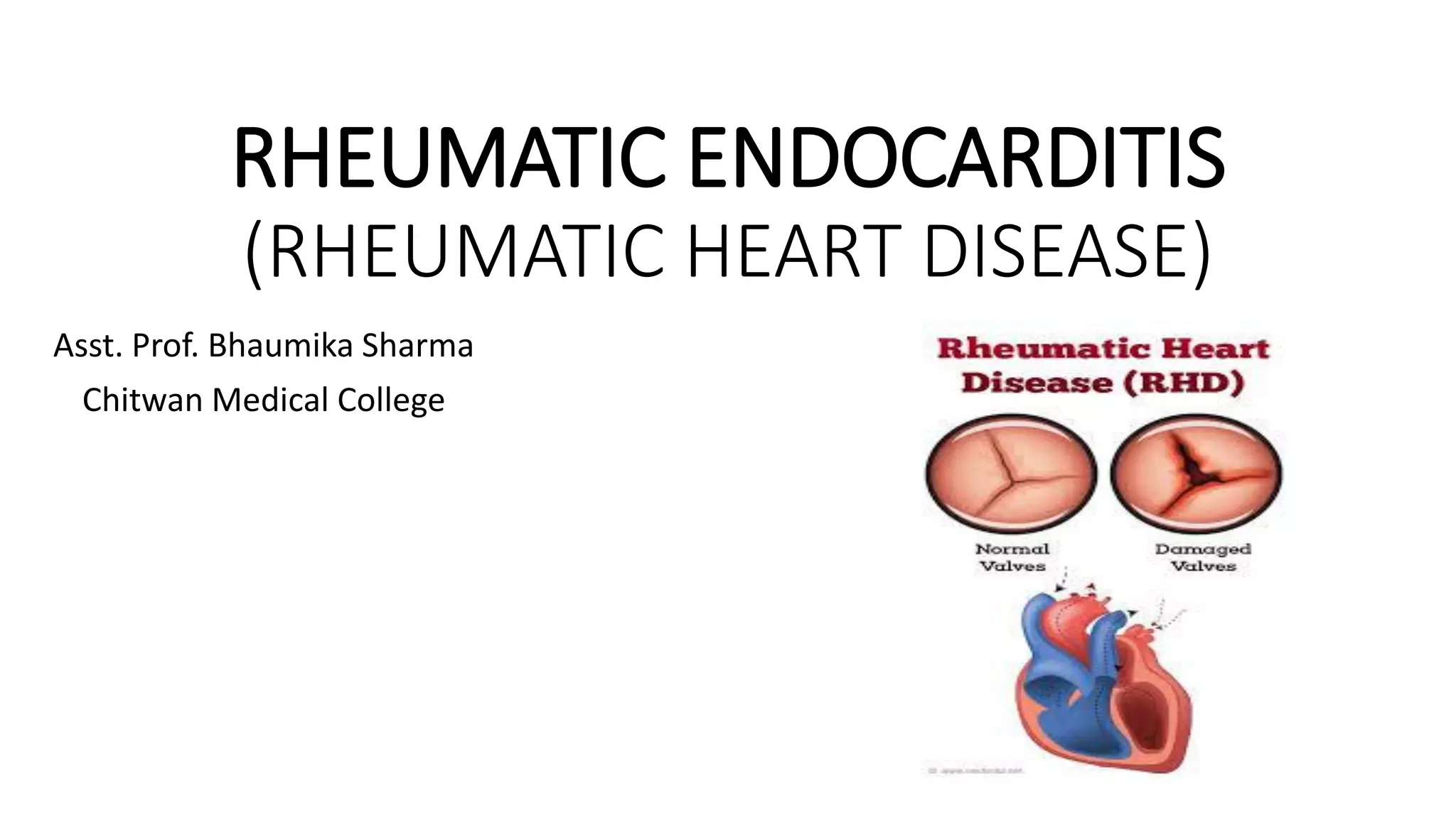
RHEUMATIC ENDOCARDITIS
(RHEUMATIC HEART DISEASE)
Asst. Prof. Bhaumika Sharma
Chitwan Medical College
RHEUMATIC HEART DISEASE
• Rheumatic heart disease is cardiac inflammation and scarring
triggered by an autoimmune reaction to infection with group
A streptococci.
• In the acute stage, this condition consists of pancarditis,
involving inflammation of the myocardium, endocardium,
and epicardium.
• Chronic disease is manifested by valvular fibrosis, resulting in
stenosis and/or insufficiency.
Rheumatic Endocarditis
• Rheumatic endocarditis is an acute, recurrent inflammatory
disease that causes damage to the heart as a sequela to
group A beta-hemolytic streptococcal infection, particularly
the valves, resulting in valve leakage (insufficiency) and/or
obstruction (narrowing or stenosis).
• There are associated compensatory changes in the size of
the heart's chambers and the thickness of chamber walls.
Etiology and Pathophysiology
• Rheumatic fever is a late inflammatory, nonsuppurative complication
of pharyngitis that is caused by group A-hemolytic streptococci.
• Rheumatic fever results from humoral and cellular-mediated immune
responses occurring 1-3 weeks after the onset of streptococcal
pharyngitis.
• Streptococcal proteins display molecular mimicry recognized by the
immune system, especially bacterial M-proteins and human cardiac
antigens such as myosin and valvular endothelium.
• Antimyosin antibody recognizes laminin, an extracellular matrix
alpha-helix coiled protein, which is part of the valve basement
membrane structure.
Contd.
• The valves most affected by rheumatic fever, in order,
are the mitral, aortic, tricuspid, and pulmonary valves.
• In most cases, the mitral valve is involved with 1 or
more of the other 3.
• In acute disease, small thrombi form along the lines of
valve closure.
• In chronic disease, there is thickening and fibrosis of
the valve resulting in stenosis, or less commonly,
regurgitation.
• T-cells that are responsive to the streptococcal M-protein
infiltrate the valve through the valvular endothelium,
activated by the binding of antistreptococcal carbohydrates
with release or tumor necrosis factor (TNF) and interleukins.
• Carditis occurs in approximately 40-50% of patients on the
first attack.
Molecular/Genetic Factors
• Familial studies of rheumatic heart disease suggest a
vulnerable population with increased risk.
• Relationships between the development of rheumatic
fever and human leukocyte antigen (HLA)-DR subtypes
have been found.
Introduction contd.
• Acute rheumatic fever is a multisystem disease characterized
by involvement of the heart, joints, central nervous system
(CNS), subcutaneous tissues, and skin.
• Mitral stenosis, Mitral insufficiency
• Severe pulmonary hypertension
• Aortic stenosis, aortic insufficiency
• Thrombotic vegetation
Contd.
• In the acute phase, verrucous endocarditis consists of small,
uniformly sized thrombotic vegetations that do not produce
valve destruction.
• The chronically scarred, inflamed, and neovascularized valve
leads to commissural fusion, valve thickening, and
calcification.
• The appearance of the mitral valve with shortened, fused
chordae and scarred commissures has been likened to the
mouth of a catfish (fish-mouth deformity).
• There is also diffuse scarring of the aortic valve, with
commissural fibrosis and thickening of the leaflets.
Contd.
• Aschoff bodies are specific for post-rheumatic carditis,
whereas Anitschkow cells can be seen in a variety of
conditions.
Aschoff nodule. The acute phase of rheumatic heart disease, or
rheumatic carditis, is characterized by the Aschoff nodule, which is an
interstitial aggregate of macrophages and lymphocytes, with necrotic
collagen, in an area of interstitial fibrosis.
Anitschkow cell. In the center of Aschoff nodules are Anitschkow or caterpillar cells,
characterized by linear condensation of nuclear chromatin with undulations
reminiscent of a caterpillar (arrows). These cells are not specific for rheumatic fever
but are seen in various conditions. In Aschoff nodules, the Anitschkow cells are
macrophages, although the same nuclear change can occur in myocytes and other
connective tissue cells.
Pathophysiology and Etiology
1. Rheumatic fever is a sequela to group A streptococcal infection that occurs
in about 3% of untreated infections. It is a preventable disease through the
detection and adequate treatment of streptococcal pharyngitis.
2. Connective tissue of the heart, blood vessels, joints, and subcutaneous
tissues can be affected.
3. Lesions in connective tissue are known as Aschoff bodies, which are
localized areas of tissue necrosis surrounded by immune cells.
4. Heart valves are affected, resulting in valve leakage and narrowing.
5. Compensatory changes in the chamber sizes and thickness of chamber
walls occur.
6. Heart involvement (carditis) also includes pericarditis, myocarditis, and
endocarditis.
Clinical Manifestations
1. Symptoms of streptococcal pharyngitis may precede
rheumatic symptoms
• Sudden onset of sore throat; throat reddened with
exudate
• Swollen, tender lymph nodes at angle of jaw
• Headache and fever 101° to 104° F (38.9° to 40° C)
• Abdominal pain (children)
• Some cases of streptococcal throat infection are relatively
asymptomatic
Contd.
2. Warm and swollen joints (polyarthritis)
3. Chorea (irregular, jerky, involuntary, unpredictable muscular
movements)
4. Erythema marginatum (transient meshlike macular rash on trunk
and extremities in about 10% of patients)
5. Subcutaneous nodules (hard, painless nodules over extensor
surfaces of extremities; rare)
6. Fever
7. Prolonged PR interval demonstrated by ECG
8. Heart murmurs; pleural and pericardial rubs
Diagnostic Evaluation
• Throat culture to determine presence of streptococcal
organisms
• Sedimentation rate, WBC count and differential, and CRP
increased during acute phase of infection
• Elevated antistreptolysin-O (ASO) titer
• ECG-prolonged PR interval or heart block
• Echocardiogram (echo)
• Chest X-ray
• Cardiac MRI
Management
• Antimicrobial therapy: penicillin is the drug of choice
• Note that missed doses of antibiotics due to the patient's
unavailability while off the unit for diagnostic tests are
given after return to the unit.
• Missed antibiotic doses may have irreversible deleterious
consequences.
• Notify health care provider if doses will be missed to make
sure that appropriate alternative measures are taken.
• Rest: to maintain optimal cardiac function.
Contd.
• Salicylates or NSAIDs: to control fever and pain
• Prevention of recurrent episodes through long-term
penicillin therapy for 5 years after initial attack in most
adults; periodic prophylaxis throughout life if valvular
damage
• In severe cases, treatment may include surgery to
replace or repair a badly damaged valve.
Complications
•Valvular heart disease
•Cardiomyopathy
•Heart failure
Heart Valves
• Heart valve prostheses were introduced more than 40 years ago.
• Prosthetic valves: needs life-long anticoagulation with warfarin which may
increase risk of injury and haemorrhage.
• Warfarin use must be carefully monitored as measured by the INR.
• But in mechanical valves there is risk for thrombus formation and
embolization and need for repeat operation.
• Also there is chances of design malfunction which may lead to failures and
even financial and litigation burdens.
• Also may develop prosthetic valve IE esp. with staphylococcal infections
leading to perivalvular abscess formation and perivalvular leak and
premature repeat valve replacement.
Contd.
• Tissue Valves (Bio-prostheses): for tissue valves implantation requires
more skill and operative time.
• These valves are of limited availability and not widely use.
• Most appropriate with presence of acute IE of native or prosthetic aortic
valve.
• The most commonly used bio-prostheses are the stented porcine or
pericardial valves, which are glutaraldehyde-treated tissues that are
mounted on a prosthetic frame.
• Do not require anticoagulation, but they are not as durable as mechanical
valves. Average life is 10-15 years; not indicated for young patients.
Aortic Bioprosthesis
Nursing Assessment
• Ask patient about symptoms of fever or throat or joint pain.
• Ask patient about chest pain, dyspnea, fatigue.
• Observe for skin lesions or rash on trunk and extremities.
• Palpate for firm, nontender movable nodules near tendons or
joints.
• Auscultate heart sounds for murmurs and/or rubs.
• Check for ECG and Echo., chest X-ray and angiocardiography
Nursing Diagnoses
• Hyperthermia related to disease process
• Decreased Cardiac Output related to decreased cardiac
contractility
• Activity Intolerance related to joint pain and easy fatigability
Nursing Interventions
Reducing Fever
• Administer penicillin therapy as prescribed to eradicate
hemolytic streptococcus; an alternative drug may be prescribed
if patient is allergic to penicillin, or sensitivity testing and
desensitization may be done.
• Give salicylates or NSAIDs as prescribed to suppress rheumatic
activity by controlling toxic manifestations, to reduce fever, and
to relieve joint pain.
• Assess for effectiveness of drug therapy.
• Take and record temperature every 3 hours.
• Evaluate patient's comfort level every 3 hours.
Maintaining Adequate Cardiac Output
• Assess for signs and symptoms of acute rheumatic carditis.
• for development of second-degree heart block or Wenckebach's disease
• Be alert to patient's complaints of chest pain, palpitations, and/or
precordial tightness.
• Monitor for tachycardia (usually persistent when patient sleeps) or
bradycardia.
• Be alert ECG (acute rheumatic carditis causes PR interval prolongation).
• Auscultate heart sounds every 4 hours.
• Document presence of murmur or pericardial friction rub.
• Document extra heart sounds (S3 gallop, S4 gallop).
• Monitor for development of chronic rheumatic endocarditis,
which may include valvular disease and heart failure.
Maintaining Activity
• Maintain bed rest for duration of fever or if signs of active
carditis are present.
• Provide ROM exercise program.
• Provide diversional activities that prevent exertion.
• Discuss need for tutorial services with parents to help child
keep up with school work.
Patient Education and Health Maintenance
•Counsel patient to maintain good nutrition.
•Counsel patient on hygienic practices.
a) Discuss proper handwashing, disposal of tissues, laundering of
handkerchiefs (decrease risk of exposure to microbes).
b) Discuss importance of using patient's own toothbrush, soap,
and washcloths when living in group situations.
•Counsel patient on importance of receiving adequate rest.
Contd.
• Instruct patient to seek treatment immediately should sore throat
occur.
• Support patients in long-term antibiotic therapy to prevent relapse (5
years for most adults).
• Instruct patient with valvular disease to use prophylactic penicillin
therapy before certain procedures and surgery.
• Explore with patient his ability to pay for medical treatment. If
appropriate, contact social services for patient. (Financial difficulties
may inhibit patient from seeking early treatment of symptoms.)
Management for Valvular Replacement
Medical Therapy
• Antibiotic prophylaxis for endocarditis before invasive
procedures indicated in most cases.
• Treatment of heart failure diuretics, sodium
restriction, vasodilators, cardiac glycosides, as
indicated.
Surgical Intervention
1) Care of the patient undergoing heart surgery.
2) For mitral stenosis:
• Closed mitral valvotomy: introduction of a dilator through the
mitral valve to split its commissures.
• Open mitral valvotomy: direct incision of the commissures.
• Mitral valve replacement.
• Balloon valvuloplasty: a balloon-tipped catheter is
percutaneously inserted, threaded to the affected valve, and
positioned across the narrowed orifice. The balloon is inflated
and deflated, causing a cracking of the calcified commissures and
enlargement of the valve orifice.
Contd.
3) For mitral insufficiency: mitral valve replacement or
annuloplasty (retailoring of the valve ring)
4) For aortic stenosis or insufficiency:
a. Replacement of aortic valve with prosthetic or tissue
valves.
b. Balloon valvuloplasty (aortic stenosis).
5) For tricuspid stenosis or insufficiency: valvuloplasty
or replacement may be done at time of surgical
intervention for associated rheumatic mitral or
aortic disease
Complications
• Left-sided heart failure
• Possible right-sided heart failure
• Dysrhythmias
Nursing Assessment
Mitral Stenosis
• Auscultate for accentuated first heart sound, usually
accompanied by an “opening snap” due to sudden tensing of
valve leaflets) at apex with diaphragm of stethoscope.
• Place the patient in left lateral recumbent position. With bell
of stethoscope at apex, auscultate for a low-pitched diastolic
murmur (rumbling murmur). Note duration of murmur (long
duration is indicative of significant stenosis).
Mitral Insufficiency
•Auscultate for diminished first heart sound.
•Auscultate for systolic murmur (prominent finding),
commencing immediately after first heart sound at apex, and
note radiation of sound to axilla and left intrascapular area.
•Mild insufficiency may produce a pansystolic murmur (little
connection between severity of mitral insufficiency and
intensity of murmur auscultated).
Aortic Stenosis
•Auscultate for prominent fourth heart sound and possible
paradoxical splitting of second heart sound (suggestive of
associated left ventricular dysfunction). First heart sound is
normal.
•Auscultate for a mid-systolic murmur at the base of the
heart (heard best) and at the apex of heart. Note harsh and
rasping quality at base of heart and a higher pitch at apex
of heart.
Aortic Insufficiency
•Auscultate for soft first heart sound.
•Place the patient in sitting position, leaning forward.
•Place diaphragm of stethoscope along left sternal border at
the third and fourth intercostal space and then along the
right sternal border.
•Auscultate for a high-pitched diastolic murmur. To increase
audibility of murmur, ask the patient to hold breath at end
of deep expiration.
•Re-auscultate for murmur.
Tricuspid Stenosis
• Auscultate for a blowing diastolic murmur at the lower left
sternal border (increases with inspiration).
• Tricuspid Insufficiency
• Auscultate for a third heart sound (may be accentuated by
inspiration).
• Auscultate for a pansystolic murmur in the parasternal
region at the fourth intercostal space. Murmur is usually high
pitched.
Nursing Diagnoses
• Decreased Cardiac Output related to altered preload,
afterload, or contractility
• Activity Intolerance related to reduced oxygen supply
• Ineffective Coping related to acute or chronic illness
Nursing Interventions
Maintaining Adequate Cardiac Output
•Assess frequently for change in existing murmur
or new murmur.
•Assess for signs of left- or right-sided heart
failure.
•Monitor and treat dysrhythmias as ordered.
•Prepare the patient for surgical intervention
(pre-operative).
Improving Tolerance
•Maintain bed rest while symptoms of heart failure are
present.
•Allow patient to rest between interventions.
•Begin activities gradually (e.g. chair sitting for brief
periods).
•Assist with or perform hygiene needs for patient to reserve
strength for ambulation.
Strengthening Coping Abilities
• Instruct the patient about specific valvular dysfunction, possible etiology,
and therapies implemented to relieve symptoms.
• Include family members in discussions with the patient.
• Stress the importance of adapting lifestyle to cope with illness.
• Discuss with the patient surgical intervention as the treatment modality,
if applicable.
• Assess the patient's use of appropriate coping mechanisms.
• Refer the patient to appropriate counseling services, if indicated
(vocational, social work, cardiac rehabilitation).
Patient Education and Health Maintenance
•Review activity restriction and schedule with patient and
family.
•Instruct patient to report signs of impending or worsening
heart failure: dyspnea, cough, increased fatigue, ankle
swelling.
•Review sodium and fluid restrictions.
•Review medications: purpose, action, schedule and
adverse effects.
Evaluation: Expected Outcomes
• Afebrile
• Denies chest pain; normal sinus rhythm
• Maintains bed rest while febrile
• BP and heart rate within normal limits
• Tolerates chair sitting for 15 minutes every 2 hours
• Discusses ways to cope with lifestyle and activity changes
Any queries ?

Rheumatic heart disease

  • 1.
    RHEUMATIC ENDOCARDITIS (RHEUMATIC HEARTDISEASE) Asst. Prof. Bhaumika Sharma Chitwan Medical College
  • 2.
    RHEUMATIC HEART DISEASE •Rheumatic heart disease is cardiac inflammation and scarring triggered by an autoimmune reaction to infection with group A streptococci. • In the acute stage, this condition consists of pancarditis, involving inflammation of the myocardium, endocardium, and epicardium. • Chronic disease is manifested by valvular fibrosis, resulting in stenosis and/or insufficiency.
  • 3.
    Rheumatic Endocarditis • Rheumaticendocarditis is an acute, recurrent inflammatory disease that causes damage to the heart as a sequela to group A beta-hemolytic streptococcal infection, particularly the valves, resulting in valve leakage (insufficiency) and/or obstruction (narrowing or stenosis). • There are associated compensatory changes in the size of the heart's chambers and the thickness of chamber walls.
  • 4.
    Etiology and Pathophysiology •Rheumatic fever is a late inflammatory, nonsuppurative complication of pharyngitis that is caused by group A-hemolytic streptococci. • Rheumatic fever results from humoral and cellular-mediated immune responses occurring 1-3 weeks after the onset of streptococcal pharyngitis. • Streptococcal proteins display molecular mimicry recognized by the immune system, especially bacterial M-proteins and human cardiac antigens such as myosin and valvular endothelium. • Antimyosin antibody recognizes laminin, an extracellular matrix alpha-helix coiled protein, which is part of the valve basement membrane structure.
  • 5.
    Contd. • The valvesmost affected by rheumatic fever, in order, are the mitral, aortic, tricuspid, and pulmonary valves. • In most cases, the mitral valve is involved with 1 or more of the other 3. • In acute disease, small thrombi form along the lines of valve closure. • In chronic disease, there is thickening and fibrosis of the valve resulting in stenosis, or less commonly, regurgitation.
  • 6.
    • T-cells thatare responsive to the streptococcal M-protein infiltrate the valve through the valvular endothelium, activated by the binding of antistreptococcal carbohydrates with release or tumor necrosis factor (TNF) and interleukins. • Carditis occurs in approximately 40-50% of patients on the first attack.
  • 7.
    Molecular/Genetic Factors • Familialstudies of rheumatic heart disease suggest a vulnerable population with increased risk. • Relationships between the development of rheumatic fever and human leukocyte antigen (HLA)-DR subtypes have been found.
  • 8.
    Introduction contd. • Acuterheumatic fever is a multisystem disease characterized by involvement of the heart, joints, central nervous system (CNS), subcutaneous tissues, and skin. • Mitral stenosis, Mitral insufficiency • Severe pulmonary hypertension • Aortic stenosis, aortic insufficiency • Thrombotic vegetation
  • 9.
    Contd. • In theacute phase, verrucous endocarditis consists of small, uniformly sized thrombotic vegetations that do not produce valve destruction. • The chronically scarred, inflamed, and neovascularized valve leads to commissural fusion, valve thickening, and calcification. • The appearance of the mitral valve with shortened, fused chordae and scarred commissures has been likened to the mouth of a catfish (fish-mouth deformity). • There is also diffuse scarring of the aortic valve, with commissural fibrosis and thickening of the leaflets.
  • 10.
    Contd. • Aschoff bodiesare specific for post-rheumatic carditis, whereas Anitschkow cells can be seen in a variety of conditions.
  • 11.
    Aschoff nodule. Theacute phase of rheumatic heart disease, or rheumatic carditis, is characterized by the Aschoff nodule, which is an interstitial aggregate of macrophages and lymphocytes, with necrotic collagen, in an area of interstitial fibrosis.
  • 12.
    Anitschkow cell. Inthe center of Aschoff nodules are Anitschkow or caterpillar cells, characterized by linear condensation of nuclear chromatin with undulations reminiscent of a caterpillar (arrows). These cells are not specific for rheumatic fever but are seen in various conditions. In Aschoff nodules, the Anitschkow cells are macrophages, although the same nuclear change can occur in myocytes and other connective tissue cells.
  • 13.
    Pathophysiology and Etiology 1.Rheumatic fever is a sequela to group A streptococcal infection that occurs in about 3% of untreated infections. It is a preventable disease through the detection and adequate treatment of streptococcal pharyngitis. 2. Connective tissue of the heart, blood vessels, joints, and subcutaneous tissues can be affected. 3. Lesions in connective tissue are known as Aschoff bodies, which are localized areas of tissue necrosis surrounded by immune cells. 4. Heart valves are affected, resulting in valve leakage and narrowing. 5. Compensatory changes in the chamber sizes and thickness of chamber walls occur. 6. Heart involvement (carditis) also includes pericarditis, myocarditis, and endocarditis.
  • 15.
    Clinical Manifestations 1. Symptomsof streptococcal pharyngitis may precede rheumatic symptoms • Sudden onset of sore throat; throat reddened with exudate • Swollen, tender lymph nodes at angle of jaw • Headache and fever 101° to 104° F (38.9° to 40° C) • Abdominal pain (children) • Some cases of streptococcal throat infection are relatively asymptomatic
  • 16.
    Contd. 2. Warm andswollen joints (polyarthritis) 3. Chorea (irregular, jerky, involuntary, unpredictable muscular movements) 4. Erythema marginatum (transient meshlike macular rash on trunk and extremities in about 10% of patients) 5. Subcutaneous nodules (hard, painless nodules over extensor surfaces of extremities; rare) 6. Fever 7. Prolonged PR interval demonstrated by ECG 8. Heart murmurs; pleural and pericardial rubs
  • 17.
    Diagnostic Evaluation • Throatculture to determine presence of streptococcal organisms • Sedimentation rate, WBC count and differential, and CRP increased during acute phase of infection • Elevated antistreptolysin-O (ASO) titer • ECG-prolonged PR interval or heart block • Echocardiogram (echo) • Chest X-ray • Cardiac MRI
  • 18.
    Management • Antimicrobial therapy:penicillin is the drug of choice • Note that missed doses of antibiotics due to the patient's unavailability while off the unit for diagnostic tests are given after return to the unit. • Missed antibiotic doses may have irreversible deleterious consequences. • Notify health care provider if doses will be missed to make sure that appropriate alternative measures are taken. • Rest: to maintain optimal cardiac function.
  • 19.
    Contd. • Salicylates orNSAIDs: to control fever and pain • Prevention of recurrent episodes through long-term penicillin therapy for 5 years after initial attack in most adults; periodic prophylaxis throughout life if valvular damage • In severe cases, treatment may include surgery to replace or repair a badly damaged valve.
  • 21.
  • 22.
    Heart Valves • Heartvalve prostheses were introduced more than 40 years ago. • Prosthetic valves: needs life-long anticoagulation with warfarin which may increase risk of injury and haemorrhage. • Warfarin use must be carefully monitored as measured by the INR. • But in mechanical valves there is risk for thrombus formation and embolization and need for repeat operation. • Also there is chances of design malfunction which may lead to failures and even financial and litigation burdens. • Also may develop prosthetic valve IE esp. with staphylococcal infections leading to perivalvular abscess formation and perivalvular leak and premature repeat valve replacement.
  • 24.
    Contd. • Tissue Valves(Bio-prostheses): for tissue valves implantation requires more skill and operative time. • These valves are of limited availability and not widely use. • Most appropriate with presence of acute IE of native or prosthetic aortic valve. • The most commonly used bio-prostheses are the stented porcine or pericardial valves, which are glutaraldehyde-treated tissues that are mounted on a prosthetic frame. • Do not require anticoagulation, but they are not as durable as mechanical valves. Average life is 10-15 years; not indicated for young patients.
  • 25.
  • 26.
    Nursing Assessment • Askpatient about symptoms of fever or throat or joint pain. • Ask patient about chest pain, dyspnea, fatigue. • Observe for skin lesions or rash on trunk and extremities. • Palpate for firm, nontender movable nodules near tendons or joints. • Auscultate heart sounds for murmurs and/or rubs. • Check for ECG and Echo., chest X-ray and angiocardiography
  • 27.
    Nursing Diagnoses • Hyperthermiarelated to disease process • Decreased Cardiac Output related to decreased cardiac contractility • Activity Intolerance related to joint pain and easy fatigability
  • 28.
    Nursing Interventions Reducing Fever •Administer penicillin therapy as prescribed to eradicate hemolytic streptococcus; an alternative drug may be prescribed if patient is allergic to penicillin, or sensitivity testing and desensitization may be done. • Give salicylates or NSAIDs as prescribed to suppress rheumatic activity by controlling toxic manifestations, to reduce fever, and to relieve joint pain. • Assess for effectiveness of drug therapy. • Take and record temperature every 3 hours. • Evaluate patient's comfort level every 3 hours.
  • 29.
    Maintaining Adequate CardiacOutput • Assess for signs and symptoms of acute rheumatic carditis. • for development of second-degree heart block or Wenckebach's disease • Be alert to patient's complaints of chest pain, palpitations, and/or precordial tightness. • Monitor for tachycardia (usually persistent when patient sleeps) or bradycardia. • Be alert ECG (acute rheumatic carditis causes PR interval prolongation). • Auscultate heart sounds every 4 hours. • Document presence of murmur or pericardial friction rub. • Document extra heart sounds (S3 gallop, S4 gallop). • Monitor for development of chronic rheumatic endocarditis, which may include valvular disease and heart failure.
  • 30.
    Maintaining Activity • Maintainbed rest for duration of fever or if signs of active carditis are present. • Provide ROM exercise program. • Provide diversional activities that prevent exertion. • Discuss need for tutorial services with parents to help child keep up with school work.
  • 31.
    Patient Education andHealth Maintenance •Counsel patient to maintain good nutrition. •Counsel patient on hygienic practices. a) Discuss proper handwashing, disposal of tissues, laundering of handkerchiefs (decrease risk of exposure to microbes). b) Discuss importance of using patient's own toothbrush, soap, and washcloths when living in group situations. •Counsel patient on importance of receiving adequate rest.
  • 32.
    Contd. • Instruct patientto seek treatment immediately should sore throat occur. • Support patients in long-term antibiotic therapy to prevent relapse (5 years for most adults). • Instruct patient with valvular disease to use prophylactic penicillin therapy before certain procedures and surgery. • Explore with patient his ability to pay for medical treatment. If appropriate, contact social services for patient. (Financial difficulties may inhibit patient from seeking early treatment of symptoms.)
  • 33.
    Management for ValvularReplacement Medical Therapy • Antibiotic prophylaxis for endocarditis before invasive procedures indicated in most cases. • Treatment of heart failure diuretics, sodium restriction, vasodilators, cardiac glycosides, as indicated.
  • 34.
    Surgical Intervention 1) Careof the patient undergoing heart surgery. 2) For mitral stenosis: • Closed mitral valvotomy: introduction of a dilator through the mitral valve to split its commissures. • Open mitral valvotomy: direct incision of the commissures. • Mitral valve replacement. • Balloon valvuloplasty: a balloon-tipped catheter is percutaneously inserted, threaded to the affected valve, and positioned across the narrowed orifice. The balloon is inflated and deflated, causing a cracking of the calcified commissures and enlargement of the valve orifice.
  • 35.
    Contd. 3) For mitralinsufficiency: mitral valve replacement or annuloplasty (retailoring of the valve ring) 4) For aortic stenosis or insufficiency: a. Replacement of aortic valve with prosthetic or tissue valves. b. Balloon valvuloplasty (aortic stenosis). 5) For tricuspid stenosis or insufficiency: valvuloplasty or replacement may be done at time of surgical intervention for associated rheumatic mitral or aortic disease
  • 36.
    Complications • Left-sided heartfailure • Possible right-sided heart failure • Dysrhythmias
  • 37.
    Nursing Assessment Mitral Stenosis •Auscultate for accentuated first heart sound, usually accompanied by an “opening snap” due to sudden tensing of valve leaflets) at apex with diaphragm of stethoscope. • Place the patient in left lateral recumbent position. With bell of stethoscope at apex, auscultate for a low-pitched diastolic murmur (rumbling murmur). Note duration of murmur (long duration is indicative of significant stenosis).
  • 38.
    Mitral Insufficiency •Auscultate fordiminished first heart sound. •Auscultate for systolic murmur (prominent finding), commencing immediately after first heart sound at apex, and note radiation of sound to axilla and left intrascapular area. •Mild insufficiency may produce a pansystolic murmur (little connection between severity of mitral insufficiency and intensity of murmur auscultated).
  • 39.
    Aortic Stenosis •Auscultate forprominent fourth heart sound and possible paradoxical splitting of second heart sound (suggestive of associated left ventricular dysfunction). First heart sound is normal. •Auscultate for a mid-systolic murmur at the base of the heart (heard best) and at the apex of heart. Note harsh and rasping quality at base of heart and a higher pitch at apex of heart.
  • 40.
    Aortic Insufficiency •Auscultate forsoft first heart sound. •Place the patient in sitting position, leaning forward. •Place diaphragm of stethoscope along left sternal border at the third and fourth intercostal space and then along the right sternal border. •Auscultate for a high-pitched diastolic murmur. To increase audibility of murmur, ask the patient to hold breath at end of deep expiration. •Re-auscultate for murmur.
  • 41.
    Tricuspid Stenosis • Auscultatefor a blowing diastolic murmur at the lower left sternal border (increases with inspiration). • Tricuspid Insufficiency • Auscultate for a third heart sound (may be accentuated by inspiration). • Auscultate for a pansystolic murmur in the parasternal region at the fourth intercostal space. Murmur is usually high pitched.
  • 42.
    Nursing Diagnoses • DecreasedCardiac Output related to altered preload, afterload, or contractility • Activity Intolerance related to reduced oxygen supply • Ineffective Coping related to acute or chronic illness
  • 43.
    Nursing Interventions Maintaining AdequateCardiac Output •Assess frequently for change in existing murmur or new murmur. •Assess for signs of left- or right-sided heart failure. •Monitor and treat dysrhythmias as ordered. •Prepare the patient for surgical intervention (pre-operative).
  • 44.
    Improving Tolerance •Maintain bedrest while symptoms of heart failure are present. •Allow patient to rest between interventions. •Begin activities gradually (e.g. chair sitting for brief periods). •Assist with or perform hygiene needs for patient to reserve strength for ambulation.
  • 45.
    Strengthening Coping Abilities •Instruct the patient about specific valvular dysfunction, possible etiology, and therapies implemented to relieve symptoms. • Include family members in discussions with the patient. • Stress the importance of adapting lifestyle to cope with illness. • Discuss with the patient surgical intervention as the treatment modality, if applicable. • Assess the patient's use of appropriate coping mechanisms. • Refer the patient to appropriate counseling services, if indicated (vocational, social work, cardiac rehabilitation).
  • 46.
    Patient Education andHealth Maintenance •Review activity restriction and schedule with patient and family. •Instruct patient to report signs of impending or worsening heart failure: dyspnea, cough, increased fatigue, ankle swelling. •Review sodium and fluid restrictions. •Review medications: purpose, action, schedule and adverse effects.
  • 47.
    Evaluation: Expected Outcomes •Afebrile • Denies chest pain; normal sinus rhythm • Maintains bed rest while febrile • BP and heart rate within normal limits • Tolerates chair sitting for 15 minutes every 2 hours • Discusses ways to cope with lifestyle and activity changes Any queries ?