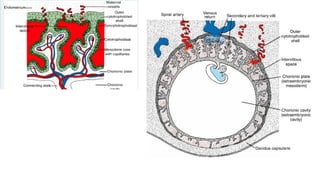
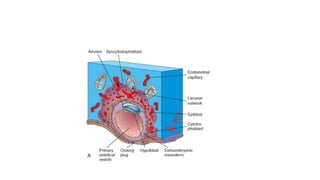
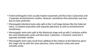
PLACENTA AND MEMBRANES
Dr. Mbira
• From the 3rd
-8th
week there is a rapid increase in the number of tertiary villi, the
blood vessels in the mesoderm of the connecting stalk, the tertiary villi and the
capillaries in the cytotrophoblastic outer shell interconnect to form the
extraembryonic circulation.
• With time, the cytotrophoblastic shell will thin out bringing the maternal
capillary wall closer to the syncytiotrophoblast for easier diffusion of nutrients
and metabolic products to and from the embryonic circulation.
• This is achieved by cytotrophoblastic cells invading maternal capillary
walls(endothelium) and replacing some of the maternal endothelial cells such
that the maternal capillaries will now have elements of (cyto)trophoblastic cells
within their wall and will transform into wide low resistance vessels for easier
diffusion of substances.
• Initially the whole chorionic surface is covered by villi, but as the pregnancy progresses, the
villi on the embryonic pole continue to grow as they interact with the decidua basalis and
look bushy hence the name chorion frondosum.
• Meanwhile, the villi on the abembryonic side flatten out and degenerate such that by the 4th
month the chorion is smooth and thus is termed chorion laeve.
• The decidua basalis becomes firmly attached to the villi on the embryonic pole and is known
as the decidual plate while that on the abembryonic pole is known as decidua capsularis.
• As the embryo grows, the decidua capsularis will degenerate and the pregnancy will grow to
the point where it will fill the whole uterine lumen and touch the other walls of the uterus
known as decidua parietalis. At this point, the chorion laeve and the decidua parietalis will
fuse.
• Also, the embryo will grow such that the chorionic space will be obliterated and the amnion
will fuse with the chorion to form the amniochorionic membrane.
• The placenta has a fetal and maternal components. On the fetal side is the chorionic
plate and on the maternal side is the maternal plate.
• Between the 2 plates is a network of intervillous lacunae filled with maternal blood.
• Always there is a barrier of syncytial cells between the maternal blood and the fetus.
• In the 5th
month, the decidua projects some incomplete septae towards the
chorionic plate but not reaching it and this ends up dividing the placenta into
cotyledons which are visible at birth as 15-20 bulges when the placenta is delivered.
• The placenta continues to increase in surface area and thickness as the pregnancy
progresses and covers 15-30% of the internal surface of the uterus.
• At term, the placenta has a diameter of 15-25cm, is about 3cm thick
and weighs about 500g.
• The placenta is expelled after birth and viewed from the maternal side
are visible cotyledons, from the fetal side is seen a smooth amnion
covering the chorion and vessels are seen coming from the placenta
and converging towards the cord.
• Functions of the placenta:
• Production of hormones
• Exchange of metabolic products, gases, nutrients, electrolytes.
• Transmission of maternal antibodies (IgG) from around week 14.
• The umbilical ring will initially contain the connecting stalk with blood
vessels, yolk sac as it is being “pinched” off by the expanding amniotic
cavity, and within this yolk sac will be vitelline vessels.
• By the 5th
week, the primitive umbilical ring will contain:
• Connecting stalk within which are the umbilical vessels( 2 arteries and a vein)
and the allantois.
• Yolk stalk(vitelline duct) accompanied by vitelline vessels.
• Canal connecting the intraembryonic and extraembryonic cavities.
• At some point up to the 12th
week, there is temporary protrusion of intestines into the primitive
umbilical cord before eventual withdrawal back into the abdomen.
• By the end of the 3rd
month, there is obliteration of the allantois, the yolk stalk and vitelline
vessels leaving only the umbilical vessels surrounded by Wharton’s jelly.
• The amniotic cavity is filled with amniotic fluid which at term is about a
litre of clear watery fluid derived partly from amniotic cells but mostly
from maternal blood.
• Amniotic fluid is swallowed by the foetus from around 5 months about
400ml daily which is subsequently urinated. This helps in the development
and maturation of the GIT and urinary systems.
• Functions of amniotic fluid:
• Shock absorber
• Prevents fetus from sticking to amniotic membrane.
• Allows free movement of the foetus.
• Forms a hydrostatic wedge that helps dilate the cervix during childbirth.
• Fraternal/dizygotic twins usually implant separately and thus have 2 placentae and
2 separate amniochorionic cavities. However, sometimes their placentae may fuse
due to close proximity.
• Monozygotic/identical twins who split at the 2 cell stage behave like the fraternal
twins, i.e implant separately with separate placentae as well as amniochorionic
membranes.
• Monozygotic twins who split at the blastocyst stage end up with 2 embryos within
the same blastocystic cavity and thus have 1 placenta, 1 chorionic cavity but 2
separate amniotic cavities.
• Monozygotic twins who result from splitting of the embryo at the bilaminar germ
disc stage end up with the same placenta, same chorionic cavity and same
amniotic cavity.
• During birth, the cervix begins to dilate and uterine contractions push
the foetus gradually downwards through the dilating cervix, the
membranes rupture pouring out the amniotic fluid, baby is born,
placenta then follows a variable while later(max. 30 min later)
accompanied by shedding of the decidual layer of the uterus.
*Preeclampsia/eclampsia
*Velamentous cord insertion
*Rhesus incompartibility
*Oligohydramnios/polyhydramnios
*Parasitic twin/twin transfusion syndrome
*Conjoined twins
*Placenta praevia
*Placenta accreta

The Placenta and membranes development ppt

  • 1.
  • 2.
    • From the3rd -8th week there is a rapid increase in the number of tertiary villi, the blood vessels in the mesoderm of the connecting stalk, the tertiary villi and the capillaries in the cytotrophoblastic outer shell interconnect to form the extraembryonic circulation. • With time, the cytotrophoblastic shell will thin out bringing the maternal capillary wall closer to the syncytiotrophoblast for easier diffusion of nutrients and metabolic products to and from the embryonic circulation. • This is achieved by cytotrophoblastic cells invading maternal capillary walls(endothelium) and replacing some of the maternal endothelial cells such that the maternal capillaries will now have elements of (cyto)trophoblastic cells within their wall and will transform into wide low resistance vessels for easier diffusion of substances.
  • 9.
    • Initially thewhole chorionic surface is covered by villi, but as the pregnancy progresses, the villi on the embryonic pole continue to grow as they interact with the decidua basalis and look bushy hence the name chorion frondosum. • Meanwhile, the villi on the abembryonic side flatten out and degenerate such that by the 4th month the chorion is smooth and thus is termed chorion laeve. • The decidua basalis becomes firmly attached to the villi on the embryonic pole and is known as the decidual plate while that on the abembryonic pole is known as decidua capsularis. • As the embryo grows, the decidua capsularis will degenerate and the pregnancy will grow to the point where it will fill the whole uterine lumen and touch the other walls of the uterus known as decidua parietalis. At this point, the chorion laeve and the decidua parietalis will fuse. • Also, the embryo will grow such that the chorionic space will be obliterated and the amnion will fuse with the chorion to form the amniochorionic membrane.
  • 12.
    • The placentahas a fetal and maternal components. On the fetal side is the chorionic plate and on the maternal side is the maternal plate. • Between the 2 plates is a network of intervillous lacunae filled with maternal blood. • Always there is a barrier of syncytial cells between the maternal blood and the fetus. • In the 5th month, the decidua projects some incomplete septae towards the chorionic plate but not reaching it and this ends up dividing the placenta into cotyledons which are visible at birth as 15-20 bulges when the placenta is delivered. • The placenta continues to increase in surface area and thickness as the pregnancy progresses and covers 15-30% of the internal surface of the uterus.
  • 13.
    • At term,the placenta has a diameter of 15-25cm, is about 3cm thick and weighs about 500g. • The placenta is expelled after birth and viewed from the maternal side are visible cotyledons, from the fetal side is seen a smooth amnion covering the chorion and vessels are seen coming from the placenta and converging towards the cord.
  • 15.
    • Functions ofthe placenta: • Production of hormones • Exchange of metabolic products, gases, nutrients, electrolytes. • Transmission of maternal antibodies (IgG) from around week 14.
  • 16.
    • The umbilicalring will initially contain the connecting stalk with blood vessels, yolk sac as it is being “pinched” off by the expanding amniotic cavity, and within this yolk sac will be vitelline vessels. • By the 5th week, the primitive umbilical ring will contain: • Connecting stalk within which are the umbilical vessels( 2 arteries and a vein) and the allantois. • Yolk stalk(vitelline duct) accompanied by vitelline vessels. • Canal connecting the intraembryonic and extraembryonic cavities.
  • 17.
    • At somepoint up to the 12th week, there is temporary protrusion of intestines into the primitive umbilical cord before eventual withdrawal back into the abdomen. • By the end of the 3rd month, there is obliteration of the allantois, the yolk stalk and vitelline vessels leaving only the umbilical vessels surrounded by Wharton’s jelly.
  • 18.
    • The amnioticcavity is filled with amniotic fluid which at term is about a litre of clear watery fluid derived partly from amniotic cells but mostly from maternal blood. • Amniotic fluid is swallowed by the foetus from around 5 months about 400ml daily which is subsequently urinated. This helps in the development and maturation of the GIT and urinary systems. • Functions of amniotic fluid: • Shock absorber • Prevents fetus from sticking to amniotic membrane. • Allows free movement of the foetus. • Forms a hydrostatic wedge that helps dilate the cervix during childbirth.
  • 19.
    • Fraternal/dizygotic twinsusually implant separately and thus have 2 placentae and 2 separate amniochorionic cavities. However, sometimes their placentae may fuse due to close proximity. • Monozygotic/identical twins who split at the 2 cell stage behave like the fraternal twins, i.e implant separately with separate placentae as well as amniochorionic membranes. • Monozygotic twins who split at the blastocyst stage end up with 2 embryos within the same blastocystic cavity and thus have 1 placenta, 1 chorionic cavity but 2 separate amniotic cavities. • Monozygotic twins who result from splitting of the embryo at the bilaminar germ disc stage end up with the same placenta, same chorionic cavity and same amniotic cavity.
  • 22.
    • During birth,the cervix begins to dilate and uterine contractions push the foetus gradually downwards through the dilating cervix, the membranes rupture pouring out the amniotic fluid, baby is born, placenta then follows a variable while later(max. 30 min later) accompanied by shedding of the decidual layer of the uterus.
  • 23.
    *Preeclampsia/eclampsia *Velamentous cord insertion *Rhesusincompartibility *Oligohydramnios/polyhydramnios *Parasitic twin/twin transfusion syndrome *Conjoined twins *Placenta praevia *Placenta accreta