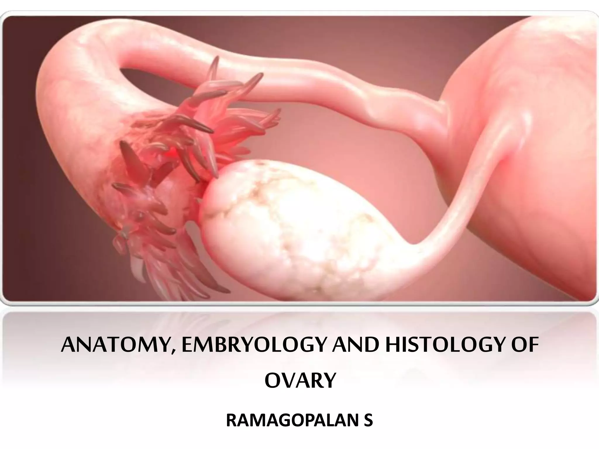
ovary - Anatomy, histology and embryological development of ovary
The document provides a comprehensive overview of the anatomy, embryology, and histology of the ovary, detailing its location, structure, blood supply, and nerve supply. It explains how the ovary develops from the coelomic epithelium and forms primordial follicles through a series of cellular interactions. Additionally, histological features such as the cortex and medulla of the ovary are described, including the different phases of ovarian follicles.
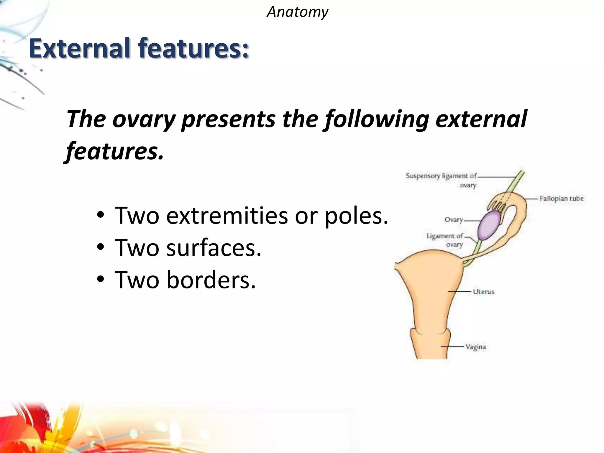
Anatomy
• The ovaryis ovoid or almond
in shape.
• It is about 3 cm long, 2 cm wide and
1 cm thick
• Ovaries lie on each side of the uterus, almost
vertically.
• Suspended in the pelvic cavity by a double fold
of peritoneum.
Location:
5.
Peritoneal relation:
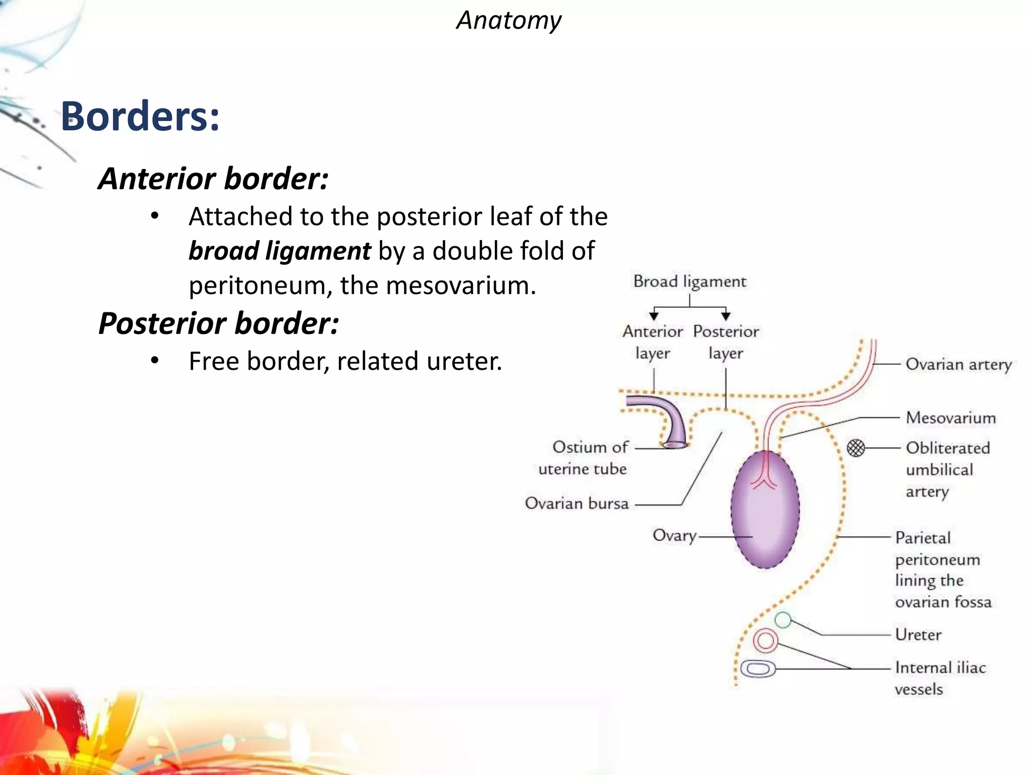
Anatomy
Each ovaryis attached to the
posterior surface of the broad
ligament by a short peritoneal
fold called mesovarium.
Each ovary is almost entirely
covered by the peritoneum
except along the mesovarian.
The mesovarium acts as a
hilum of the ovary and
conveys blood vessels and
nerves to the ovary.
Poles:
Anatomy
Upper pole:
• Itis broader than the lower pole
• Suspensory ligament of ovary
Lower pole:
• Connected to the lateral angle of
the uterus by ligament of ovary.
Surfaces:
Later surface:
• lies in the angle between the internal
and external iliac vessels.
Medial surface:
• mainly related to the uterine tube.
8.
Anatomy
Anterior border:
• Attachedto the posterior leaf of the
broad ligament by a double fold of
peritoneum, the mesovarium.
Posterior border:
• Free border, related ureter.
Borders:
9.
Anatomy
Blood supply:
• Arterialsupply: mainly, ovarian artery which arises from the
aorta at the level of L1 vertebra.
• Venous drainage:The right ovarian vein drains into the inferior
vena cava while the left ovarian vein drains into the left renal
vein.
Lymphatics drainage:
• Pre-aortic and para-aortic lymph nodes
Nerve supply:
• Postganglionic sympathetic (T10, T11) and parasympathetic (S2,
S3, S4) fibres, derived from abdominal autonomic plexuses.
• The visceral afferent fibres from the ovary run along the
sympathetic pathways to the spinal segments T10, T11.
Embryology
The coelomic epitheliumon the medial side of the mesonephros
becomes thickened to form genital ridges.
Primordial germs cells, that are formed in relation to yolk sac, migrate
to the region of developing ovary, and give rise to oocytes.
Embryology
The sex cordsbecome broken up into small masses. The cells of each mass
surround one primordial germ cell, or oocyte, to form a primordial follicle.
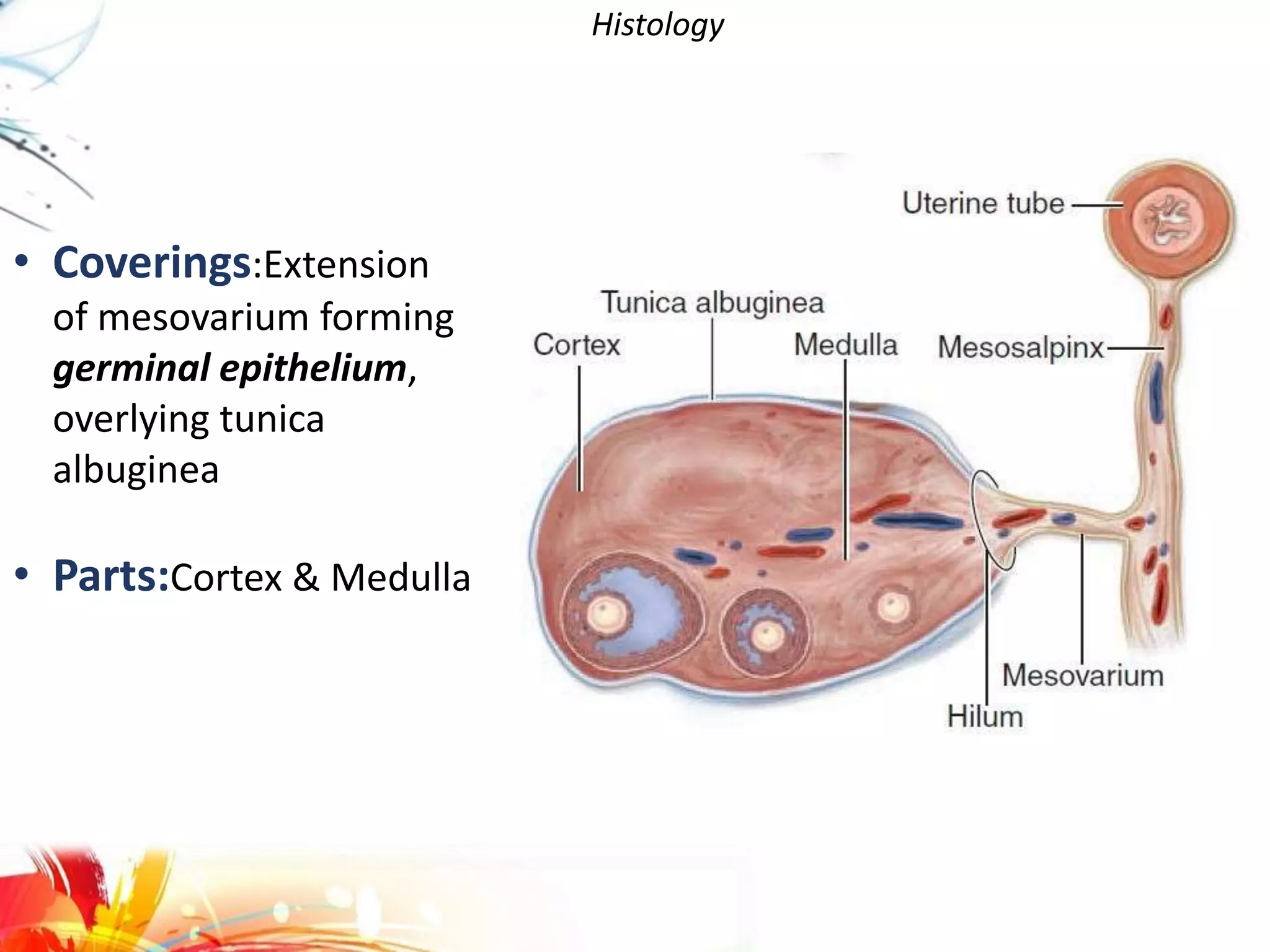
Histology
Different phases ofovarian follicles:
Primordial follicles Primary follicles Secondary follicles
Graafian follicles Corpus luteum Corpus albicans
19.
Histology
Medulla: Most
internal partof the
ovary, consists of loose
connective tissue and
blood vessels entering
through hilum from
mesenteries.
Cortex
medulla
20.
Reference:
• Human Anatomy,Fourth Edition - Saladin, Kenneth. S
• JUNQUEIRA's Basic Histology 13th Edition
• Last’s Anatomy Regional and Applied 12th edition
• Gray's Anatomy The Anatomical Basis of Clinical
Practice, 40th Edition