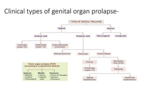
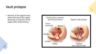
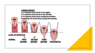
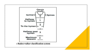
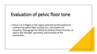
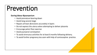
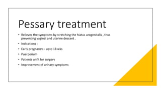
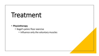
This document discusses displacement of the uterus. It begins by defining version and flexion of the uterus. It then discusses genital prolapse, describing the three levels of support for the uterus. It outlines the clinical types of genital organ prolapse including anterior and posterior vaginal wall prolapse as well as vault prolapse. It discusses the etiology, symptoms, diagnostic approach, and treatment options which include pessary, physiotherapy, and various surgical procedures. Ayurvedic view on pathogenesis and treatment involving local application of ghrita, swedana, and bandaging is also summarized.
Please find the power point on Utero-Vaginal Prolapse. I tried to present it on understandable way and all the contents are reviewed by experts and from very reliable references. Thank you
Pelvic organ prolapse
Etiology of pelvic organ prolapse
Vaginal vault prolapse
Etiological factors of vault prolapse
Signs and symptoms of vaginal vault prolapse
Diagnosis of vaginal vault prolapse
Treatment measures
Pelvic organ prolapse
Pelvic Organ Prolapse
Most common gynaecological problem.
Amongst parous women.
A form of hernia.
Anatomy of Uterus
Anteversion and anteflexion position.
Lies between rectum and bladder.
Cervix pierces the vagina at the right angle to the axis of vagina.
Supports of Uterus
Uterus is held in position by 3 tier support system.
Upper tier
Middle tier
Inferior tier.
Upper tier
Primarily, maintains the uterus in anteverted position.
The structures responsible are:
Endopelvic fascia.
Round ligaments.
Broad ligaments.
Middle tier
Constitutes the strongest support of uterus.
Responsible structures are:
Pericervical ring.
Pelvic cellular tissues.
Inferior tier
Indirect support of uterus.
Responsible structures are pelvic floor muscles including:
Levator ani
Endopelvic fascia
Levator plate
Perineal body
Urogenital diaphragm
Anatomical Factors
Gravitational stress.
Parturition stress.
Pelvic floor weakness.
Inherent weakness of supporting structures.
Acquired Predisposing Factors
Trauma of vaginal delivery causing injury :
Ligaments
Endopelvic fascia
Levator muscle
Perineal body
Pudendal nerve and muscle damage due to
repeated child birth.
Congenital Predisposing Factors
Inborn weakness of supporting structure.
Aggravating factors
Post menopausal atrophy
Poor collagen tissue repair with age.
Increased intra abdominal pressure.
Occupational hazards
Asthenia
Obesity
Fibroid/Polyp
Clinical Degrees Of Uterine Prolapse
Symptoms
Feeling of something coming out per vaginum.
Backache or dragging pain in pelvis
Dyspareunia
Urinary symptoms
Bowel symptoms
Clinical examination
Inspection and palpation.
General examination.
Pelvic examination
Uterine prolapse
Management of Prolapse
Preventive
Conservative
Surgery
Preventive Measures
Pelvic floor exercise during puerperium.
Avoid strenuous activities.
Avoiding prolonged cough.
Avoiding constipation.
Avoiding heavy weight lifting.
Avoiding future pregnancy too early.
Conservative Management
Indications :
Asymptomatic women
Mild degree prolapse
POP in early pregnancy
Treatment :
Oestrogen replacement therapy.
Kegel exercise
Pessary treatment
Surgical Management of Prolapse
Restorative
Extirpative
Obliterative
Genital prolapse is the descent of one or more of the genital organ (urethra, bladder, uterus, rectum or Douglas pouch or rectouterine pouch”) through the fasciomuscular pelvic floor below their normal level.
Explore natural remedies for syphilis treatment in Singapore. Discover alternative therapies, herbal remedies, and lifestyle changes that may complement conventional treatments. Learn about holistic approaches to managing syphilis symptoms and supporting overall health.
Ozempic: Preoperative Management of Patients on GLP-1 Receptor Agonists Saeid Safari
Preoperative Management of Patients on GLP-1 Receptor Agonists like Ozempic and Semiglutide
ASA GUIDELINE
NYSORA Guideline
2 Case Reports of Gastric Ultrasound
Couples presenting to the infertility clinic- Do they really have infertility...Sujoy Dasgupta
Dr Sujoy Dasgupta presented the study on "Couples presenting to the infertility clinic- Do they really have infertility? – The unexplored stories of non-consummation" in the 13th Congress of the Asia Pacific Initiative on Reproduction (ASPIRE 2024) at Manila on 24 May, 2024.
Report Back from SGO 2024: What’s the Latest in Cervical Cancer?bkling
Are you curious about what’s new in cervical cancer research or unsure what the findings mean? Join Dr. Emily Ko, a gynecologic oncologist at Penn Medicine, to learn about the latest updates from the Society of Gynecologic Oncology (SGO) 2024 Annual Meeting on Women’s Cancer. Dr. Ko will discuss what the research presented at the conference means for you and answer your questions about the new developments.
Title: Sense of Taste
Presenter: Dr. Faiza, Assistant Professor of Physiology
Qualifications:
MBBS (Best Graduate, AIMC Lahore)
FCPS Physiology
ICMT, CHPE, DHPE (STMU)
MPH (GC University, Faisalabad)
MBA (Virtual University of Pakistan)
Learning Objectives:
Describe the structure and function of taste buds.
Describe the relationship between the taste threshold and taste index of common substances.
Explain the chemical basis and signal transduction of taste perception for each type of primary taste sensation.
Recognize different abnormalities of taste perception and their causes.
Key Topics:
Significance of Taste Sensation:
Differentiation between pleasant and harmful food
Influence on behavior
Selection of food based on metabolic needs
Receptors of Taste:
Taste buds on the tongue
Influence of sense of smell, texture of food, and pain stimulation (e.g., by pepper)
Primary and Secondary Taste Sensations:
Primary taste sensations: Sweet, Sour, Salty, Bitter, Umami
Chemical basis and signal transduction mechanisms for each taste
Taste Threshold and Index:
Taste threshold values for Sweet (sucrose), Salty (NaCl), Sour (HCl), and Bitter (Quinine)
Taste index relationship: Inversely proportional to taste threshold
Taste Blindness:
Inability to taste certain substances, particularly thiourea compounds
Example: Phenylthiocarbamide
Structure and Function of Taste Buds:
Composition: Epithelial cells, Sustentacular/Supporting cells, Taste cells, Basal cells
Features: Taste pores, Taste hairs/microvilli, and Taste nerve fibers
Location of Taste Buds:
Found in papillae of the tongue (Fungiform, Circumvallate, Foliate)
Also present on the palate, tonsillar pillars, epiglottis, and proximal esophagus
Mechanism of Taste Stimulation:
Interaction of taste substances with receptors on microvilli
Signal transduction pathways for Umami, Sweet, Bitter, Sour, and Salty tastes
Taste Sensitivity and Adaptation:
Decrease in sensitivity with age
Rapid adaptation of taste sensation
Role of Saliva in Taste:
Dissolution of tastants to reach receptors
Washing away the stimulus
Taste Preferences and Aversions:
Mechanisms behind taste preference and aversion
Influence of receptors and neural pathways
Impact of Sensory Nerve Damage:
Degeneration of taste buds if the sensory nerve fiber is cut
Abnormalities of Taste Detection:
Conditions: Ageusia, Hypogeusia, Dysgeusia (parageusia)
Causes: Nerve damage, neurological disorders, infections, poor oral hygiene, adverse drug effects, deficiencies, aging, tobacco use, altered neurotransmitter levels
Neurotransmitters and Taste Threshold:
Effects of serotonin (5-HT) and norepinephrine (NE) on taste sensitivity
Supertasters:
25% of the population with heightened sensitivity to taste, especially bitterness
Increased number of fungiform papillae
- Video recording of this lecture in English language: https://youtu.be/lK81BzxMqdo
- Video recording of this lecture in Arabic language: https://youtu.be/Ve4P0COk9OI
- Link to download the book free: https://nephrotube.blogspot.com/p/nephrotube-nephrology-books.html
- Link to NephroTube website: www.NephroTube.com
- Link to NephroTube social media accounts: https://nephrotube.blogspot.com/p/join-nephrotube-on-social-media.html
Tom Selleck Health: A Comprehensive Look at the Iconic Actor’s Wellness Journeygreendigital
Tom Selleck, an enduring figure in Hollywood. has captivated audiences for decades with his rugged charm, iconic moustache. and memorable roles in television and film. From his breakout role as Thomas Magnum in Magnum P.I. to his current portrayal of Frank Reagan in Blue Bloods. Selleck's career has spanned over 50 years. But beyond his professional achievements. fans have often been curious about Tom Selleck Health. especially as he has aged in the public eye.
Follow us on: Pinterest
Introduction
Many have been interested in Tom Selleck health. not only because of his enduring presence on screen but also because of the challenges. and lifestyle choices he has faced and made over the years. This article delves into the various aspects of Tom Selleck health. exploring his fitness regimen, diet, mental health. and the challenges he has encountered as he ages. We'll look at how he maintains his well-being. the health issues he has faced, and his approach to ageing .
Early Life and Career
Childhood and Athletic Beginnings
Tom Selleck was born on January 29, 1945, in Detroit, Michigan, and grew up in Sherman Oaks, California. From an early age, he was involved in sports, particularly basketball. which played a significant role in his physical development. His athletic pursuits continued into college. where he attended the University of Southern California (USC) on a basketball scholarship. This early involvement in sports laid a strong foundation for his physical health and disciplined lifestyle.
Transition to Acting
Selleck's transition from an athlete to an actor came with its physical demands. His first significant role in "Magnum P.I." required him to perform various stunts and maintain a fit appearance. This role, which he played from 1980 to 1988. necessitated a rigorous fitness routine to meet the show's demands. setting the stage for his long-term commitment to health and wellness.
Fitness Regimen
Workout Routine
Tom Selleck health and fitness regimen has evolved. adapting to his changing roles and age. During his "Magnum, P.I." days. Selleck's workouts were intense and focused on building and maintaining muscle mass. His routine included weightlifting, cardiovascular exercises. and specific training for the stunts he performed on the show.
Selleck adjusted his fitness routine as he aged to suit his body's needs. Today, his workouts focus on maintaining flexibility, strength, and cardiovascular health. He incorporates low-impact exercises such as swimming, walking, and light weightlifting. This balanced approach helps him stay fit without putting undue strain on his joints and muscles.
Importance of Flexibility and Mobility
In recent years, Selleck has emphasized the importance of flexibility and mobility in his fitness regimen. Understanding the natural decline in muscle mass and joint flexibility with age. he includes stretching and yoga in his routine. These practices help prevent injuries, improve posture, and maintain mobilit
MANAGEMENT OF ATRIOVENTRICULAR CONDUCTION BLOCK.pdfJim Jacob Roy
Cardiac conduction defects can occur due to various causes.
Atrioventricular conduction blocks ( AV blocks ) are classified into 3 types.
This document describes the acute management of AV block.
Title: Sense of Smell
Presenter: Dr. Faiza, Assistant Professor of Physiology
Qualifications:
MBBS (Best Graduate, AIMC Lahore)
FCPS Physiology
ICMT, CHPE, DHPE (STMU)
MPH (GC University, Faisalabad)
MBA (Virtual University of Pakistan)
Learning Objectives:
Describe the primary categories of smells and the concept of odor blindness.
Explain the structure and location of the olfactory membrane and mucosa, including the types and roles of cells involved in olfaction.
Describe the pathway and mechanisms of olfactory signal transmission from the olfactory receptors to the brain.
Illustrate the biochemical cascade triggered by odorant binding to olfactory receptors, including the role of G-proteins and second messengers in generating an action potential.
Identify different types of olfactory disorders such as anosmia, hyposmia, hyperosmia, and dysosmia, including their potential causes.
Key Topics:
Olfactory Genes:
3% of the human genome accounts for olfactory genes.
400 genes for odorant receptors.
Olfactory Membrane:
Located in the superior part of the nasal cavity.
Medially: Folds downward along the superior septum.
Laterally: Folds over the superior turbinate and upper surface of the middle turbinate.
Total surface area: 5-10 square centimeters.
Olfactory Mucosa:
Olfactory Cells: Bipolar nerve cells derived from the CNS (100 million), with 4-25 olfactory cilia per cell.
Sustentacular Cells: Produce mucus and maintain ionic and molecular environment.
Basal Cells: Replace worn-out olfactory cells with an average lifespan of 1-2 months.
Bowman’s Gland: Secretes mucus.
Stimulation of Olfactory Cells:
Odorant dissolves in mucus and attaches to receptors on olfactory cilia.
Involves a cascade effect through G-proteins and second messengers, leading to depolarization and action potential generation in the olfactory nerve.
Quality of a Good Odorant:
Small (3-20 Carbon atoms), volatile, water-soluble, and lipid-soluble.
Facilitated by odorant-binding proteins in mucus.
Membrane Potential and Action Potential:
Resting membrane potential: -55mV.
Action potential frequency in the olfactory nerve increases with odorant strength.
Adaptation Towards the Sense of Smell:
Rapid adaptation within the first second, with further slow adaptation.
Psychological adaptation greater than receptor adaptation, involving feedback inhibition from the central nervous system.
Primary Sensations of Smell:
Camphoraceous, Musky, Floral, Pepperminty, Ethereal, Pungent, Putrid.
Odor Detection Threshold:
Examples: Hydrogen sulfide (0.0005 ppm), Methyl-mercaptan (0.002 ppm).
Some toxic substances are odorless at lethal concentrations.
Characteristics of Smell:
Odor blindness for single substances due to lack of appropriate receptor protein.
Behavioral and emotional influences of smell.
Transmission of Olfactory Signals:
From olfactory cells to glomeruli in the olfactory bulb, involving lateral inhibition.
Primitive, less old, and new olfactory systems with different path
micro teaching on communication m.sc nursing.pdfAnurag Sharma
Microteaching is a unique model of practice teaching. It is a viable instrument for the. desired change in the teaching behavior or the behavior potential which, in specified types of real. classroom situations, tends to facilitate the achievement of specified types of objectives.
These simplified slides by Dr. Sidra Arshad present an overview of the non-respiratory functions of the respiratory tract.
Learning objectives:
1. Enlist the non-respiratory functions of the respiratory tract
2. Briefly explain how these functions are carried out
3. Discuss the significance of dead space
4. Differentiate between minute ventilation and alveolar ventilation
5. Describe the cough and sneeze reflexes
Study Resources:
1. Chapter 39, Guyton and Hall Textbook of Medical Physiology, 14th edition
2. Chapter 34, Ganong’s Review of Medical Physiology, 26th edition
3. Chapter 17, Human Physiology by Lauralee Sherwood, 9th edition
4. Non-respiratory functions of the lungs https://academic.oup.com/bjaed/article/13/3/98/278874
ARTIFICIAL INTELLIGENCE IN HEALTHCARE.pdfAnujkumaranit
Artificial intelligence (AI) refers to the simulation of human intelligence processes by machines, especially computer systems. It encompasses tasks such as learning, reasoning, problem-solving, perception, and language understanding. AI technologies are revolutionizing various fields, from healthcare to finance, by enabling machines to perform tasks that typically require human intelligence.
Lung Cancer: Artificial Intelligence, Synergetics, Complex System Analysis, S...Oleg Kshivets
RESULTS: Overall life span (LS) was 2252.1±1742.5 days and cumulative 5-year survival (5YS) reached 73.2%, 10 years – 64.8%, 20 years – 42.5%. 513 LCP lived more than 5 years (LS=3124.6±1525.6 days), 148 LCP – more than 10 years (LS=5054.4±1504.1 days).199 LCP died because of LC (LS=562.7±374.5 days). 5YS of LCP after bi/lobectomies was significantly superior in comparison with LCP after pneumonectomies (78.1% vs.63.7%, P=0.00001 by log-rank test). AT significantly improved 5YS (66.3% vs. 34.8%) (P=0.00000 by log-rank test) only for LCP with N1-2. Cox modeling displayed that 5YS of LCP significantly depended on: phase transition (PT) early-invasive LC in terms of synergetics, PT N0—N12, cell ratio factors (ratio between cancer cells- CC and blood cells subpopulations), G1-3, histology, glucose, AT, blood cell circuit, prothrombin index, heparin tolerance, recalcification time (P=0.000-0.038). Neural networks, genetic algorithm selection and bootstrap simulation revealed relationships between 5YS and PT early-invasive LC (rank=1), PT N0—N12 (rank=2), thrombocytes/CC (3), erythrocytes/CC (4), eosinophils/CC (5), healthy cells/CC (6), lymphocytes/CC (7), segmented neutrophils/CC (8), stick neutrophils/CC (9), monocytes/CC (10); leucocytes/CC (11). Correct prediction of 5YS was 100% by neural networks computing (area under ROC curve=1.0; error=0.0).
CONCLUSIONS: 5YS of LCP after radical procedures significantly depended on: 1) PT early-invasive cancer; 2) PT N0--N12; 3) cell ratio factors; 4) blood cell circuit; 5) biochemical factors; 6) hemostasis system; 7) AT; 8) LC characteristics; 9) LC cell dynamics; 10) surgery type: lobectomy/pneumonectomy; 11) anthropometric data. Optimal diagnosis and treatment strategies for LC are: 1) screening and early detection of LC; 2) availability of experienced thoracic surgeons because of complexity of radical procedures; 3) aggressive en block surgery and adequate lymph node dissection for completeness; 4) precise prediction; 5) adjuvant chemoimmunoradiotherapy for LCP with unfavorable prognosis.
2. Background
• The uterus is normally anteverted, anteflexed
• Version: is the angle between the longitudinal axis of cervix,
and that of the vagina
• Flexion: is the angle between the longitudinal axis of the
uterus, and that of the cervix
3. Genital Prolapse
• Genital prolapse is the descent of one or more of the genital
organ (urethra, bladder, uterus, rectum or Pouch of Douglas
or rectouterine pouch) through the fasciomuscular pelvic
floor below their normal level.
• Vaginal prolapse can occur without uterine prolapse but the
uterus cannot descend without carrying the vagina with it.
4.
5. Three level of Supports of
Uterus
• Level I: The cardinal uterosacral ligament complex
• Level II: The pubo- cervical and recto-vaginal fascia
• Level III: The pubo-urethral ligaments anteriorly & the
perineal body posteriorly
6. Supports of Uterus -
• Upper tier – maintain the uterus in anteverted position.
• Endopelvic fascia covering the uterus
• Round ligaments
• Broad ligaments
• Middle tier – strongest support of the uterus
1. Peri cervical ring – fibroelastic connective tissues encircling the supravaginal cervix,
stabilizes the cervix at the level of interspinous diameter ,connected with
• Pubocervical ligaments
• vesicovaginal septum
• Cardinal ligaments
• Uterosacral ligaments
• Rectovaginal septum
7. 2. Pelvic cellular tissue
• Inferior tier – gives indirect support to the uterus ,
• Pelvic floor muscles (levator ani)
• Endopelvic fascia
• Levator plate
• Perineal body
• Urogenital diaphragm
9. Anterior vaginal wall prolapse
Prolapse of the upper 2/3 part of the anterior vaginal wall with
the base of the bladder is called cystocele
Prolapse
Prolapse of the lower 1/3 part of the anterior vaginal wall with
the urethra is called urethrocele.
Prolapse
Complete anterior vaginal wall prolapse is called cysto-
urethrocele.
Complete
10. Anterior vaginal wall prolapse
• Weakness in the
• Supports of the bladder neck
• Urethero vesical junction
• Proximal urethra
• Caused by
• Weakness of pubocervical fascia and pubourethral
ligaments
11. Posterior vaginal wall prolapse
a) It is called rectocele if there is laxity of the middle third of the posterior
vaginal wall.
b) Relaxed perineum
VAULT PROLAPSE -It is called enterocele if there is laxity of the upper third of
the posterior vaginal wall results in herniation of POD.
12.
13. Uterine descent
• Utero-vaginal (the uterus descends first followed by the
vagina): This usually occurs in cases of virginal and nulliparous
prolapse due to congenital weakness of the cervical
ligaments.
• Vagino-uterine (the vagina descends first followed by the
uterus):This usually occurs in cases of prolapse resulting from
obstetric trauma.
14. Vault prolapse
• Descent of the vaginal vault,
where the top of the vagina
descends or inversion of the
vagina after hysterectomy.
18. Precipitating factors
• Acute and chronic trauma of vaginal delivery
• Aging ( post menopausal atrophy )
• Estrogen deprivation
• Intrinsic collagen abnormalities
• Multiparity
• Iatrogenic
19. ↑ intra abdominal pressure
↑ weight of the uterus
Traction of the uterus by vaginal prolapse or by a large cervical polyp
Obesity(40%--75%)
Smoking
Pulmonary disease (chronic coughing)
Constipation (chronic straining)
Recreational or occupational activities
(frequent or heavy lifting)
20. Symptoms of Prolapse
• Pelvic floor disorders become symptomatic through either of two
mechanisms:
1. Mechanical difficulties produced by the actual prolapse,
2. Bladder or bowel dysfunction, disrupting either storage or emptying.
21. Clinical presentation
• Before actual prolapse. the patient feels a sensation of weakness in the perineum. particularly towards the end
of the day
• Later the patient notices a mass which appears on straining. and disappears when she lies down
• Urinary symptoms are common and trouble some even with slight prolapse:
a) Urgency and frequency by day
b) Stress incontinence
c) Inability to micturate unless the anterior vaginal wall is pushed upwards by the patient's fingers
d) Frequency when cystitis develops
22. • Rectal symptoms are not so marked. The patient always feels heaviness in
the rectum and a constant desire to defaecate. Piles develop from
straining.
• Backache, congestive dysmenorrhoea and menorrhagia are common.
• Leucorrhoea is caused by the congestion and associated by chronic
cervicitis.
• Associated decubitus ulcer may result in discharge which may be purulent
or blood stained.
23. Diagnostic approach
• Beginning with a careful inspection of the vulva and vagina to
identify erosions, ulcerations, or other lesions
• The extent of prolapse should be systematically assessed
• Suspicious lesions should be biopsied
24. Examination
Local examination
Per speculum
examination
Per vaginal/
Bimanual
examination
Bonney’s stress test
Evaluation of tone
of pelvic muscles
Recto vaginal
examination
Position of patient
for examination
• - standing & straining
• - dorsal lithotomy
25. Diagnostic approach
• The maximal extent of prolapse is demonstrated with a
standing straining examination when the bladder is empty
• Pelvic muscle function should be assessed after the bimanual
examination → palpate the pelvic muscles a few centimeters
inside the hymen, along pelvic sidewalls at the 4 & 8 o’clock
• Resting tone & voluntary contraction of the anal sphincters
should be assessed during rectovaginal examination
26. Evaluation of pelvic floor tone
• Place 1 or 2 fingers in the vagina and instruct the patient to
contract her pelvic floor muscles (i.e., the levator ani
muscles). Then gauge her ability to contract these muscles, as
well as the strength, symmetry, and duration of the
contraction.
27. Prevention
During labour &puerperium
• Avoid premature bearing down
• Avoid long second stage
• Repairs all tears &incisions accurately in layers
• Do not express the uterus when attempting to deliver placenta
• Encourage pelvic floor exercise
• Avoid puerperal constipation
• To avoid strenuous activities for at least 6 months following delivery.
• To avoid further pregnancy too soon with help of contraceptive practice.
28. Pessary treatment
• Relieves the symptoms by stretching the hiatus urogenitalis , thus
preventing vaginal and uterine descent .
• Indications :
• Early pregnancy – upto 18 wks
• Puerperium
• Patients unfit for surgery
• Improvement of urinary symptoms
30. Principles of Management
• Physical examination must not be used in isolation to develop
treatment strategy.
• Any decision for surgical intervention should take account of
how prolapse is affecting lifestyle.
31. Types of Operations
• Anterior colporrhaphy
• Perineorrhaphy
• Colpoperineorrhaphy
• Pelvic floor repair
• Fothergill’s or Manchester operation
• Hysterectomy
• Cervicopexy or sling operation (Purandare’s operation)
33. Pathogenesis according to ayurveda–
• Aggravation of nidana’s will result in apana vata vikruti or vata pitta dushti, this
will further affect garbhasaya gata mamsa dhatu which will cause khavaigunyata
in yoni and there will be shithilata and sramsa on pelvic organs (vagina/uterus).
35. • According to Susrutha, treatment for this condition is given as Abyangam of the
Yoni with Ghrita and then applying Swedana with milk. It should be inserted into
the vagina canal with the hand. Veshawara pinda made up of Sunti, Maricha,
Dhanyaka, Ajaji, Dadima and Pippali moola is kept inside and to keep the organ
in place and to exert Gophana bhandha should be applied up to the next Mutra
vega. Re-bandaging is necessary.