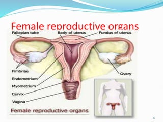
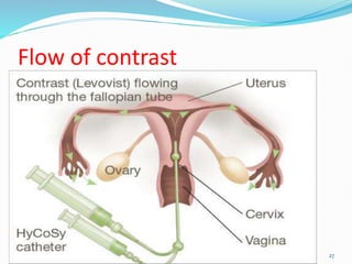
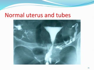
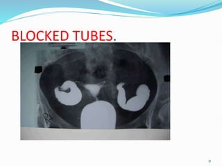
ITM.
MODULE1 3
1
THE ANATOMY .
 Ability to reproduce off-springs of the same
kind (one of the characteristics of living things).
 Two systems are;
a. Female reproductive systems.
b. Male reproductive systems.
FEMALE REPRODUCTIVE SYSTEMS.
 Divide into 2 groups.
a. Internal organs of reproduction.
b. External organs of reproduction.
2
THE ANATOMY .
FEMALE REPRODUCTIVE SYSTEMS.
a. Internal organs of reproduction.
 Situated in the internal pelvic cavity.
 These are;
i. Ovaries. 2.
ii. Uterine tubes/fallopian tubes. 2.
iii. Uterus. 1.
iv. Vagina. 1.
b. External female organs of reproduction.
 These are;
1) Mons pubis-covered by hair, rounded
eminence in front of symphysis.
3
THE ANATOMY .
b. External female organs of reproduction
cont;
1) Labia majora- forms vulva.
2) Labia minora.
3) Vestibule.
4) Greater vestibular glands.
5) Clitoris- erectile structure i.e. as in penis.
6) Vaginal orifice (Median slit).
7) Hymen (Mucous membrane).
8) External urethral orifice.
4
Fig. Uterus and fallopian tubes: coronal section to
show blood supply and ureter relative to uterine
artery, cervix and vaginal fornices.
5
Female reproductive organs
6
Fig. Female pelvis: sagittal section showing
pelvic floor.
7
HYSTEROSALPINGOGRAPHY (H.S.G).
Definition;
 Is the radiographic examination of the uterus
and uterine tubes following injection of
contrast medium through the cervix.
Indications.
1. Infertility primary or secondary.
2. Recurrent abortions.
3. Following tubal surgery (tubal ligation).
8
HYSTEROSALPINGOGRAPHY (H.S.G).
Contraindication.
a) Pregnancy.
b) A purulent discharge.
c) Recent dilatation and curettage. D&C.
d) Recent abortion.
e) Immediately post-menstruation 10days.
Contrast Medium.
 Water soluble (non-ionic) e.g.
i. Urografin.
ii. Ultravists.
iii. Uromiro 300 etc.
 Dosage 10 – 20mls.
9
Performing HSG
10
Physician and chaperon
11
HYSTEROSALPINGOGRAPHY.
Equipment and basic trolley settings.
 Fluoroscopy unit with spot film device.
 Vaginal speculum, vulselum forceps & uterine
cannula.
 Uterine sound, sponge-holding & tissue forceps.
 Lotion bowl, gallipot, towels, gauze swabs,
gown, sterile rubber gloves. 10mls syringe.
Lower shelf.
 Suitable Cleanser/clean gloves/sanitary pad.
 Contrast medium in bowl of warm water.
 File for opening ampoules.
 Emergency drugs.
12
HYSTEROSALPINGOGRAPHY (H.S.G).
Patient Preparation.
 The examination is booked between the 4th
and the 10th day post-menstruation for a
patient with regular 28-days cycle.
 If the cycle is not regular, then the patient
should avoid sexual intercourse between
booking and appointment time unless she uses
reliable contraceptives methods.
 Any abnormal discharge should be treated
before the examination.
13
HYSTEROSALPINGOGRAPHY (H.S.G).
Patient Preparation.
 Apprehensive patient may need
premedication Paracetamol 1gm, 1 hr prior to
the examination..
 Shaving of the pubic areas.
 The patient must undress completely and
wear a front-open gown.
 She should micturate before the examination.
14
HYSTEROSALPINGOGRAPHY (H.S.G).
Preliminary Film.
 Coned Postero-Anterior (P.A) view of the
uterine cavity.
Radiation Protection.
 Since it is impossible to protect the gonads
from the radiation some means of reducing
radiation dose to a minimum is employed.
 Rare earth or very fast tungstate screens are
used.
15
HYSTEROSALPINGOGRAPHY (H.S.G).
Radiation Protection.
 High KV technique is used.
 Image intensification should be employed.
 Screening time kept as low as possible
consistent with good visualization.
 The hands of the
gynecologist/radiologist/radiographer giving
injection should be protected.
16
HYSTEROSALPINGOGRAPHY (H.S.G).
Radiation Protection cont;
 Such protection may consist of a large sheet of
lead rubber placed under the patient legs, With
its upper border below the symphysis pubis.
 Alternatively a lead lined box may be placed
between the patient thigh and the instrument
inserted through it.
 A lid being placed on top when the instrument
are in position.
17
HYSTEROSALPINGOGRAPHY (H.S.G).
Technique.
 The patient lie supine on the table with the knees
and hips flexed and legs abducted.
(lithotomic position).
 She should be covered as much as possible to
afford privacy which is an important part of the
examination.
 Using a septic technique, the gynecologist/
radiologists/radiographer inserts the speculum
and clean the vagina and cervix with a suitable
cleanser. 18
HYSTEROSALPINGOGRAPHY (H.S.G).
Technique cont;
 The anterior lip of cervix is gripped and
steadied by the vulsellum forceps.
 A uterine sound may be inserted to show the
length and direction of the cervical canal.
 Uterine cannula is inserted into the cervical
canal and a syringe containing contrast medium
is connected in place.
19
HYSTEROSALPINGOGRAPHY (H.S.G).
Technique cont;
 Fluoroscopy with image intensification is
commenced and the injection of contrast
medium is observed.( 10 mls)
 Spasms of the uterine cornu might occur
preventing tubal filling, This may be relieved
by inhalation of octyl-nitrite.
20
Filming ……both AP pelvis done.
 1 st film… Using the undercouch tube, as the uterus begins to
fill. ( after introducing 10 mls)
 2 nd film…When the tubes begin to fill and peritoneal spill
has occurred.( after introduction of another 10 mls).
 After spillage all the instruments are removed.
21
Introducing dye.
22
2 nd dose of contrast
23
HYSTEROSALPINGOGRAPHY (H.S.G).
After Care.
 Observe the patient hygiene.
 It must be ensured that the patient has no serious
discomfort nor has significant bleeding before
she leaves.
 A sanitary pad should be provided.
 She should be advised that there may be
bleeding per vagina for 1–2 days.
 Analgesics may be necessary for up to 1week.
24
25
HYSTEROSALPINGOGRAM.
1. Body of uterus. 2. Fundus of uterus.
3. Uterine cornu . 4. Isthmus of fallopian tube.
5. Ampulla of fallopian tube. 6. Peritoneal spill of contrast.
Hysterosalpingogram.
26
Flow of contrast
27
Septated uterus
28
Normal uterus and tubes
29
Bilaterally blocked tubes.
30
BLOCKED TUBES.
31
Benefits of HSG.
 Hysterosalpingography is a minimally invasive
procedure with rare complications.
 Hysterosalpingography is a relatively short procedure
that can provide valuable information on a variety of
abnormalities that cause infertility or problems
carrying a fetus to term.
32
 Hysterosalpingography can occasionally open fallopian
tubes that are blocked allowing the patient to become
pregnant afterwards.
 No radiation remains in a patient's body after an x-ray
examination.
 X-rays usually have minimal side effects in the typical
diagnostic range for this exam.( Minimal radiation
dose when meticulous technique is applied)
33
Risks
 There is always a slight chance of cancer from excessive exposure to
radiation.
 However, the benefit of an accurate diagnosis far outweighs the risk.
 In the event of a chronic PID, pelvic infection or untreated sexually
transmitted disease, notify the physician before the procedure to avoid
worsening of infection.
 Women should always inform their physician if there is any possibility
that they are pregnant.
34
GYNAECOGRAPHY (PELVIC
PNEUMOGRAPHY).
Definition;
 This is a radiographic examination of the uterus
and ovaries following instillation of Carbon
dioxide (CO²) into the peritoneal cavity.
Indication.
• Primary/secondary amenorrhea.
• Ovarian agenesis.
• Ovarian neoplasm and cysts.
35
GYNAECOGRAPHY (PELVIC
PNEUMOGRAPHY).
Note.
 Ultrasound and laparatomy are now the tools of
choice.
 Gynaecography is of historical interests only.
Additional obstetric examinations;
1) Ultrasound.
2) Erect Lateral Pelvimetry (E.L.P).
3) Plain abdominal films.
4) Placentography.
36
THE MALE
REPRODUCTIVE TRACT.
Anatomy.
37
MALE REPRODUCTIVE TRACTS.
 These Organs are;
a) Testis. 2.
b) Deferent ducts (vas-deferens). 2.
c) Seminal vesicles. 2.
d) Ejaculatory ducts. 2.
e) Prostate gland. 1.
f) Epididymis. 2.
g) Penis. 1.
38
organs
39
Fig. Testis and epididymis:
(a) internal architecture.
40
Fig. Testis and epididymis:
(b) blood supply;
41
VESICULOGRAPHY.
Defination.
 Radiographic examination of the vasa-
diferentia, seminal vesicles and ejaculatory
ducts following injection of contrast medium
into the ducts.
Indication.
1. Differential diagnosis between prostatic
hyperplasia and carcinoma.
2. Investigation of epididymo-orchitis.
3. Diagnosis of seminal ductal system disorders.
4. To confirm or excludes ejaculatory duct
obstruction.
42
VESICULOGRAPHY.
Note.
 The examination is carried out following
vasectomy.
 The radiographs are either done in theatre
using mobile unit or the patient is taken to
radiology department with catheter in situ.
Contrast Medium.
 Any of non-ionic contrast medium.
 Dosage 1.5–2.0mls/side/injection.
43
VESICULOGRAPHY.
Preliminary Film.
 Antero-Posterior view of bladder with the
patient supine.
 Using a 30x40cm cassette.
 With upper border at the level of the Anterior
Superior Iliac Spine (ASIS).
 Centre in the midline at the level of Anterior
Superior Iliac Spine (ASIS), With central ray
angled 15°caudad.
44
Vesiculography
45
VESICULOGRAPHY.
Technique.
 Catheter is inserted into the ducts during
operation.
 Contrast medium is injected into each duct
simultaneously for the Antero-Posterior (A.P)
view.
 Into each duct separately for the oblique’s
views.
 Exposure must be taken during injection when
there is maximum filling of the ducts.
46
TRUS- vesiculography
47
VESICULOGRAPHY.
Films.
a) Antero-Posterior AP view as for preliminary.
b) Posterior oblique's.
Posterior Oblique VIEW.
 These are necessary when the ducts are
obscured by overflow of contrast medium into
the prostatic urethra on the 1st injection.
 From the supine position, the patient is rotated
to each side in turn and an exposure is made in
each position.
48
VESICULOGRAPHY.
Posterior Oblique VIEW.
 The right posterior oblique view is done to
demonstrate the left ducts and vice-versa.
 Centre 2.5cm medially to the Anterior Superior
Iliac Spine (ASIS) of the raised side.
After Care.
 As for the theatre patient.
49
50
Terminologies.
Aspermia. Absence of semen.
Azoospermia. Absence of sperms.
Hypospermia. Low semen volume.
Oligozoospermia. Low sperm counts.
Asthenozoospermia. Poor sperm motility.
Teratozoospermia. Sperm carry more morphological
defects than usual.
Necrozoospermia. All sperm in the ejaculate are dead.
END OF REPRODUCTIVE
SYSTEM.
51

REPRODUCTIVE SYSTEM EXAM.pptx

  • 1.
  • 2.
    THE ANATOMY . Ability to reproduce off-springs of the same kind (one of the characteristics of living things).  Two systems are; a. Female reproductive systems. b. Male reproductive systems. FEMALE REPRODUCTIVE SYSTEMS.  Divide into 2 groups. a. Internal organs of reproduction. b. External organs of reproduction. 2
  • 3.
    THE ANATOMY . FEMALEREPRODUCTIVE SYSTEMS. a. Internal organs of reproduction.  Situated in the internal pelvic cavity.  These are; i. Ovaries. 2. ii. Uterine tubes/fallopian tubes. 2. iii. Uterus. 1. iv. Vagina. 1. b. External female organs of reproduction.  These are; 1) Mons pubis-covered by hair, rounded eminence in front of symphysis. 3
  • 4.
    THE ANATOMY . b.External female organs of reproduction cont; 1) Labia majora- forms vulva. 2) Labia minora. 3) Vestibule. 4) Greater vestibular glands. 5) Clitoris- erectile structure i.e. as in penis. 6) Vaginal orifice (Median slit). 7) Hymen (Mucous membrane). 8) External urethral orifice. 4
  • 5.
    Fig. Uterus andfallopian tubes: coronal section to show blood supply and ureter relative to uterine artery, cervix and vaginal fornices. 5
  • 6.
  • 7.
    Fig. Female pelvis:sagittal section showing pelvic floor. 7
  • 8.
    HYSTEROSALPINGOGRAPHY (H.S.G). Definition;  Isthe radiographic examination of the uterus and uterine tubes following injection of contrast medium through the cervix. Indications. 1. Infertility primary or secondary. 2. Recurrent abortions. 3. Following tubal surgery (tubal ligation). 8
  • 9.
    HYSTEROSALPINGOGRAPHY (H.S.G). Contraindication. a) Pregnancy. b)A purulent discharge. c) Recent dilatation and curettage. D&C. d) Recent abortion. e) Immediately post-menstruation 10days. Contrast Medium.  Water soluble (non-ionic) e.g. i. Urografin. ii. Ultravists. iii. Uromiro 300 etc.  Dosage 10 – 20mls. 9
  • 10.
  • 11.
  • 12.
    HYSTEROSALPINGOGRAPHY. Equipment and basictrolley settings.  Fluoroscopy unit with spot film device.  Vaginal speculum, vulselum forceps & uterine cannula.  Uterine sound, sponge-holding & tissue forceps.  Lotion bowl, gallipot, towels, gauze swabs, gown, sterile rubber gloves. 10mls syringe. Lower shelf.  Suitable Cleanser/clean gloves/sanitary pad.  Contrast medium in bowl of warm water.  File for opening ampoules.  Emergency drugs. 12
  • 13.
    HYSTEROSALPINGOGRAPHY (H.S.G). Patient Preparation. The examination is booked between the 4th and the 10th day post-menstruation for a patient with regular 28-days cycle.  If the cycle is not regular, then the patient should avoid sexual intercourse between booking and appointment time unless she uses reliable contraceptives methods.  Any abnormal discharge should be treated before the examination. 13
  • 14.
    HYSTEROSALPINGOGRAPHY (H.S.G). Patient Preparation. Apprehensive patient may need premedication Paracetamol 1gm, 1 hr prior to the examination..  Shaving of the pubic areas.  The patient must undress completely and wear a front-open gown.  She should micturate before the examination. 14
  • 15.
    HYSTEROSALPINGOGRAPHY (H.S.G). Preliminary Film. Coned Postero-Anterior (P.A) view of the uterine cavity. Radiation Protection.  Since it is impossible to protect the gonads from the radiation some means of reducing radiation dose to a minimum is employed.  Rare earth or very fast tungstate screens are used. 15
  • 16.
    HYSTEROSALPINGOGRAPHY (H.S.G). Radiation Protection. High KV technique is used.  Image intensification should be employed.  Screening time kept as low as possible consistent with good visualization.  The hands of the gynecologist/radiologist/radiographer giving injection should be protected. 16
  • 17.
    HYSTEROSALPINGOGRAPHY (H.S.G). Radiation Protectioncont;  Such protection may consist of a large sheet of lead rubber placed under the patient legs, With its upper border below the symphysis pubis.  Alternatively a lead lined box may be placed between the patient thigh and the instrument inserted through it.  A lid being placed on top when the instrument are in position. 17
  • 18.
    HYSTEROSALPINGOGRAPHY (H.S.G). Technique.  Thepatient lie supine on the table with the knees and hips flexed and legs abducted. (lithotomic position).  She should be covered as much as possible to afford privacy which is an important part of the examination.  Using a septic technique, the gynecologist/ radiologists/radiographer inserts the speculum and clean the vagina and cervix with a suitable cleanser. 18
  • 19.
    HYSTEROSALPINGOGRAPHY (H.S.G). Technique cont; The anterior lip of cervix is gripped and steadied by the vulsellum forceps.  A uterine sound may be inserted to show the length and direction of the cervical canal.  Uterine cannula is inserted into the cervical canal and a syringe containing contrast medium is connected in place. 19
  • 20.
    HYSTEROSALPINGOGRAPHY (H.S.G). Technique cont; Fluoroscopy with image intensification is commenced and the injection of contrast medium is observed.( 10 mls)  Spasms of the uterine cornu might occur preventing tubal filling, This may be relieved by inhalation of octyl-nitrite. 20
  • 21.
    Filming ……both APpelvis done.  1 st film… Using the undercouch tube, as the uterus begins to fill. ( after introducing 10 mls)  2 nd film…When the tubes begin to fill and peritoneal spill has occurred.( after introduction of another 10 mls).  After spillage all the instruments are removed. 21
  • 22.
  • 23.
    2 nd doseof contrast 23
  • 24.
    HYSTEROSALPINGOGRAPHY (H.S.G). After Care. Observe the patient hygiene.  It must be ensured that the patient has no serious discomfort nor has significant bleeding before she leaves.  A sanitary pad should be provided.  She should be advised that there may be bleeding per vagina for 1–2 days.  Analgesics may be necessary for up to 1week. 24
  • 25.
  • 26.
    1. Body ofuterus. 2. Fundus of uterus. 3. Uterine cornu . 4. Isthmus of fallopian tube. 5. Ampulla of fallopian tube. 6. Peritoneal spill of contrast. Hysterosalpingogram. 26
  • 27.
  • 28.
  • 29.
  • 30.
  • 31.
  • 32.
    Benefits of HSG. Hysterosalpingography is a minimally invasive procedure with rare complications.  Hysterosalpingography is a relatively short procedure that can provide valuable information on a variety of abnormalities that cause infertility or problems carrying a fetus to term. 32
  • 33.
     Hysterosalpingography canoccasionally open fallopian tubes that are blocked allowing the patient to become pregnant afterwards.  No radiation remains in a patient's body after an x-ray examination.  X-rays usually have minimal side effects in the typical diagnostic range for this exam.( Minimal radiation dose when meticulous technique is applied) 33
  • 34.
    Risks  There isalways a slight chance of cancer from excessive exposure to radiation.  However, the benefit of an accurate diagnosis far outweighs the risk.  In the event of a chronic PID, pelvic infection or untreated sexually transmitted disease, notify the physician before the procedure to avoid worsening of infection.  Women should always inform their physician if there is any possibility that they are pregnant. 34
  • 35.
    GYNAECOGRAPHY (PELVIC PNEUMOGRAPHY). Definition;  Thisis a radiographic examination of the uterus and ovaries following instillation of Carbon dioxide (CO²) into the peritoneal cavity. Indication. • Primary/secondary amenorrhea. • Ovarian agenesis. • Ovarian neoplasm and cysts. 35
  • 36.
    GYNAECOGRAPHY (PELVIC PNEUMOGRAPHY). Note.  Ultrasoundand laparatomy are now the tools of choice.  Gynaecography is of historical interests only. Additional obstetric examinations; 1) Ultrasound. 2) Erect Lateral Pelvimetry (E.L.P). 3) Plain abdominal films. 4) Placentography. 36
  • 37.
  • 38.
    MALE REPRODUCTIVE TRACTS. These Organs are; a) Testis. 2. b) Deferent ducts (vas-deferens). 2. c) Seminal vesicles. 2. d) Ejaculatory ducts. 2. e) Prostate gland. 1. f) Epididymis. 2. g) Penis. 1. 38
  • 39.
  • 40.
    Fig. Testis andepididymis: (a) internal architecture. 40
  • 41.
    Fig. Testis andepididymis: (b) blood supply; 41
  • 42.
    VESICULOGRAPHY. Defination.  Radiographic examinationof the vasa- diferentia, seminal vesicles and ejaculatory ducts following injection of contrast medium into the ducts. Indication. 1. Differential diagnosis between prostatic hyperplasia and carcinoma. 2. Investigation of epididymo-orchitis. 3. Diagnosis of seminal ductal system disorders. 4. To confirm or excludes ejaculatory duct obstruction. 42
  • 43.
    VESICULOGRAPHY. Note.  The examinationis carried out following vasectomy.  The radiographs are either done in theatre using mobile unit or the patient is taken to radiology department with catheter in situ. Contrast Medium.  Any of non-ionic contrast medium.  Dosage 1.5–2.0mls/side/injection. 43
  • 44.
    VESICULOGRAPHY. Preliminary Film.  Antero-Posteriorview of bladder with the patient supine.  Using a 30x40cm cassette.  With upper border at the level of the Anterior Superior Iliac Spine (ASIS).  Centre in the midline at the level of Anterior Superior Iliac Spine (ASIS), With central ray angled 15°caudad. 44
  • 45.
  • 46.
    VESICULOGRAPHY. Technique.  Catheter isinserted into the ducts during operation.  Contrast medium is injected into each duct simultaneously for the Antero-Posterior (A.P) view.  Into each duct separately for the oblique’s views.  Exposure must be taken during injection when there is maximum filling of the ducts. 46
  • 47.
  • 48.
    VESICULOGRAPHY. Films. a) Antero-Posterior APview as for preliminary. b) Posterior oblique's. Posterior Oblique VIEW.  These are necessary when the ducts are obscured by overflow of contrast medium into the prostatic urethra on the 1st injection.  From the supine position, the patient is rotated to each side in turn and an exposure is made in each position. 48
  • 49.
    VESICULOGRAPHY. Posterior Oblique VIEW. The right posterior oblique view is done to demonstrate the left ducts and vice-versa.  Centre 2.5cm medially to the Anterior Superior Iliac Spine (ASIS) of the raised side. After Care.  As for the theatre patient. 49
  • 50.
    50 Terminologies. Aspermia. Absence ofsemen. Azoospermia. Absence of sperms. Hypospermia. Low semen volume. Oligozoospermia. Low sperm counts. Asthenozoospermia. Poor sperm motility. Teratozoospermia. Sperm carry more morphological defects than usual. Necrozoospermia. All sperm in the ejaculate are dead.
  • 51.

Editor's Notes

  • #41 Parameters. 1. sperm count concentration= 20m/1mlts. 40m/ejaculates. 2. motility , 60% live of those observed qty n qnty. 3,morphology, small,large, double head, tail, no head or tail. 4. volume (seminal fluid) 1.0 ml – 6.5 ml normal. 5. fructose level in semen 3mg/ml(an absence of fructose indicates a problem with seminal vesicle). 6. ph value = 7.1 – 8.0 WHO, 7.2 – 7.8.
  • #42 Terminologies. Aspermia. Absence of semen. Azoospermia. Absence of sperms. Hypospermia. Low semen volume. Oligozoospermia. Low sperm counts. Asthenozoospermia. Poor sperm motility. Teratozoospermia. Sperm carry more morphological defects than usual. Necrozoospermia. All sperm in the ejaculate are dead.