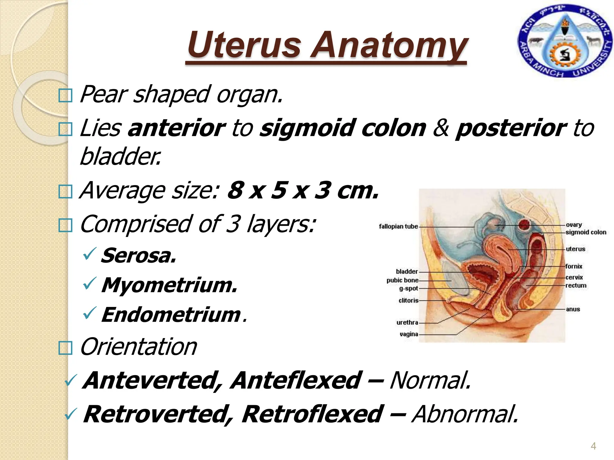
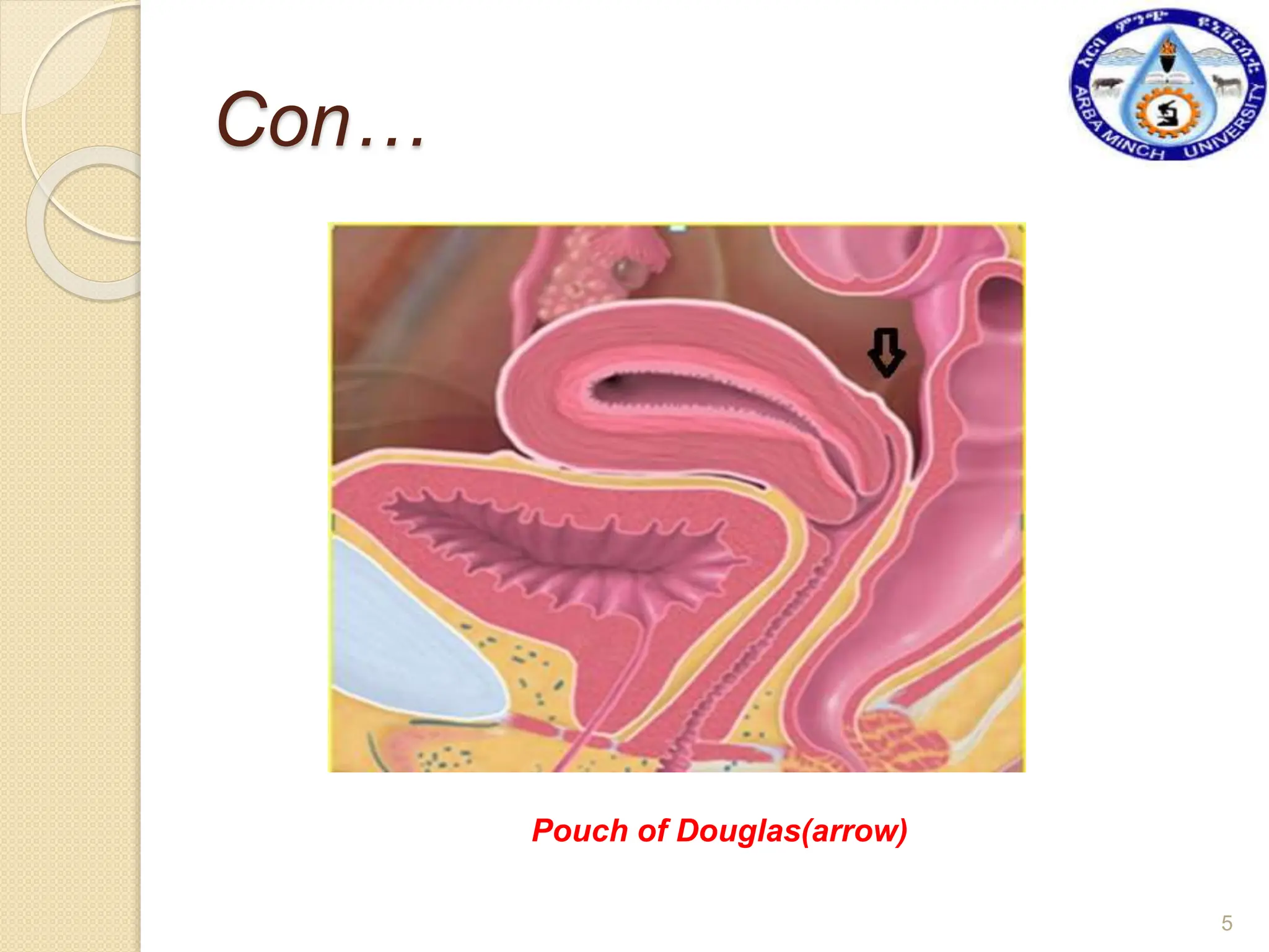
Arba Minch University
College of Medicine and Health Sciences
Department of MRT
1
Chapter – Three
Hysterosalpingography
2
OBJECTIVES:
 After completing this chapter, the student will be able
to:
 Identify major components of female genital tract ,to
include their location & function.
 Discuss radiographic examination of female genital
tract, including pre- & post procedural patient care for
hysterosalpingography (HSG).
 List & describe equipment commonly used for HSG.
 Define terminology associated with female genital tract
& special radiographic procedures, to include anatomy,
& pathology.
FEMALE GENITAL TRACT
3
Uterus Anatomy
 Pear shaped organ.
 Lies anterior to sigmoid colon & posterior to
bladder.
 Average size: 8 x 5 x 3 cm.
 Comprised of 3 layers:
Serosa.
Myometrium.
Endometrium.
 Orientation
 Anteverted, Anteflexed – Normal.
 Retroverted, Retroflexed – Abnormal.
4
Con…
Pouch of Douglas(arrow)
5
The Uterus
 The uterus is subdivided into four divisions:
(1) the fundus,
(2) the corpus (body),
(3) the isthmus, and
(4) the cervix (neck).
6
 Orientation
 The uterine fundus may point anteriorly toward the
navel, anteversion, or posteriorly toward the spine,
retroversion.
 Long axis of the uterus lies horizontally in sagittal
plan at 90 degree with the vagina, Anteverted.
 The fundus is flexed anteriorly in relation to the cervix
with 125 degree, Anteflexed.
7
8
 Retroversion & Retroflexion are opposite of
anteverted and anteflexed respectively.
Adenexal Anatomy
Fallopian Tubes
 Located below fundus
 Average size: 1- 4 mm in diameter and 10-
12cm long
 Four segments
 Isthmus.
 Ampulla.
Infundibulum.
Fimbriae.
10
Female Reproductive
System
11
HYSTEROSALPINGOGRAPHY(HS
G)
 Is radiographic demonstration of female reproductive
tract with a contrast medium.
 Best demonstrates uterine cavity and patency
(degree of openness) of uterine tubes.
 Uterine cavity is outlined by injection of a contrast
medium through the cervix.
 The shape and contour of uterine cavity are
assessed to detect any uterine pathologic process.
12
INDICATIONS for HSG
 Infertility Assessment - Most Common.
 Demonstration of intrauterine pathology:
 Lesions demonstrated can include endometrial polyps,
uterine fibroids, and intrauterine adhesions.
 HSG is also use to diagnose fistulas and congenital
defects.
 For evaluation of uterine tube after tubal ligation or
reconstructive surgery.
13
CONTRAINDICATIONS
 HSG is contraindicated with pregnancy.
 To avoid possibility that patient may be pregnant, examination
is typically performed 7 to 10 days after onset of menstruation.
 Acute pelvic inflammatory disease and
 Active uterine bleeding, immediate pre and post menses.
14
PATIENT PREPARATION
 Proper bowel preparations.
 Abstain from intercourse b/n booking the appointment and
time of exam.
 Take a mild pain reliever before the examination.
 Empty her bladder immediately before the examination.
 Informed consent obtained.
15
TROLLEY SETTING (HSG SET)
 Upper (sterile) shelf
 Vaginal speculum.
 Vulselum forceps.
 Uterine sound.
 Uterine canula.
 Sterile gauze.
 Sterile drapes.
 Sterile gloves.
 Sponge-holding forceps.
 10-ml syringes.
 Basin.
 Cotton balls.
Lower(unsterile)
shelf
• Antiseptic solution.
• Contrast media.
• Emergency drugs.
• Obstetric cream.
(e.g. Hibitane 1%)
• Masks.
16
 CONTRAST MEDIA
 Water-soluble iodinated CM.
 DOSAGE
 10-20 ml is used
 Approximately 5ml is necessary to fill uterine cavity &
 An additional 5ml is needed to demonstrate uterine tube
patency.
 If uterine tubes are patent (open), contrast media will
flow from distal ends of the tubes into peritoneal cavity.
17
PROCEDURE
 Empty bladder immediately before the examination.
 AP scout image may be taken on a 24x30 cm film to
look for calcified masses.
 Central ray: 5cm superior to the symphysis pubis.
18
19
Cont .
CANNULA/CATHETER PLACEMENT AND INJECTION PROCESS
 The patient lies supine on the table in the LITHOTOMY position.
 The patient bends her knees and places her feet at the end of the
table.
 The patient is draped with sterile towels.
 With sterile technique a vaginal speculum is inserted.
 The vaginal walls and cervix are cleansed with an antiseptic
solution.
 The anterior lip of the cervix is grasped by vulsellum forceps.
 A uterine sound is inserted to show the length and direction of the
uterus.
 Injection cannula is inserted in to the cervical canal.
 Speculum is removed.
 A syringe filled with CM is attached to the cannula.
 Under fluoroscopic guidance CM is injected. 20
Lithotomy position
Trendelenburg position
(15-300 feet raised )
21
22
Cont.
 A tenaculum (vulsellem forceps) may be necessary to aid in the
insertion and fixation of the cannula or catheter.
 Once cervical placement of the cannula or catheter is obtained, the
speculum is removed.
 The patient is placed in a slight Trendelenburg position. This position
facilitates the flow of contrast media into the uterine cavity.
 A syringe filled with contrast is attached to the cannula or balloon catheter.
 Using fluoroscopy, the gynecologist slowly injects the contrast medium
into the uterine cavity.
23
Vaginal Speculum
Vaginal Speculum, Forceps & Uterine Sound
24
Tenaculum (vulsellem forceps)
25
Uterine Cannula & Syringe
26
Uterine Dilator
27
TECHNEQUE
 Fluoroscopy, conventional radiography, or a
combination of both may be used.
FLUOROSCOPY/SPOT FILMING
 During the injection of the CM, a series of AP images
are taken.
 After injection of the CM, an additional image are
taken.
 Slight LPO or RPO may be taken.
28
CONVENTIONAL RADIOGRAPHY
 If fluoroscopy is unavailable, fractional injection of the
CM is implemented.
 Overhead radiographs are taken after each fraction.
 Additional images may include LPO or RPO.
29
Fig. HSG demonstrating uterus
and fallopian tube (white arrows)
are normal spillages seen.
Radiographic
Criteria
 The pelvic AP projection
should be centered within
collimation field.
 The cannula or balloon
catheter should be
demonstrated within the cervix.
 An opacified uterine cavity
& uterine tubes are
demonstrated centered to the
image. 30
Cont.
 Contrast medium is seen within peritoneum if one or
both uterine tubes are patent.
 Appropriate density & short-scale contrast demonstrate
anatomy & contrast medium.
 The patient ID marker should be clear, and
 R or L marker should be visualized without superimposing
anatomy.
31
AFTER CARE OF THE PATIENT
 A recovery room should be available.
Make sure that:
 There is no serious discomfort.
 No significant bleeding.
Advise the patient that:
 She may have bleeding for 1-2 days.
 Pain may persist for 2 weeks.
32
- Normal hysterosalpingogram
picture.
- A smooth triangular uterine
cavity and spill from both tubes.
- The bones of the pelvis are seen
on the x-ray around edges of
image
- HSG showing a normal uterus and
blocked tubes.
- No "spill" of dye is seen at the ends
of the tubes.
- Both tubes are slightly dilated and
fluid filled - Hydrosalpinx
33
Complications associated with a HSG are rare.
Possible risks include:
 Allergic reaction to the dye, which is uncommon. This usually
causes a rash, but can rarely be more serious.
 Endometrial (uterine lining) or fallopian tube infection.
 Injury to the uterus, such as perforation.
 If a woman has multiple sexual partners or is otherwise at
risk for sexually transmitted diseases, she might be screened
with cervical cultures before doing an HSG.
 Some physicians prescribe several days of antibiotics to
reduce risk of infection after HSG.
34
HSG on assessment of Infertility
 Infertility may be caused by:
 Structural abnormalities in the uterus, which may be
congenital or acquired.
 Blockage of the fallopian tubes.
 Scar tissue in the uterus.
 Uterine fibroids.
 Uterine tumors or polyps.
35
- HSG showing multiple "filling
defects" in uterine cavity.
- These represent numerous
endometrial polyps.
- The polyps were then removed
by hysteroscopic resection.
- Abnormal study with a collection of
dye in a "pocket" at the end of the
left tube.
- Scar tissue (adhesions) are
holding the dye in the pocket.
- Right tube was previously
removed at surgery for a tubal
pregnancy.
36
- HSG picture showing uterus with a fibroid that is pushing in to the
cavity &
Another fibroid on the outside of uterus is circumscribed by dye
along red line.
- Fibroids inside the cavity can cause infertility, miscarriage or
preterm birth.
37
Sample Report
Name: Age: yr Sex: Female
Examination: – HSG under Fluoroscopy
 Findings
- Cervix is normal and no erosion seen (speculum examination).
- Uterine cavity is normal.
- No filling defect.
- Both tubes are visualized with peritoneal spillage.
CONCLUSION:- Normal HSG.
38
INFERTILITY
DEFINITION
 Infertility is defined as the inability of couples to
conceive after 12 or more months of regular
intercourse without contraception.
Classification
 Primary Infertility - applies to those who have never
conceived.
 Secondary Infertility - designates those who have conceived
at some time in the past.
 Infertility affects about 10% to 15% of reproductive–age
couples.
39
Factors Essential for Conception
 Healthy spermatozoa should be deposited high
in the vagina at or near the cervix (male factor).
 Spermatozoa should undergo changes
(capacitation, acrosome reaction) and acquire
motility(cervical factor).
 Motile spermatozoa should ascend through the
cervix into the uterine cavity and the fallopian
tubes.
 There should be ovulation (ovarian factor).
 The fallopian tubes should be patent and the
oocyte should be picked up by the fimbriae end
of the tube (tubal factor).
 The spermatozoa should fertilize the oocyte at
the ampulla of the tube.
40
 The embryo should reach the uterine cavity.
 The endometrium should be receptive (by
estrogen, progesterone, IGF-l, cytokines, integrins)
for implantation, and the corpus luteum should
function adequately.
41
Factors Essential for Conception
42
Etiology of Infertility
 Successful pregnancy requires a complex
sequence of events including ovulation,
ovum pick-up by a fallopian tube,
fertilization, transport of a fertilized ovum
into the uterus, and implantation into a
receptive uterine cavity.
 With male infertility, sperm of adequate
number and quality must be deposited at the
cervix near the time of ovulation.
 Remembering these critical events can help
direct a clinician to develop an appropriate
evaluation and treatment strategy.
43
Causes
44
Faults in the Male
 Defective spermatogenesis.
 Obstruction of the efferent duct system.
 Failure to deposit sperm high in the
vagina.
 Errors in the seminal fluid.
45
Female Causes
Ovulatory
 Premature Ovarian Failure.
 Viral infection of the Ovary.
 Chemotherapy.
 Surgery.
46
Tubal Causes
 Symptoms such as chronic pelvic pain or
dysmenorrhea may suggest the presence of tubal
obstruction or pelvic adhesions or both.
 Adhesions can prevent normal tubal movement, ovum
pick-up, and transport of the fertilized egg into the uterus.
 A wide variety of etiologies may contribute to tubal
disease, including pelvic infection, endometriosis, and
prior pelvic surgery.
 A history of pelvic inflammatory disease (PID) is
highly suspicious for damage to the fallopian tubes or the
presence of pelvic adhesions.
47
 The most common causes of tubal
disease are infection with Chlamydia
trachomatis or Neisseria gonorrhoeae,
whereas tuberculosis is a common
cause of both tubal and intrauterine
disease in countries with endemic
infection and should be considered in
immigrant populations.
 Tubal infertility has been estimated to
follow in 12 percent, 23 percent, and 54
percent of women following one, two, or
three cases of PID, respectively.
48
Uterine Abnormalities
 Congenital Anomalies.
 Endometrial polps.
 Myoma.
 Asherman’s syndrome (The presence of
intrauterine adhesions).
49
The End !!!
3/6/2024 50

Special radiographic procedure -Hysterosalpingography.pptx

  • 1.
    Arba Minch University Collegeof Medicine and Health Sciences Department of MRT 1
  • 2.
  • 3.
    OBJECTIVES:  After completingthis chapter, the student will be able to:  Identify major components of female genital tract ,to include their location & function.  Discuss radiographic examination of female genital tract, including pre- & post procedural patient care for hysterosalpingography (HSG).  List & describe equipment commonly used for HSG.  Define terminology associated with female genital tract & special radiographic procedures, to include anatomy, & pathology. FEMALE GENITAL TRACT 3
  • 4.
    Uterus Anatomy  Pearshaped organ.  Lies anterior to sigmoid colon & posterior to bladder.  Average size: 8 x 5 x 3 cm.  Comprised of 3 layers: Serosa. Myometrium. Endometrium.  Orientation  Anteverted, Anteflexed – Normal.  Retroverted, Retroflexed – Abnormal. 4
  • 5.
  • 6.
    The Uterus  Theuterus is subdivided into four divisions: (1) the fundus, (2) the corpus (body), (3) the isthmus, and (4) the cervix (neck). 6
  • 7.
     Orientation  Theuterine fundus may point anteriorly toward the navel, anteversion, or posteriorly toward the spine, retroversion.  Long axis of the uterus lies horizontally in sagittal plan at 90 degree with the vagina, Anteverted.  The fundus is flexed anteriorly in relation to the cervix with 125 degree, Anteflexed. 7
  • 8.
    8  Retroversion &Retroflexion are opposite of anteverted and anteflexed respectively.
  • 9.
    Adenexal Anatomy Fallopian Tubes Located below fundus  Average size: 1- 4 mm in diameter and 10- 12cm long  Four segments  Isthmus.  Ampulla. Infundibulum. Fimbriae. 10
  • 10.
  • 11.
    HYSTEROSALPINGOGRAPHY(HS G)  Is radiographicdemonstration of female reproductive tract with a contrast medium.  Best demonstrates uterine cavity and patency (degree of openness) of uterine tubes.  Uterine cavity is outlined by injection of a contrast medium through the cervix.  The shape and contour of uterine cavity are assessed to detect any uterine pathologic process. 12
  • 12.
    INDICATIONS for HSG Infertility Assessment - Most Common.  Demonstration of intrauterine pathology:  Lesions demonstrated can include endometrial polyps, uterine fibroids, and intrauterine adhesions.  HSG is also use to diagnose fistulas and congenital defects.  For evaluation of uterine tube after tubal ligation or reconstructive surgery. 13
  • 13.
    CONTRAINDICATIONS  HSG iscontraindicated with pregnancy.  To avoid possibility that patient may be pregnant, examination is typically performed 7 to 10 days after onset of menstruation.  Acute pelvic inflammatory disease and  Active uterine bleeding, immediate pre and post menses. 14
  • 14.
    PATIENT PREPARATION  Properbowel preparations.  Abstain from intercourse b/n booking the appointment and time of exam.  Take a mild pain reliever before the examination.  Empty her bladder immediately before the examination.  Informed consent obtained. 15
  • 15.
    TROLLEY SETTING (HSGSET)  Upper (sterile) shelf  Vaginal speculum.  Vulselum forceps.  Uterine sound.  Uterine canula.  Sterile gauze.  Sterile drapes.  Sterile gloves.  Sponge-holding forceps.  10-ml syringes.  Basin.  Cotton balls. Lower(unsterile) shelf • Antiseptic solution. • Contrast media. • Emergency drugs. • Obstetric cream. (e.g. Hibitane 1%) • Masks. 16
  • 16.
     CONTRAST MEDIA Water-soluble iodinated CM.  DOSAGE  10-20 ml is used  Approximately 5ml is necessary to fill uterine cavity &  An additional 5ml is needed to demonstrate uterine tube patency.  If uterine tubes are patent (open), contrast media will flow from distal ends of the tubes into peritoneal cavity. 17
  • 17.
    PROCEDURE  Empty bladderimmediately before the examination.  AP scout image may be taken on a 24x30 cm film to look for calcified masses.  Central ray: 5cm superior to the symphysis pubis. 18
  • 18.
  • 19.
    Cont . CANNULA/CATHETER PLACEMENTAND INJECTION PROCESS  The patient lies supine on the table in the LITHOTOMY position.  The patient bends her knees and places her feet at the end of the table.  The patient is draped with sterile towels.  With sterile technique a vaginal speculum is inserted.  The vaginal walls and cervix are cleansed with an antiseptic solution.  The anterior lip of the cervix is grasped by vulsellum forceps.  A uterine sound is inserted to show the length and direction of the uterus.  Injection cannula is inserted in to the cervical canal.  Speculum is removed.  A syringe filled with CM is attached to the cannula.  Under fluoroscopic guidance CM is injected. 20
  • 20.
  • 21.
  • 22.
    Cont.  A tenaculum(vulsellem forceps) may be necessary to aid in the insertion and fixation of the cannula or catheter.  Once cervical placement of the cannula or catheter is obtained, the speculum is removed.  The patient is placed in a slight Trendelenburg position. This position facilitates the flow of contrast media into the uterine cavity.  A syringe filled with contrast is attached to the cannula or balloon catheter.  Using fluoroscopy, the gynecologist slowly injects the contrast medium into the uterine cavity. 23
  • 23.
    Vaginal Speculum Vaginal Speculum,Forceps & Uterine Sound 24
  • 24.
  • 25.
  • 26.
  • 27.
    TECHNEQUE  Fluoroscopy, conventionalradiography, or a combination of both may be used. FLUOROSCOPY/SPOT FILMING  During the injection of the CM, a series of AP images are taken.  After injection of the CM, an additional image are taken.  Slight LPO or RPO may be taken. 28
  • 28.
    CONVENTIONAL RADIOGRAPHY  Iffluoroscopy is unavailable, fractional injection of the CM is implemented.  Overhead radiographs are taken after each fraction.  Additional images may include LPO or RPO. 29
  • 29.
    Fig. HSG demonstratinguterus and fallopian tube (white arrows) are normal spillages seen. Radiographic Criteria  The pelvic AP projection should be centered within collimation field.  The cannula or balloon catheter should be demonstrated within the cervix.  An opacified uterine cavity & uterine tubes are demonstrated centered to the image. 30
  • 30.
    Cont.  Contrast mediumis seen within peritoneum if one or both uterine tubes are patent.  Appropriate density & short-scale contrast demonstrate anatomy & contrast medium.  The patient ID marker should be clear, and  R or L marker should be visualized without superimposing anatomy. 31
  • 31.
    AFTER CARE OFTHE PATIENT  A recovery room should be available. Make sure that:  There is no serious discomfort.  No significant bleeding. Advise the patient that:  She may have bleeding for 1-2 days.  Pain may persist for 2 weeks. 32
  • 32.
    - Normal hysterosalpingogram picture. -A smooth triangular uterine cavity and spill from both tubes. - The bones of the pelvis are seen on the x-ray around edges of image - HSG showing a normal uterus and blocked tubes. - No "spill" of dye is seen at the ends of the tubes. - Both tubes are slightly dilated and fluid filled - Hydrosalpinx 33
  • 33.
    Complications associated witha HSG are rare. Possible risks include:  Allergic reaction to the dye, which is uncommon. This usually causes a rash, but can rarely be more serious.  Endometrial (uterine lining) or fallopian tube infection.  Injury to the uterus, such as perforation.  If a woman has multiple sexual partners or is otherwise at risk for sexually transmitted diseases, she might be screened with cervical cultures before doing an HSG.  Some physicians prescribe several days of antibiotics to reduce risk of infection after HSG. 34
  • 34.
    HSG on assessmentof Infertility  Infertility may be caused by:  Structural abnormalities in the uterus, which may be congenital or acquired.  Blockage of the fallopian tubes.  Scar tissue in the uterus.  Uterine fibroids.  Uterine tumors or polyps. 35
  • 35.
    - HSG showingmultiple "filling defects" in uterine cavity. - These represent numerous endometrial polyps. - The polyps were then removed by hysteroscopic resection. - Abnormal study with a collection of dye in a "pocket" at the end of the left tube. - Scar tissue (adhesions) are holding the dye in the pocket. - Right tube was previously removed at surgery for a tubal pregnancy. 36
  • 36.
    - HSG pictureshowing uterus with a fibroid that is pushing in to the cavity & Another fibroid on the outside of uterus is circumscribed by dye along red line. - Fibroids inside the cavity can cause infertility, miscarriage or preterm birth. 37
  • 37.
    Sample Report Name: Age:yr Sex: Female Examination: – HSG under Fluoroscopy  Findings - Cervix is normal and no erosion seen (speculum examination). - Uterine cavity is normal. - No filling defect. - Both tubes are visualized with peritoneal spillage. CONCLUSION:- Normal HSG. 38
  • 38.
    INFERTILITY DEFINITION  Infertility isdefined as the inability of couples to conceive after 12 or more months of regular intercourse without contraception. Classification  Primary Infertility - applies to those who have never conceived.  Secondary Infertility - designates those who have conceived at some time in the past.  Infertility affects about 10% to 15% of reproductive–age couples. 39
  • 39.
    Factors Essential forConception  Healthy spermatozoa should be deposited high in the vagina at or near the cervix (male factor).  Spermatozoa should undergo changes (capacitation, acrosome reaction) and acquire motility(cervical factor).  Motile spermatozoa should ascend through the cervix into the uterine cavity and the fallopian tubes.  There should be ovulation (ovarian factor).  The fallopian tubes should be patent and the oocyte should be picked up by the fimbriae end of the tube (tubal factor).  The spermatozoa should fertilize the oocyte at the ampulla of the tube. 40
  • 40.
     The embryoshould reach the uterine cavity.  The endometrium should be receptive (by estrogen, progesterone, IGF-l, cytokines, integrins) for implantation, and the corpus luteum should function adequately. 41 Factors Essential for Conception
  • 41.
  • 42.
    Etiology of Infertility Successful pregnancy requires a complex sequence of events including ovulation, ovum pick-up by a fallopian tube, fertilization, transport of a fertilized ovum into the uterus, and implantation into a receptive uterine cavity.  With male infertility, sperm of adequate number and quality must be deposited at the cervix near the time of ovulation.  Remembering these critical events can help direct a clinician to develop an appropriate evaluation and treatment strategy. 43
  • 43.
  • 44.
    Faults in theMale  Defective spermatogenesis.  Obstruction of the efferent duct system.  Failure to deposit sperm high in the vagina.  Errors in the seminal fluid. 45
  • 45.
    Female Causes Ovulatory  PrematureOvarian Failure.  Viral infection of the Ovary.  Chemotherapy.  Surgery. 46
  • 46.
    Tubal Causes  Symptomssuch as chronic pelvic pain or dysmenorrhea may suggest the presence of tubal obstruction or pelvic adhesions or both.  Adhesions can prevent normal tubal movement, ovum pick-up, and transport of the fertilized egg into the uterus.  A wide variety of etiologies may contribute to tubal disease, including pelvic infection, endometriosis, and prior pelvic surgery.  A history of pelvic inflammatory disease (PID) is highly suspicious for damage to the fallopian tubes or the presence of pelvic adhesions. 47
  • 47.
     The mostcommon causes of tubal disease are infection with Chlamydia trachomatis or Neisseria gonorrhoeae, whereas tuberculosis is a common cause of both tubal and intrauterine disease in countries with endemic infection and should be considered in immigrant populations.  Tubal infertility has been estimated to follow in 12 percent, 23 percent, and 54 percent of women following one, two, or three cases of PID, respectively. 48
  • 48.
    Uterine Abnormalities  CongenitalAnomalies.  Endometrial polps.  Myoma.  Asherman’s syndrome (The presence of intrauterine adhesions). 49
  • 49.

Editor's Notes

  • #11 The adenexa are comprised of the ovaries and fallopian tubes. The fallopian tubes attach to the uterus below the fundus and are comprised of four segments. Due to their small diameter, they will only appear visible on US when they are dilated. The ovaries may difficult to find due to their position in the pelvis or overlying bowel gas. They are usually elliptical in shape and located anterior to the internal iliac artery. Depending on the phases of the woman’s cycle, the ovary may contain many small cysts or just one large corpus luteum cyst.