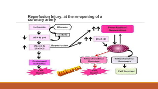
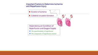
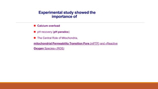
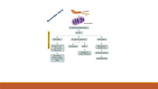
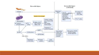
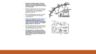
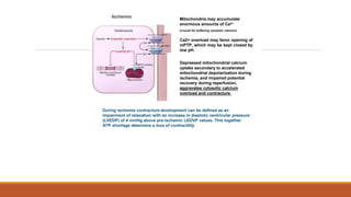
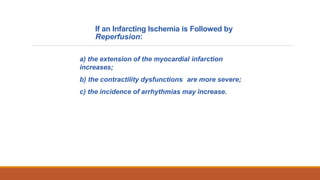
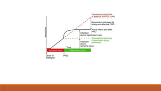
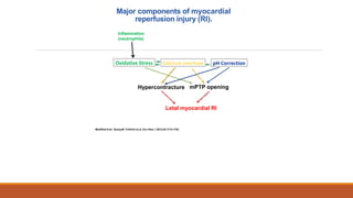
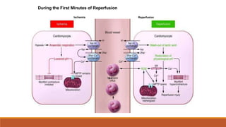
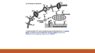
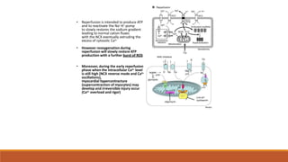
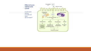
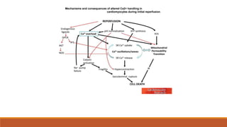
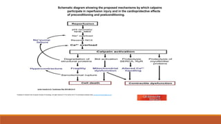
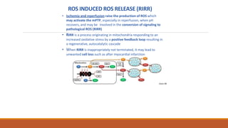
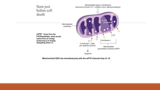
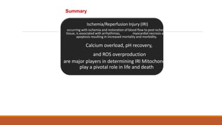
REPERFUSION
INJURY
MANU JACOB
PERFUSIONIST
Reperfusion injury occurs after tissues undergo a period of reduced oxygenation caused by poor
perfusion.
During the ischemic period anaerobic metabolism causes metabolic acids to build up in tissue.
Poor perfusion also inhibits the removal of carbon dioxide which builds up in the tissues.
The resulting intracellular acidosis deactivates the antioxidants. When perfusion is restored,
oxygen and calcium enter the weakened cells and cause reperfusion injury before the
intracellular pH can be normalized and the antioxidants reactivated.
The oxygen stress injury occurs even when the perfused oxygen levels are low. The use of
elevated oxygen levels to reperfuse ischemic tissues can further magnify the damage.
The best way to avoid reperfusion injury is to avoid ischemia. However when this is not possible,
such as when the heart is cross clamped or during deep hypothermic arrest, the perfusionist
should employ a reperfusion strategy to minimize the damage.
Why Reperfusion Injury?
Reperfusion Injury: at the re-opening of a
coronary artery
Important Factors to Determine Ischemia
and Reperfusion Injury
 Duration of ischemia
 Collateral circulation formation
Dependency on Condition of
Reperfusion and Oxygen Supply
 The speed/modality of reperfusion
 The components of reperfusion solution
t
IS
Experimental study showed the
importance of
 Calcium overload
 pH recovery (pH paradox)
 The Central Role of Mitochondria,
mitochondrial Permeability Transition Pore (mPTP) and «Reactive
Oxygen Species» (ROS)
Ischemia:
the occlusion of a coronary
artery
Alteration
of
Ion
exchanges
Troponin
Alteration of Ion exchanges
• Cessation of oxygen supply in ischemia
leads to a loss of ATP production and an
increase of reactive oxygen species (ROS) in
the mitochondria
• Reduced activity of the ATP consuming Na+-
K+-pump leads to Na+ accumulation in the
myocyte and the resting membrane
potential is lowered
• With the development of acidosis, the Na+-
H+-exchanger (NHX) further increases
intracellular Na+
• Under these conditions the 3Na+-1Ca2+-
exchanger (NCX) slows down due to
acidotic pH and intracellular accumulation
of Na+ or may even operate in the reverse
mode, letting Ca2+ into the cell
• Ca2+ also enters through the sarcolemmal L-
type voltage-gated Ca2+ -channel (L) as the
resting membrane potential is low
• The increased Ca2+ is taken up into the
sarcoplasmic reticulum (SR) by the SR Ca2+
pump SERCA2 (P) and released (leak) from
there via RYR, leading to contraction and
contracture.
Ischemia Mitochondria may accumulate
enormous amounts of Ca2+
(crucial for buffering cytosolic calcium).
Ca2+ overload may favor opening of
mPTP, which may be kept closed by
low pH.
Depressed mitochondrial calcium
uptake secondary to accelerated
mitochondrial depolarization during
ischemia, and impaired potential
recovery during reperfusion,
aggravates cytosolic calcium
overload and contracture.
During ischemia contracture development can be defined as an
impairment of relaxation with an increase in diastolic ventricular pressure
(LVEDP) of 4 mmHg above pre-ischemic LEDVP values. This together
ATP shortage determine a loss of contractility
Reperfusion Injury:
at the re-opening of a coronary artery
If an Infarcting Ischemia is Followed by
Reperfusion:
a) the extension of the myocardial infarction
increases;
b) the contractility dysfunctions are more severe;
c) the incidence of arrhythmias may increase.
Major components of myocardial
reperfusion injury (RI).
Modified from Georg M. Fröhlich et al. Eur Hear J 2013;34:1714-1722
Oxidative Stress Calcium overload pH Correction
Hypercontracture mPTP opening
Letal myocardial RI
Inflammation
(neutrophils)
Ischemia Reperfusion
3-1
During the First Minutes of Reperfusion
1) robust excretion of H+ due to prompt recovery of extracellular pH, 2) “reverse
mode” excretion of accumulated Na+ and Ca2+ influx in turn, and 3)
reexcretion of Ca2+ followed by recovery of ATP synthesis.
.
Ion exchanges at reperfusion:
• Reperfusion is intended to produce ATP
and to reactivate the Na+-K+-pump
to slowly restores the sodium gradient
leading to normal cation fluxes
with the NCX eventually extruding the
excess of cytosolic Ca2+
• However reoxygenation during
reperfusion will slowly restore ATP
production with a further burst of ROS
• Moreover, during the early reperfusion
phase when the intracellular Ca2+ level
is still high (NCX reverse mode and Ca2+
oscillations),
myocardial hypercontracture
(supercontraction of myocytes) may
develop and irreversible injury occur
(Ca2+ overload and rigor)
Murphy
The consequences of
Calcium overload
Effects of excess
calcium in the
cytosol
Cut-off point
between
reversible cell
injury
and cell death??
Mechanisms and consequences of altered Ca2+ handling in
cardiomyocytes during initial reperfusion.
Schematic diagram showing the proposed mechanisms by which calpains
participate in reperfusion injury and in the cardioprotective effects
of preconditioning and postconditioning.
Javier Inserte et al. Cardiovasc Res 2012;96:23-31
Published on behalf of the European Society of Cardiology. All rights reserved. © The Author 2012. For permissions please email: journals.permissions@oup.com
ROS are double-edged swords
(They can be good or bad)
The bad-ugly consequences of
ROS overproduction
In the cardio-circulatory system, ROS can be
generated by
• cardiomyocytes,
• endothelial cells, and
• neutrophils in inflammatory processes.
Different enzyme/mechanisms can produce ROS,
including
• electrons leaked from mitochondrial complexes,
• NADPH oxidase,
• xanthine dehydrogenase/xanthine oxidase,
• lipoxygenases,
• cyclooxygenases,
• peroxidases, and
• uncoupled nitric oxide synthase (NOS).
Sources of ROS
Mitochondria represent 36-40% of cardiomyocytes mass
H2O
catalase
Reperfusion
injury
ROS INDUCED ROS RELEASE (RIRR)
• Ischemia and reperfusion raise the production of ROS which
may activate the mPTP, especially in reperfusion, when pH
recovers, and may be involved in the conversion of signaling to
pathological ROS (RIRR)
• RIRR is a process originating in mitochondria responding to an
increased oxidative stress by a positive feedback loop resulting in
a regenerative, autocatalytic cascade
• When RIRR is inappropriately not terminated, it may lead to
unwanted cell loss such as after myocardial infarction
Zorov DB
Seen just
before cell
death
Mitochondrial ROS rise simultaneously with the mPTP-induced drop of ∆Ψ
mPTP : forms from the
FATPsynthase, which would
switch from an energy-
producing to an energy-
dissipating dimer (?)
■Mechanisms of ROS-induced Ischemia/Reperfusion Injury
ROS are extremely reactive to interact with
lipids, proteins and nucleic acids.
The increase of membrane lipid peroxidation (MLP)
ROS interact with non-saturated fatty acids from
membrane lipids and further induce lipid peroxidation
reaction, which results in the structural alteration and
dysfunction of membrane.
ROS induces oxidation of lips, proteins and nucleic
acid.
ROS-mediated Membrane damage
The integrity, permeability and function of membrane are
impaired during ischemia-reperfusion due to ROS-
induced MLP.
– Deactivation and malfunction of membrane receptors and
ionic pumps
– Mitochondrial dysfunction and further decreases ATP
generation
These damages do not occur only in sarcolemma, but also in
sarcoplasmic reticulum, mitochondria, lysosomes and other
intracellular membranes.
Therefore, Ca2+ can flow into the cytoplasm through damaged
membrane according to the gradient.
Ca2+ increase Further damage
During the Subsequent Hours of
Reperfusion
• With the reperfusion, the endothelial and vessels
become permeable, thus causing interstitial edema
• Activated Endothelial cells in reperfused myocardium
express adhesion proteins, release cytokines, and
reduce production of NO
• These promote adherence, activation, and accumulation
of neutrophils and monocytes in the ischemic-reperfused
tissue
Piper HM, et al. Ann Thorac Surg 75 (2003), 644-8
• The release of reactive oxygen species and proteolytic enzymes from
these activated leukocytes can contribute to the damage of myocytes and
vascular cells
• Vascular plugging by adherent leukocytes and aggregated platelets can also
promote a slow- or no-reflow phenomenon, already favored by tissue
contracture and increased pressure of interstitial edema
• These additional reperfusion-induced noxes contribute to infarct development
predominantly during the first 2 hours of reperfusion, as myocardial necrosis
almost reaches its final size during this period
Summary
Ischemia/Reperfusion Injury (IRI)
occurring with ischemia and restoration of blood flow to post-ischemic
tissue, is associated with arrhythmias, myocardial necrosis and
apoptosis resulting in increased mortality and morbidity.
Calcium overload, pH recovery,
and ROS overproduction
are major players in determining IRI Mitochondria
play a pivotal role in life and death
REPERFUSION INJURY----------------------

REPERFUSION INJURY----------------------

  • 1.
  • 3.
    Reperfusion injury occursafter tissues undergo a period of reduced oxygenation caused by poor perfusion. During the ischemic period anaerobic metabolism causes metabolic acids to build up in tissue. Poor perfusion also inhibits the removal of carbon dioxide which builds up in the tissues. The resulting intracellular acidosis deactivates the antioxidants. When perfusion is restored, oxygen and calcium enter the weakened cells and cause reperfusion injury before the intracellular pH can be normalized and the antioxidants reactivated. The oxygen stress injury occurs even when the perfused oxygen levels are low. The use of elevated oxygen levels to reperfuse ischemic tissues can further magnify the damage.
  • 4.
    The best wayto avoid reperfusion injury is to avoid ischemia. However when this is not possible, such as when the heart is cross clamped or during deep hypothermic arrest, the perfusionist should employ a reperfusion strategy to minimize the damage.
  • 5.
  • 6.
    Reperfusion Injury: atthe re-opening of a coronary artery
  • 7.
    Important Factors toDetermine Ischemia and Reperfusion Injury  Duration of ischemia  Collateral circulation formation Dependency on Condition of Reperfusion and Oxygen Supply  The speed/modality of reperfusion  The components of reperfusion solution t IS
  • 8.
    Experimental study showedthe importance of  Calcium overload  pH recovery (pH paradox)  The Central Role of Mitochondria, mitochondrial Permeability Transition Pore (mPTP) and «Reactive Oxygen Species» (ROS)
  • 9.
    Ischemia: the occlusion ofa coronary artery
  • 10.
  • 11.
  • 12.
    • Cessation ofoxygen supply in ischemia leads to a loss of ATP production and an increase of reactive oxygen species (ROS) in the mitochondria • Reduced activity of the ATP consuming Na+- K+-pump leads to Na+ accumulation in the myocyte and the resting membrane potential is lowered • With the development of acidosis, the Na+- H+-exchanger (NHX) further increases intracellular Na+ • Under these conditions the 3Na+-1Ca2+- exchanger (NCX) slows down due to acidotic pH and intracellular accumulation of Na+ or may even operate in the reverse mode, letting Ca2+ into the cell • Ca2+ also enters through the sarcolemmal L- type voltage-gated Ca2+ -channel (L) as the resting membrane potential is low • The increased Ca2+ is taken up into the sarcoplasmic reticulum (SR) by the SR Ca2+ pump SERCA2 (P) and released (leak) from there via RYR, leading to contraction and contracture.
  • 13.
    Ischemia Mitochondria mayaccumulate enormous amounts of Ca2+ (crucial for buffering cytosolic calcium). Ca2+ overload may favor opening of mPTP, which may be kept closed by low pH. Depressed mitochondrial calcium uptake secondary to accelerated mitochondrial depolarization during ischemia, and impaired potential recovery during reperfusion, aggravates cytosolic calcium overload and contracture. During ischemia contracture development can be defined as an impairment of relaxation with an increase in diastolic ventricular pressure (LVEDP) of 4 mmHg above pre-ischemic LEDVP values. This together ATP shortage determine a loss of contractility
  • 14.
    Reperfusion Injury: at there-opening of a coronary artery
  • 15.
    If an InfarctingIschemia is Followed by Reperfusion: a) the extension of the myocardial infarction increases; b) the contractility dysfunctions are more severe; c) the incidence of arrhythmias may increase.
  • 17.
    Major components ofmyocardial reperfusion injury (RI). Modified from Georg M. Fröhlich et al. Eur Hear J 2013;34:1714-1722 Oxidative Stress Calcium overload pH Correction Hypercontracture mPTP opening Letal myocardial RI Inflammation (neutrophils)
  • 18.
    Ischemia Reperfusion 3-1 During theFirst Minutes of Reperfusion
  • 19.
    1) robust excretionof H+ due to prompt recovery of extracellular pH, 2) “reverse mode” excretion of accumulated Na+ and Ca2+ influx in turn, and 3) reexcretion of Ca2+ followed by recovery of ATP synthesis. . Ion exchanges at reperfusion:
  • 20.
    • Reperfusion isintended to produce ATP and to reactivate the Na+-K+-pump to slowly restores the sodium gradient leading to normal cation fluxes with the NCX eventually extruding the excess of cytosolic Ca2+ • However reoxygenation during reperfusion will slowly restore ATP production with a further burst of ROS • Moreover, during the early reperfusion phase when the intracellular Ca2+ level is still high (NCX reverse mode and Ca2+ oscillations), myocardial hypercontracture (supercontraction of myocytes) may develop and irreversible injury occur (Ca2+ overload and rigor) Murphy
  • 21.
  • 22.
    Effects of excess calciumin the cytosol Cut-off point between reversible cell injury and cell death??
  • 23.
    Mechanisms and consequencesof altered Ca2+ handling in cardiomyocytes during initial reperfusion.
  • 24.
    Schematic diagram showingthe proposed mechanisms by which calpains participate in reperfusion injury and in the cardioprotective effects of preconditioning and postconditioning. Javier Inserte et al. Cardiovasc Res 2012;96:23-31 Published on behalf of the European Society of Cardiology. All rights reserved. © The Author 2012. For permissions please email: journals.permissions@oup.com
  • 25.
    ROS are double-edgedswords (They can be good or bad) The bad-ugly consequences of ROS overproduction
  • 26.
    In the cardio-circulatorysystem, ROS can be generated by • cardiomyocytes, • endothelial cells, and • neutrophils in inflammatory processes. Different enzyme/mechanisms can produce ROS, including • electrons leaked from mitochondrial complexes, • NADPH oxidase, • xanthine dehydrogenase/xanthine oxidase, • lipoxygenases, • cyclooxygenases, • peroxidases, and • uncoupled nitric oxide synthase (NOS). Sources of ROS
  • 27.
    Mitochondria represent 36-40%of cardiomyocytes mass H2O catalase Reperfusion injury
  • 28.
    ROS INDUCED ROSRELEASE (RIRR) • Ischemia and reperfusion raise the production of ROS which may activate the mPTP, especially in reperfusion, when pH recovers, and may be involved in the conversion of signaling to pathological ROS (RIRR) • RIRR is a process originating in mitochondria responding to an increased oxidative stress by a positive feedback loop resulting in a regenerative, autocatalytic cascade • When RIRR is inappropriately not terminated, it may lead to unwanted cell loss such as after myocardial infarction Zorov DB
  • 29.
    Seen just before cell death MitochondrialROS rise simultaneously with the mPTP-induced drop of ∆Ψ mPTP : forms from the FATPsynthase, which would switch from an energy- producing to an energy- dissipating dimer (?)
  • 30.
    ■Mechanisms of ROS-inducedIschemia/Reperfusion Injury ROS are extremely reactive to interact with lipids, proteins and nucleic acids. The increase of membrane lipid peroxidation (MLP) ROS interact with non-saturated fatty acids from membrane lipids and further induce lipid peroxidation reaction, which results in the structural alteration and dysfunction of membrane. ROS induces oxidation of lips, proteins and nucleic acid.
  • 31.
    ROS-mediated Membrane damage Theintegrity, permeability and function of membrane are impaired during ischemia-reperfusion due to ROS- induced MLP. – Deactivation and malfunction of membrane receptors and ionic pumps – Mitochondrial dysfunction and further decreases ATP generation These damages do not occur only in sarcolemma, but also in sarcoplasmic reticulum, mitochondria, lysosomes and other intracellular membranes. Therefore, Ca2+ can flow into the cytoplasm through damaged membrane according to the gradient. Ca2+ increase Further damage
  • 32.
    During the SubsequentHours of Reperfusion • With the reperfusion, the endothelial and vessels become permeable, thus causing interstitial edema • Activated Endothelial cells in reperfused myocardium express adhesion proteins, release cytokines, and reduce production of NO • These promote adherence, activation, and accumulation of neutrophils and monocytes in the ischemic-reperfused tissue Piper HM, et al. Ann Thorac Surg 75 (2003), 644-8
  • 33.
    • The releaseof reactive oxygen species and proteolytic enzymes from these activated leukocytes can contribute to the damage of myocytes and vascular cells • Vascular plugging by adherent leukocytes and aggregated platelets can also promote a slow- or no-reflow phenomenon, already favored by tissue contracture and increased pressure of interstitial edema • These additional reperfusion-induced noxes contribute to infarct development predominantly during the first 2 hours of reperfusion, as myocardial necrosis almost reaches its final size during this period
  • 34.
    Summary Ischemia/Reperfusion Injury (IRI) occurringwith ischemia and restoration of blood flow to post-ischemic tissue, is associated with arrhythmias, myocardial necrosis and apoptosis resulting in increased mortality and morbidity. Calcium overload, pH recovery, and ROS overproduction are major players in determining IRI Mitochondria play a pivotal role in life and death