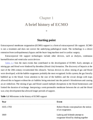
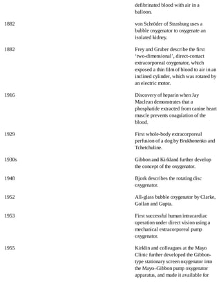
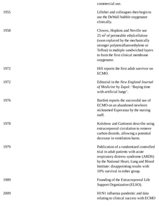
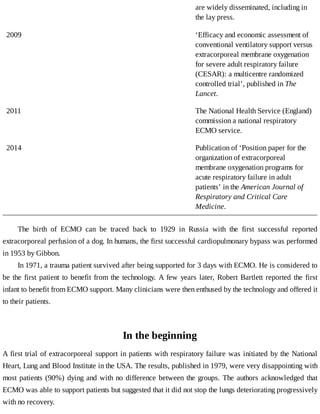
E C M O 	 I N 	 T H E 	A D U LT 	 PAT I E N T
GET MORE BOOKS AND
RESOURCES
Blog:
pharmacybr.blogspot.com
Facebook page:
www.fb.com/pharmacybr
Telegram channel:
https://t.me/pbr123GET MORE BOOKS AND RESOURCES
Blog:
pharmacybr.blogspot.com
Facebook page:
www.fb.com/pharmacybr
Telegram channel:
https://t.me/pbr123
C O R E 	 C R I T I C A L	 C A R E
Series	Editor
Dr	Alain	Vuylsteke
Papworth	Hospital
Cambridge,	UK
Other	titles	in	the	series
Intra-Abdominal	Hypertension	Manu	Malbrain	and	Jan	De	Waele	ISBN	9780521149396
Clinical	Information	Systems	in	Critical	Care	Cecilia	Morrison,	Matthew	R.	Jones	and	Julie	Bracken
ISBN	9780521156745
Delirium	in	Critical	Care,	Second	Edition	Valerie	Page	and	E.	Wesley	Ely	ISBN	9781107433656
ECMO	IN	THE	ADULT	PATIENT
Alain	Vuylsteke,	BSc,	MA,	MD,	FRCA,	FFICM
Consultant	in	Intensive	Care	and	Clinical	Director
Papworth	Hospital	Cambridge,	UK
Daniel	Brodie,	MD
Associate	Professor	of	Medicine
Columbia	University	College	of	Physicians	and	Surgeons
New	York-Presbyterian	Hospital
New	York,	NY,	USA
Alain	Combes,	MD,	PhD
Professor	of	Intensive	Care	Medicine
University	of	Paris,	Pierre	et	Marie	Curie
Senior	Intensivist	at	the	Service	de	Réanimation	Médicale	Institut	de	Cardiologie
Hôpital	Pitié-Salpêtrière
Paris,	France
Jo-anne	Fowles,	RGN
Lead	ECMO	Nurse
Papworth	Hospital
Cambridge,	UK
Giles	Peek,	MD,	FRCS	CTh,	FFICM
Professor	and	Chief	of	Pediatric	Cardiac	Surgery
ECMO	Director
The	Children’s	Hospital	of	Montefiore
New	York,	NY,	USA
University	Printing	House,	Cambridge	CB2	8BS,	United	Kingdom
Cambridge	University	Press	is	part	of	the	University	of	Cambridge.
It	furthers	the	University’s	mission	by	disseminating	knowledge	in	the	pursuit	of	education,	learning	and	research	at	the	highest
international	levels	of	excellence.
www.cambridge.org
Information	on	this	title:	www.cambridge.org/9781107681248
©	Alain	Vuylsteke,	Daniel	Brodie,	Alain	Combes,	Jo-anne	Fowles,	Giles	Peek	2017
This	publication	is	in	copyright.	Subject	to	statutory	exception	and	to	the	provisions	of	relevant	collective	licensing	agreements,	no
reproduction	of	any	part	may	take	place	without	the	written	permission	of	Cambridge	University	Press.
First	published	2017
Printed	in	the	United	Kingdom	by	TJ	International	Ltd,	Padstow,	Cornwall
A	catalogue	record	for	this	publication	is	available	from	the	British	Library
ISBN	978-1-107-68124-8	Paperback
Cambridge	University	Press	has	no	responsibility	for	the	persistence	or	accuracy	of	URLs	for	external	or	third-party	internet	websites
referred	to	in	this	publication,	and	does	not	guarantee	that	any	content	on	such	websites	is,	or	will	remain,	accurate	or	appropriate.
Every	effort	has	been	made	in	preparing	this	book	to	provide	accurate	and	up-to-date	information	which	is	in	accord	with	accepted
standards	and	practice	at	the	time	of	publication.	Although	case	histories	are	drawn	from	actual	cases,	every	effort	has	been	made	to
disguise	the	identities	of	the	individuals	involved.	Nevertheless,	the	authors,	editors	and	publishers	can	make	no	warranties	that	the
information	contained	herein	is	totally	free	from	error,	not	least	because	clinical	standards	are	constantly	changing	through	research	and
regulation.	The	authors,	editors	and	publishers	therefore	disclaim	all	liability	for	direct	or	consequential	damages	resulting	from	the	use	of
material	contained	in	this	book.	Readers	are	strongly	advised	to	pay	careful	attention	to	information	provided	by	the	manufacturer	of	any
drugs	or	equipment	that	they	plan	to	use.
Contents
Note	from	the	authors
List	of	abbreviations
A	patient	testimony:	I	survived	ECMO
1 A	brief	history	of	ECMO
2 An	ECMO	service
3 The	ECMO	circuit
4 Monitoring	the	patient	on	ECMO
5 Case	selection
6 Cannulation	and	decannulation
7 Coagulation,	blood	and	ECMO
8 Management	of	the	patient	on	veno-venous	ECMO:	general	principles
9 Management	of	the	patient	on	veno-arterial	ECMO:	general	principles
10 Patient	transfer
11 Liberation	from	ECMO
12 Specifics	of	intensive	care	management	for	the	patient	on	ECMO
13 Extracorporeal	carbon	dioxide	removal	or	ECCO2R
14 ECMO	to	support	organ	donation
15 ECMO	registries	and	research
Appendix:	The	future	of	ECMO
Index
Note	from	the	authors
This	book	is	about	ECMO	in	the	adult	patient.	The	adult	patient	can	be	defined	in	many	ways	but	we
have	arbitrarily	chosen	someone	older	than	16	years	and,	more	importantly	in	relation	to	the	discussed
technology,	heavier	than	20	kg.
We	 would	 like	 to	 acknowledge	 the	 contributions	 of:	 Dr	 Mindaugus	 Balciunas,	 UK;	 Dr	 Richard
Porter,	UK;	Dr	Mathieu	Schmidt,	France;	and	Dr	Martin	Besser,	UK.
Abbreviations
ACT
activated	coagulation	time
AKI
acute	kidney	injury
anti-Xa
anti-factor	Xa
APR
activated	prothrombin	time	ratio
aPTT
activated	prothrombin	time
ARDS
acute	respiratory	distress	syndrome
CO2
carbon	dioxide
CPR
cardiopulmonary	resuscitation
CT
computed	tomography
DCD
donation	after	cardiac	death
ECCO2R
extracorporeal	CO2	removal
ECMO
extracorporeal	membrane	oxygenation
ECMONet
International	ECMO	Network
eCPR
extracorporeal	cardiopulmonary	resuscitation	or	ECMO-assisted	cardiopulmonary	resuscitation
ELSO
Extracorporeal	Life	Support	Organization
FiO2
fraction	of	inspired	oxygen
HbS
sickle	cell	haemoglobin
HIT
heparin-induced	thrombocytopenia
HLA
human	leukocyte	antigen
ICU
intensive	care	unit
INR
international	normalized	ratio
LMWH
low-molecular-weight	heparin
O2
oxygen
PaCO2
partial	pressure	of	CO2	in	the	arterial	blood
PaO2
partial	pressure	of	O2	in	arterial	blood
PEEP
positive	end-expiratory	pressure
RRT
renal	replacement	therapy
A	patient	testimony:	I	survived	ECMO
It	is	somewhat	challenging	to	define	with	precision	what	could	facilitate	your	journey	to	recovery,	as
there	is	no	precedent	in	your	life.	You	are	intensely	searching	for	an	invisible	marker,	a	destination	you
seek	but	cannot	see	on	the	horizon.
Meeting	 with	 some	 of	 the	 doctors,	 nurses	 and	 physiotherapists	 who	 looked	 after	 me	 was	 a	 very
unique	and	special	experience.	Being	able	to	put	a	face	to	the	names	I	had	heard	of	so	often	started	to
anchor	me	in	this	part	of	my	life	I	could	not	access	before.	It	also	allowed	me	to	say	thank	you	in	person,
a	pivotal	part	of	the	healing	process,	because	I	was	physically	there,	unsupported	by	any	machine	and
quite	well	recovered	in	fact.
Although	without	any	recollection	of	the	hospital,	I	suspected	that	I	had	probably	‘recorded’	many
sounds	of	intensive	care	without	realizing	it.	This	was	confirmed	when	I	was	able	to	hear	an	ECMO
alarm:	the	reaction,	although	slow	coming,	was	strong.	This	is	my	only	memory,	my	very	own,	and	I	do
hold	it	surprisingly	dear.	It	is	an	oddly	reassuring	sensation	because	it	acts	as	the	explanation,	if	not	the
actual	validation,	of	everything	that	has	happened	since.	It	almost	gives	a	logical	meaning	to	the	last	30	or
so	months	of	my	life.
Even	more	noteworthy	was	the	utterly	inspired	decision	to	bring	me	to	the	bed	of	a	lady	undergoing
ECMO.	This	was	what	I	very	much	needed	but	was	too	shy	to	ask	for.	I	felt	overwhelmed	and	a	bit
amazed,	but	I	was	not	frightened	in	any	way.	It	made	me	realize	how	far	I	had	come.
It	has	proved	to	be	a	truly	cathartic	experience	for	me.	It	is	vastly	important	to	encourage	patients	to
return,	because	it	is	a	milestone	not	only	in	their	journey	of	healing	but	in	their	quest	for	acceptance	too.
Someone	told	me	once	that	I	was	brave;	I	do	not	think	this	is	true.	You	either	sink	or	swim.	I	did	not
have	a	choice,	that	is	all.	Strangely,	it	makes	things	simpler	and	therefore	easier	to	get	on	with.
I	also	believe	that	you	never	know	what	you	are	really	capable	of	until	you	are	challenged	to	show
it.	If	you	never	are,	you	are	blessed,	truly.
If,	however,	the	dice	is	cast	the	other	way,	there	is	still	one	option	left:
Fight	back.	It	is	worth	it.
Chapter	1
A	brief	history	of	ECMO
◈
Starting	point
Extracorporeal	membrane	oxygenation	(ECMO)	support	is	a	form	of	extracorporeal	life	support.	ECMO
is	 not	 a	 treatment	 and	 does	 not	 correct	 the	 underlying	 pathological	 insult.	 The	 technology	 is	 a	 direct
extension	from	cardiopulmonary	bypass	and	the	heart–lung	machine	used	in	cardiac	surgery.
Extracorporeal	 life	 support	 technologies	 include	 other	 devices,	 such	 as	 dialysis,	 continuous
haemofiltration	and	ventricular	assist	devices
Table	 1.1	 lists	 the	 main	 events	 that	 contributed	 to	 the	 development	 of	 ECMO.	 Early	 attempts	 at
mixing	gas	and	blood	were	hindered	by	thrombus	(blood	clot)	formation.	The	discovery	of	heparin	at	the
start	of	the	20th	century	circumvented	this	obstacle.	Various	devices	to	allow	mixing	of	gas	and	blood
were	developed,	with	the	bubble	oxygenator	probably	the	most	recognized.	In	this	system,	the	gas	literally
bubbled	 up	 in	 the	 blood.	 Great	 attention	 to	 the	 size	 of	 the	 bubbles	 and	 the	 circuit	 design	 with	 traps
allowed	this	to	happen	without	the	air	bubbles	being	entrained	into	the	patient’s	bloodstream	and	causing
an	air	embolism.	The	mixing	of	gas	and	blood	caused	multiple	disruptions	to	the	blood	homeostasis	and
limited	the	duration	of	exchange.	Interposing	a	semi-permeable	membrane	between	the	air	and	the	blood
was	a	key	development	that	allowed	longer	periods	of	support.
Table	1.1	Milestones	in	the	history	of	ECMO	support
Year Event
1635–1703 Robert	Hooke	conceptualizes	the	notion
of	an	oxygenator.
1869 Ludwig	and	Schmidt	attempt	to
oxygenate	blood	by	shaking	together
defibrinated	blood	with	air	in	a
balloon.
1882 von	Schröder	of	Strasburg	uses	a
bubble	oxygenator	to	oxygenate	an
isolated	kidney.
1882 Frey	and	Gruber	describe	the	first
‘two-dimensional’,	direct-contact
extracorporeal	oxygenator,	which
exposed	a	thin	film	of	blood	to	air	in	an
inclined	cylinder,	which	was	rotated	by
an	electric	motor.
1916 Discovery	of	heparin	when	Jay
Maclean	demonstrates	that	a
phosphatide	extracted	from	canine	heart
muscle	prevents	coagulation	of	the
blood.
1929 First	whole-body	extracorporeal
perfusion	of	a	dog	by	Brukhonenko	and
Tchetchuline.
1930s Gibbon	and	Kirkland	further	develop
the	concept	of	the	oxygenator.
1948 Bjork	describes	the	rotating	disc
oxygenator.
1952 All-glass	bubble	oxygenator	by	Clarke,
Gollan	and	Gupta.
1953 First	successful	human	intracardiac
operation	under	direct	vision	using	a
mechanical	extracorporeal	pump
oxygenator.
1955 Kirklin	and	colleagues	at	the	Mayo
Clinic	further	developed	the	Gibbon-
type	stationary	screen	oxygenator	into
the	Mayo–Gibbon	pump	oxygenator
apparatus,	and	made	it	available	for
commercial	use.
1955 Lillehei	and	colleagues	then	begin	to
use	the	DeWall	bubble	oxygenator
clinically.
1958 Clowes,	Hopkins	and	Neville	use
25	m2	of	permeable	ethylcellulose
(soon	replaced	by	the	mechanically
stronger	polytetrafluoroethylene	or
Teflon)	in	multiple	sandwiched	layers
to	form	the	first	clinical	membrane
oxygenator.
1972 Hill	reports	the	first	adult	survivor	on
ECMO.
1972 Editorial	in	the	New	England	Journal
of	Medicine	by	Zapol:	‘Buying	time
with	artificial	lungs’.
1976 Bartlett	reports	the	successful	use	of
ECMO	on	an	abandoned	newborn
nicknamed	Esperanza	by	the	nursing
staff.
1978 Kolobow	and	Gattinoni	describe	using
extracorporeal	circulation	to	remove
carbon	dioxide,	allowing	a	potential
decrease	in	ventilation	harm.
1979 Publication	of	a	randomized	controlled
trial	in	adult	patients	with	acute
respiratory	distress	syndrome	(ARDS)
by	the	National	Heart,	Lung	and	Blood
Institute:	disappointing	results	with
10%	survival	in	either	group.
1989 Founding	of	the	Extracorporeal	Life
Support	Organization	(ELSO).
2009 H1N1	influenza	pandemic	and	data
relating	to	clinical	success	with	ECMO
are	widely	disseminated,	including	in
the	lay	press.
2009 ‘Efficacy	and	economic	assessment	of
conventional	ventilatory	support	versus
extracorporeal	membrane	oxygenation
for	severe	adult	respiratory	failure
(CESAR):	a	multicentre	randomized
controlled	trial’,	published	in	The
Lancet.
2011 The	National	Health	Service	(England)
commission	a	national	respiratory
ECMO	service.
2014 Publication	of	‘Position	paper	for	the
organization	of	extracorporeal
membrane	oxygenation	programs	for
acute	respiratory	failure	in	adult
patients’	in	the	American	Journal	of
Respiratory	and	Critical	Care
Medicine.
The	 birth	 of	 ECMO	 can	 be	 traced	 back	 to	 1929	 in	 Russia	 with	 the	 first	 successful	 reported
extracorporeal	perfusion	of	a	dog.	In	humans,	the	first	successful	cardiopulmonary	bypass	was	performed
in	1953	by	Gibbon.
In	1971,	a	trauma	patient	survived	after	being	supported	for	3	days	with	ECMO.	He	is	considered	to
be	the	first	patient	to	benefit	from	the	technology.	A	few	years	later,	Robert	Bartlett	reported	the	first
infant	to	benefit	from	ECMO	support.	Many	clinicians	were	then	enthused	by	the	technology	and	offered	it
to	their	patients.
In	the	beginning
A	first	trial	of	extracorporeal	support	in	patients	with	respiratory	failure	was	initiated	by	the	National
Heart,	Lung	and	Blood	Institute	in	the	USA.	The	results,	published	in	1979,	were	very	disappointing	with
most	patients	(90%)	dying	and	with	no	difference	between	the	groups.	The	authors	acknowledged	that
ECMO	was	able	to	support	patients	but	suggested	that	it	did	not	stop	the	lungs	deteriorating	progressively
with	no	recovery.
As	a	result,	most	clinicians	stopped	offering	ECMO.
A	minority	continued	to	improve	the	technique.	Others	worked	on	modifying	other	aspects	of	the
support	 of	 respiratory	 failure	 patients.	 Clinicians	 understood	 that	 lungs	 were	 being	 damaged	 by
mechanical	 ventilation	 with	 positive	 pressure.	 Methods	 to	 decrease	 this	 mechanical	 insult	 were
developed.	The	so-called	protective	ventilation	strategies	are	in	fact	the	least-damaging	lung	ventilation
techniques.	 One	 promising	 method	 was	 to	 combine	 therapies	 using	 ECMO	 to	 remove	 carbon	 dioxide
(CO2)	to	reduce	the	amount	of	ventilation	required	with	mechanical	ventilation.	However,	there	was	no
evidence	in	comparative	studies	that	using	ECMO	led	to	better	outcomes	than	conventional	therapy.
Clinicians	using	ECMO	to	support	infants	were	convinced	that	they	were	saving	lives,	and	several
trials	and	case	series	proved	this	to	be	correct.	Paediatric	ECMO	developed	and	was	embraced	by	many.
Paediatric	 centres	 continued	 to	 accumulate	 expertise	 and	 experience.	 But	 this	 book	 is	 about	 the	 adult
patient…
Enthusiast	 clinicians	 teamed	 together	 and	 founded	 the	 Extracorporeal	 Life	 Support	 Organization
(ELSO)	 in	 1989,	 justified	 mainly	 by	 the	 successes	 observed	 in	 paediatric	 support.	 This	 network
understood	 the	 importance	 of	 sharing	 practice	 and	 collecting	 data	 from	 all	 participating	 centres.	 Data
about	paediatric	and	adult	ECMO	were	progressively	accumulated	to	inform	practice	around	the	world.
Moving	forward
At	the	beginning	of	the	21st	century,	technology	had	advanced	with	the	development	and	optimization	of
devices	 used	 in	 cardiopulmonary	 bypass.	 The	 bubble	 oxygenator	 had	 long	 been	 forgotten	 and	 the
membrane	oxygenator	was	being	used	by	all	(allowing	separation	of	blood	and	gas	by	a	semi-permeable
membrane).	This,	combined	with	the	advent	of	centrifugal	pumps,	improved	the	biocompatibility	of	the
whole	 process.	 Although	 still	 not	 harmless,	 the	 technique	 was	 becoming	 simpler.	 Improved	 design
removed	many	mechanical	issues.	The	introduction	of	smaller,	less-intrusive	circuits	allowed	portability.
The	 changes	 were	 such	 that	 this	 era	 can	 be	 referred	 to	 as	 the	 start	 of	 the	 next	 generation	 of	 ECMO
(informally	called	ECMO	v2.0).
Specialist	centres	had	started	using	ECMO	in	specific	patients,	such	as	after	lung	transplantation.
Others	 were	 exploring	 ECMO	 to	 support	 the	 heart	 and	 lungs.	 The	 technique	 was	 confined	 to	 highly
specialized	centres	and	the	occasional	patient.
In	2009,	when	confronted	with	a	new	subtype	of	influenza	virus	(H1N1)	that	targeted	mainly	young
people,	the	clinical	community	used	ECMO	with	success	to	support	many	patients	to	full	recovery.	While
it	is	questionable	that	ECMO	made	a	difference	in	outcome	(some	are	convinced	it	did,	but	the	data	have
been	the	subject	of	many	debates),	this	experience	led	to	a	widespread	use	of	the	practice.	Noteworthy
was	the	fact	that	ECMO	was	offered	to	a	large	number	of	very	sick	patients	during	the	pandemic,	without
modern	health	services	imploding,	albeit	stretching	available	resources.
Simultaneous	to	the	pandemic,	the	results	of	a	large	prospective	trial	of	the	use	of	ECMO	in	patients
with	acute	respiratory	distress	syndrome	(ARDS)	was	published	in	The	Lancet	(the	CESAR	trial)	and
fuelled	 both	 the	 controversy	 about	 and	 the	 use	 of	 the	 technology.	 This	 trial	 showed	 that	 transferring
patients	with	ARDS	to	a	specialist	centre	that	could	offer	ECMO	if	required	led	to	a	better	outcome.	It
did	not	show	that	ECMO	itself	helped.
As	a	result	of	the	pandemic,	and	supported	by	the	published	evidence,	clinicians	started	to	consider
ECMO	earlier	and	many	providers	started	to	offer	it.	Some	countries	set	up	national	networks	(e.g.	the
specifications	 of	 the	 National	 Health	 Service	 (England)	 national	 respiratory	 ECMO	 service	 can	 be
accessed	online;	see	Chapter	2).
Parallel	 to	 the	 development	 of	 ECMO	 to	 support	 respiratory	 function,	 ECMO	 has	 been	 used	 to
support	patients	with	cardiopulmonary	failure.	In	this	setting,	ECMO	can	be	seen	as	a	way	to	provide
cardiopulmonary	bypass	either	rapidly	(such	as	in	a	cardiac	arrest	situation)	or	for	several	days	(such	as
when	continuing	cardiopulmonary	bypass	after	cardiac	surgery).	Case	series	(and	numerous	case	reports)
are	supporting	the	development	of	veno-arterial	ECMO	as	a	way	to	supply	most	organs	with	a	continuous
oxygenated	 blood	 supply.	 This	 support	 is	 being	 used	 in	 an	 increasing	 number	 of	 patients,	 based	 on
clinicians’	belief	that	it	helps	on	some	occasions.	However,	scientific	evidence	is	lacking.
Key	points
To	learn	more
Gattinoni	L,	Pesenti	A,	Mascheroni	D,	et	al.	(1986).	Low-frequency	positive-pressure	ventilation	with
extracorporeal	CO2	removal	in	severe	acute	respiratory	failure.	Journal	of	the	American	Medical
Association,	256,	881–6.
This	is	a	book	about	adult	patients.
ECMO	circuits	are	now	simpler	and	safer.
ECMO	saved	lives	during	the	H1N1	influenza	pandemic.
ECMO	is	a	support	modality	and	not	a	treatment.
Lim	MW.	(2006).	The	history	of	extracorporeal	oxygenators.	Anaesthesia,	61,	984–95.
Noah	MA,	Peek	GJ,	Finney	SJ,	et	al.	(2011).	Referral	to	an	extracorporeal	membrane	oxygenation	center
and	mortality	among	patients	with	severe	2009	influenza	A(H1N1).	Journal	of	the	American	Medical
Association,	306,	1659–68.
Peek	GJ,	Mugford	M,	Tiruvoipati	R,	et	al.	(2009).	Efficacy	and	economic	assessment	of	conventional
ventilatory	support	versus	extracorporeal	membrane	oxygenation	for	severe	adult	respiratory	failure
(CESAR):	a	multicentre	randomised	controlled	trial.	Lancet,	374,	1351–63.
Chapter	2
An	ECMO	service
◈
Staffing
An	ECMO	service	is	entirely	dependent	on	its	people.	The	team	is	organized	to	provide	a	continuous
service,	24	h	a	day.	Table	2.1	provides	an	example	of	a	list	of	the	key	members	of	the	team.	This	will
vary	from	centre	to	centre,	depending	on	local	and	national	organizational	factors.
Table	2.1	Example	of	a	list	of	key	members	of	the	ECMO	service
Title Role
Lead	clinician	(ECMO
director)
Overall	responsibility	for	the	ECMO	service.	Must	practice	intensive
care	medicine	but	can	be	from	a	variety	of	backgrounds.
Attending	clinicians Round-the-clock	management	of	the	patient.	These	will	usually	be	a
mix	of	post-	and	pre-certification	doctors.
Attending	surgeons Immediate	cardiothoracic	and	vascular	surgical	support,	which	is
required	round	the	clock.
ECMO	coordinator To	coordinate	referrals	and	retrievals.	Ensures	multidisciplinary
education	and	training	requirements	are	achieved.	Develops	and
reviews	protocols	and	guidelines.
ECMO	specialists Staff	who	have	undergone	specialist	training	and	have	expert
knowledge	of	the	management	of	the	ECMO	patient	and	the	ECMO
circuit.	Will	support	the	ECMO	coordinator	in	day-to-day	coordination
of	the	service.
Attending	nurses Day-to-day	patient	care	and	bedside	monitoring.	They	are	experienced
intensive	care	unit	nurses	who	have	undergone	additional	training	in
caring	for	the	patient	on	ECMO.	They	will	be	supported	by	heath	care
support	workers.
Lead	perfusionist Perfusionist	with	specialist	ECMO	knowledge	who	supports	the
ECMO	coordinator	in	meeting	educational	and	training	requirements.
Clinical	perfusionists Provide	technical	support	in	relation	to	the	ECMO	circuit.
Physiotherapists Provide	day-to-day	rehabilitation	input.
Pharmacists Provide	day-to-day	pharmacy	input.	Will	seek	pertinent	and	updated
information	in	relation	to	the	pharmacokinetics	of	drugs	used	in	the
ECMO	patients.
Dieticians Provide	day-to-day	dietetic	input.
ECMO	secretary To	support	numerous	administrative	tasks	related	to	the	ECMO	service.
Clinical	data	analyst Collection	and	analysis	of	data,	which	is	important	to	develop	a	strong
service.	This	allows	submission	to	the	international	registry.
Other	specialities	involved
in	the	greater
multidisciplinary	team
Radiologists,	haematologists,	microbiologists,	virologists,	biochemists,
blood	bank	specialists,	cardiologists,	respiratory	physicians,
neurologists,	nephrologists,	obstetricians,	gynaecologists,	clinical
psychologists,	psychiatrists,	orthopaedic	and	trauma	surgeons,
ophthalmologists,	ear–nose–throat	(ENT)	specialists	and	palliative
care	clinicians.
Ancillary	staff Kitchen	staff,	cleaners,	porters,	drivers,	switchboard	operators,
accountants	and	technical	support.
All	members	of	the	team	require	specialist	knowledge	in	managing	the	ECMO	patient,	and	a	robust
teaching	 programme	 should	 be	 established.	 The	 volume	 of	 activity	 of	 a	 centre	 should	 be	 sufficient	 to
allow	 availability	 of	 the	 required	 resources	 for	 training.	 Staff	 training	 should	 be	 mandatory.	 Regular
refreshers	should	be	provided	and	competencies	regularly	assessed.	Table	2.2	lists	the	topics	that	the	in-
house	training	programme	should	cover.	The	ELSO	provides	regularly	updated	guidelines	and	resources
for	the	training	and	continuous	education	of	the	ECMO	specialist.
Table	2.2	Specialized	topics	to	be	covered	in	the	training	of	an	ECMO	clinician
Types	of	ECMO
Risk	and	potential	benefits	of	ECMO
Indication	and	contraindication	for	ECMO
Pathophysiology	of	the	patient	on	ECMO
ECMO	equipment	(including	circuits)
Gas	exchange	on	ECMO
ECMO	emergencies
Inserting	ECMO:	Why?	When?	How?
Liberation	from	ECMO:	Why?	When?	How?
Coagulation	management	while	on	ECMO
The	post-ECMO	patient:	immediate	and	long	term
Complications	of	ECMO
Cost	of	ECMO
Transfer	of	the	patient	on	ECMO
The	team	of	educators	must	be	allocated	time	and	resources	to	attend	state-of-the-art	conferences.
Educators	can	then	ensure	their	local	programme	is	up	to	date.	Training	 should	 include	 regular	 water
drills,	 during	 which	 a	 primed	 circuit	 is	 used	 without	 being	 connected	 to	 a	 patient.	 This	 allows
familiarization	with	the	equipment	and	identification	of	potential	issues.	Staff	are	trained	to	recognize	and
manage	specific	ECMO	emergencies,	and	then	do	it	again	and	again.
A	list	of	competencies	expected	from	each	role	for	clinicians	involved	in	the	management	of	the
patient	on	ECMO	is	useful.	These	can	be	developed	at	the	local	or	national	level,	and	the	ELSO	provides
examples	and	ready-to-use	lists.
Multidisciplinary	meetings	should	be	scheduled	on	a	regular	basis	and	newly	acquired	knowledge
and	experience	shared	to	ensure	the	whole	team	continues	to	learn	and	evolve.
Doctors
Doctors	looking	after	ECMO	patients	will	require	multiple	skills.	The	ideal	ECMOlogist	would	be	a
surgeon,	intensivist,	chest	physician	and	anaesthesiologist	combined.	This	person	should	have	acquired
the	 skills	 of	 many	 different	 specialities.	 As	 this	 is	 rather	 unusual,	 a	 good	 ECMOlogist	 will	 be	 a
‘connexist’,	i.e.	someone	who	can	recognize	his/her	own	limitations	and	call	on	others’	expertise.	The
closest	to	this	ideal	are	intensive	care	doctors	as	they	are	usually	working	in	this	manner.
The	starting	point	for	any	doctor	wishing	to	learn	ECMO	is	a	thorough	understanding	of	the	ECMO
circuit	itself	(see	Chapter	3).	The	second	key	clinical	skill	to	acquire	is	how	to	select	the	right	patients,
i.e.	patients	requiring	support	while	recovering	from	a	reversible	insult	or	eligible	for	another	type	of
long-term	support	(see	Chapter	5).
The	ECMO	community	has	a	great	tradition	of	supporting	each	other.	These	days,	multiple	courses
are	available	to	teach	the	basics	of	ECMO.	Clinicians	involved	in	ECMO	are	always	willing	to	help	each
other.
Adult	intensive	care	skills	are	central	to	the	safe	delivery	of	ECMO,	and	all	the	basics	of	intensive
care	management	should	be	adhered	to.
ECMO	is	a	complete	system	of	care,	not	just	a	bolt-on	accessory.
Junior	doctors	should	be	involved	in	all	aspects	of	the	care	of	ECMO	patients.	The	first	skill	they
will	need	to	acquire	is	to	recognize	their	limitations	and	when	to	call	for	help.
ECMO	specialist
The	ECMO	specialist	has	a	key	role.	They	are	immediately	available	in	the	clinical	area	and	are	the	first
line	between	ECMO	and	the	patient.	They	require	advanced	skills	to	ensure	no	harm	comes	to	the	patient.
They	 are	 the	 first	 line	 in	 managing	 the	 patient	 and	 circuit	 emergencies	 and,	 in	 addition	 to	 excellent
technical	skills,	should	have	effective	communication	skills	and	the	ability	to	work	in	stressful	situations.
The	ECMO	specialist	will	be	skilled	in	the	care	of	patients	on	ECMO,	and	should	have	a	strong
intensive	care	background.
The	 ECMO	 specialist	 must	 achieve	 competency	 through	 completing	 the	 necessary	 training	 and
assessment.	Practical	skill	sessions	concentrate	on	circuit	surveillance,	troubleshooting	and	emergency
procedures.	 Emergency	 procedures	 such	 as	 air	 embolus	 removal	 are	 practised	 repeatedly	 to	 ensure
competency.
The	ECMO	specialist	can	undertake	other	roles,	such	as	ensuring	appropriate	anticoagulation,	and
titrating	ECMO	flow	and	gas	to	ensure	adequate	levels	of	cardiac	and/or	respiratory	support.
ECMO	specialists	can	be	nurses	who	have	undergone	additional	training,	or	can	come	from	other
specialities,	such	as	perfusion	or	medical	backgrounds.
All	nurses	caring	for	the	ECMO	patient	must	have	basic	skills	specific	to	both	the	intensive	care	unit
(ICU)	and	the	ECMO	patient.	They	must	be	able	to	act	immediately	in	a	case	of	a	catastrophic	failure	of
the	ECMO	circuit.	An	ECMO	circuit	must	never	be	left	in	the	hands	of	a	practitioner	who	is	not	qualified
to	look	after	it.
ECMO	coordinator
The	ECMO	coordinator	usually	refers	to	a	highly	experienced	ECMO	specialist	with	clinical,	educational
and	 managerial	 responsibility	 for	 the	 ECMO	 programme.	 This	 person	 may	 be	 supported	 by	 deputy
coordinators	 and	 shift	 leaders	 who	 are	 experienced	 ECMO	 specialists	 to	 ensure	 round-the-clock
availability.	They	are	involved	in	the	development	of	all	ECMO-related	protocols.
This	 coordination	 is	 an	 essential	 part	 of	 ensuring	 the	 safety	 of	 the	 patient	 on	 ECMO.	 It	 ensures
communication	between	team	members	and	smooth	management	of	patient	referral,	transport,	cannulation
and	ongoing	care.
ECMO	director
The	 ECMO	 director	 is	 an	 ECMO	 clinician	 nominated	 to	 have	 overall	 managerial	 and	 clinical
responsibility	for	the	ECMO	programme.	The	ECMO	director	does	not	need	to	dictate	the	care	of	each
patient	but	leads	a	team	to	deliver	the	best	possible	care.
Perfusionist
Perfusionists	are	experts	in	the	management	of	extracorporeal	circuits,	and	their	main	expertise	is	with
short-term	 cardiopulmonary	 bypass	 used	 in	 the	 operating	 theatre.	 A	 perfusionist	 will	 need	 additional
training	to	gain	the	knowledge	required	to	manage	the	ECMO	circuit.
Perfusionists	are	an	integral	and	important	part	of	many	ECMO	programmes.	It	is	usual	practice	in
some	 institutions	 for	 a	 perfusionist	 to	 prime	 the	 ECMO	 circuit	 and	 initiate	 ECMO	 before	 handing
management	 of	 the	 circuit	 over	 to	 the	 ECMO	 specialist.	 Once	 the	 patient	 is	 returned	 to	 the	 ICU	 and
established	on	ECMO,	the	perfusionist’s	role	includes	circuit	or	component	replacement	and	transport	of
the	patient	to	other	areas	of	the	hospital,	such	as	the	catheter	laboratory	or	the	computed	tomography	(CT)
scanner.
A	perfusionist	who	has	developed	a	special	interest	in	ECMO	can	act	as	the	lead	of	all	perfusionists
in	an	institution	and	should	ensure	that	knowledge	and	good	practice	are	shared.
Perfusionists	should	be	involved	in	circuit	design	and	modifications,	management	of	ordering	and
stock	levels,	equipment	maintenance	and	development	of	protocols.
Transfer	team
ECMO	is	a	specialist	support	that	may	not	be	provided	in	all	institutions.	A	centre	will	need	a	minimum
number	of	cases	per	year	to	maintain	expertise	and	justify	the	set-up	expenses.	Most	patients	requiring
ECMO	will	need	to	be	transferred	from	one	centre	to	an	ECMO	centre.
Patients	 referred	 to	 the	 ECMO	 centre	 will	 often	 be	 unstable,	 and	 transfer	 on	 ECMO	 will	 be
necessary.
The	difficulty	of	commencing	ECMO	on	a	patient	outside	the	comfortable	surroundings	of	one’s	own
hospital	should	not	be	underestimated.
The	transfer	team	will	include	at	least	a	doctor	and	a	transfer-trained	ECMO	nurse.	The	team	must
be	trained	and	equipped	to	work	in	any	of	the	vehicles	and	with	all	the	equipment	that	might	be	used.
Other	members	of	the	team
Other	doctors,	nurses	and	allied	health	professionals	will	be	involved	in	the	care	of	the	ECMO	patient.	It
is	helpful	to	try	and	identify	one	or	two	individuals	in	each	department	who	are	interested	in	ECMO,	so
that	they	can	develop	specific	expertise	with	these	patients.
Table	 2.1	 lists	 the	 many	 specialties	 required.	 Surgeons	 (general,	 cardiothoracic	 and	 vascular),
physiotherapists,	cardiologists,	microbiologists,	dieticians,	haematologists,	radiologists	and	occupational
therapists	are	all	frequently	involved	in	care	of	the	adult	patient	on	ECMO.	Specific	knowledge	will	be
required	from	each	of	them.
Infrastructure
An	ECMO	centre	must	be	located	in	an	area	of	sufficient	population	density	to	ensure	that	patients	are
regularly	admitted	and	a	minimum	number	are	supported	each	year.	This	number	is	often	the	subject	of
intense	 debate,	 but	 we	 estimate	 that	 expertise	 and	 investment	 will	 be	 maintained	 if	 a	 centre	 supports
around	20	patients	per	year.	The	relationship	between	volume	and	outcome	is	recognized	in	many	clinical
specialities.
The	centre	must	be	easily	accessible	by	road,	or	by	fixed	wing	or	rotary	aircraft.	A	helipad	directly
linked	to	the	hospital	is	ideal.
The	 required	 facilities	 (Table	 2.3)	 mean	 that	 ECMO	 services	 will	 usually	 be	 based	 in	 tertiary
referral	 centres	 that	 can	 provide	 round-the-clock	 cardiothoracic	 and	 vascular	 support,	 alongside	 other
services	such	as	advanced	imaging	and	specialist	microbiology.
Table	2.3	Co-located	clinical	services
To	be	provided	on	the	same
site	and	to	be	immediately
available	round	the	clock
Competent	medical	practitioner	with	advanced	airway	skills
Endoscopy
Radiology:	CT,	ultrasound,	plain	X-ray,	echocardiography
Access	to	theatres,	including	cardiothoracic	and	vascular
Transfusion	services
Essential	haematology/biochemistry	service	and	point-of-care
service
Haemofiltration	and	plasmapheresis
Interventional	cardiology
Physiotherapy
Pharmacy
Medical	engineering	services
Informatics	support
Interdependent	services,
available	round	the	clock;
the	response	time	to	these
specialities	will	range	from
available	within	30	min	to	a
maximum	of	4	h
Interventional	vascular	and	non-vascular	radiology
Neurosurgery
Vascular	surgery
General	surgery
Nephrology
Trauma	and	orthopaedic	surgery
Plastic	surgery
Maxillofacial	surgery
Ear–nose–throat	(ENT)	surgery
Obstetrics	and	gynaecology
Organ	donation	services
Acute/early	phase	rehabilitation	services
Additional	laboratory	diagnostic	services
Interdependent	services, Occupational	therapy
available	during	daytime
hours	(Monday–Friday)
Dietetics
Speech	and	language	therapy
Bereavement	services
Patient	liaison	service
Related	services,	available
following	the	critical	care
phase	of	the	patient	pathway
Local	hospital	and	community	rehabilitation	services
Specialized	rehabilitation	services
Critical	care	follow-up
Clinical	psychology	and	psychiatry
Primary	care
Burns	services
Voluntary	support	services
Adapted	from	https://www.england.nhs.uk/commissioning/spec-services/npc-crg/group-d/d16/.
The	ECMO	service	will	often	be	part	of	the	ICU,	but	some	centres	have	dedicated	ECMO	units.
These	are	specialist	ICUs	looking	solely	after	the	ECMO	patient,	similar	to	burns	units.
Adult	 and	 paediatric	 ECMO	 services	 have	 historically	 been	 combined	 due	 to	 the	 common
technology.	 It	 is	 our	 opinion	 that	 patients	 are	 best	 served	 in	 a	 unit	 specializing	 in	 the	 original	 insult
(respiratory	or	cardiac),	rather	than	based	on	the	technology	used.	An	adult	patient	will	not	be	treated	the
same	way	if	supported	in	a	paediatric	ECMO	unit,	specialized	in	the	management	of	neonates	or	young
children	with	congenital	diseases.	Local	or	national	organization	may	influence	this.
The	 minimum	 specification	 for	 the	 bed	 space	 is	 the	 same	 as	 that	 required	 for	 an	 intensive	 care
patient.	There	must	be	sufficient	power	points	(and	these	must	be	resilient	in	case	of	power	failure).	A
patient	on	ECMO	may	require	numerous	devices	requiring	electrical	power,	as	listed	in	Table	2.4.	The
ECMO	circuit	will	require	a	gas	supply	in	addition	to	that	required	for	the	ventilator.
Table	2.4	Examples	of	electrical	devices	required	for	a	single	ECMO	patient
ECMO	console	and	back-up
Heater	and/or	cooler
Ventilator
Physiological	monitoring	equipment
Syringe	drivers	for	sedation,	pain	control	or	administration	of	vasoactive	substances
Pumps	for	feed	and	other	intravenous	drugs
Haemofiltration	or	plasmapheresis	machine
Intra-aortic	balloon	pump
Electric	bed
Electric	mattress	(to	avoid	pressure	sores)
Computer	for	clinical	information	system
Warming	devices	(body	warmer,	blood	warmer)
Hoist	and	scales
Ultrasound	machines
Television,	radio,	games	console	and	fan	(bladeless)	to	provide	comfort	to	and	occupy	the	awake
patient;	this	may	include	a	power	supply	to	recharge	their	phone,	tablet	or	computers
ECMO	provides	support	for	many	patients	with	transmissible	diseases	that	may	be	fatal,	so	it	is
important	that	isolation	facilities	are	available.	ECMO	centres	may	have	to	accept	several	patients	with
identical	issues	(e.g.	H1N1	influenza	virus	or	Legionella	 outbreak,	 or	 multiple	 casualties	 following	 a
blast	or	chemical	injuries).	The	possibility	of	grouping	patients	as	a	cohort	(i.e.	isolation	of	a	group	of
patients)	should	be	available.	The	team	should	be	trained	in	using	personal	protection	equipment,	and	the
facilities	should	allow	full	isolation.	Some	centres	use	personal	protection	equipment	and	strict	isolation
for	all	newly	admitted	patients	with	an	acute	respiratory	disease	requiring	ECMO	support.
The	patients	need	access	to	an	operating	room	24	h	a	day,	7	days	a	week,	and	this	must	be	easily
accessible	 from	 the	 ICU.	 Similarly,	 patients	 need	 round-the-clock	 access	 to	 imaging	 facilities	 (CT
scanner,	cardiac	catheter	and	cardiac	electrophysiology	laboratories).	Access	to	these	facilities	must	be
straightforward,	 with	 established	 procedures	 to	 minimize	 mishaps	 during	 transfer.	 All	 these	 facilities
should	be	located	in	the	same	building	and	ideally	on	the	same	floor	(unfortunately,	not	many	hospitals	are
designed	this	way).
ECMO	equipment	needs	to	be	stored	when	not	in	use.	This	requires	secure	facilities.	A	technical
service	must	be	readily	available.	It	is	essential	that	all	equipment	is	maintained	with	a	programme	in
place	to	enable	regular	maintenance	while	ensuring	adequate	equipment	is	available	for	clinical	use.
For	most	centres,	the	majority	of	patients	will	come	from	other	centres.	Retrieval	processes	must	be
in	place,	including	appropriate	transport	vehicles	and	trolleys,	adapted	to	the	requirements	of	the	ECMO
patient	(see	Chapter	10).
Organization
Clear	lines	of	accountability	and	good	communication	are	key	to	the	success	of	the	ECMO	service.	The
large	multidisciplinary	team	involved	in	the	management	of	these	patients	requires	excellent	coordination.
Conflicting	demands	must	be	arbitrated	by	a	clinician	with	experience	and	knowledge	in	the	management
of	 these	 patients.	 Examples	 include	 the	 general	 surgeon	 requiring	 that	 anticoagulation	 be	 discontinued
after	 a	 laparotomy	 in	 a	 patient	 with	 an	 arterial	 reperfusion	 line	 or	 the	 cardiologist	 requiring	 a	 set	 of
cardiac	outputs	via	a	pulmonary	artery	catheter.	The	ECMO	team	must	reconcile	these	differences,	assess
the	risk/benefit	ratio	between	conflictual	demands	and	sometimes	make	very	difficult	decisions.
The	ECMO	service	will	be	intrinsically	linked	to	other	services	and	must	have	access	to	a	vast
range	of	highly	specialized	experts.	For	patients	requiring	ECMO	due	to	cardiac	failure	(see	Chapter	9),
the	availability	and	involvement	of	heart	failure	physicians	and	surgeons	is	indispensable.	These	patients
may	only	be	able	to	progress	to	a	permanent	ventricular	assist	device	or	heart	transplantation.	For	patients
with	 respiratory	 failure	 and	 requiring	 ECMO,	 the	 availability	 and	 involvement	 of	 physicians	 with	 a
variety	 of	 interests	 are	 required.	 These	 interests	 include	 lung	 transplantation,	 interstitial	 lung	 disease,
chronic	obstructive	pulmonary	disease	and	vasculitis.
Systemic	review	of	patients	on	ECMO	by	a	multidisciplinary	team	happens	in	the	same	way	as	for
other	critically	ill	patients.	Dedicated	clinical	multidisciplinary	meetings	are	essential	and	must	be	part	of
the	routine	of	the	unit.
As	most	patients	will	be	referred	from	other	centres,	systems	should	be	in	place	to	allow	for	the
triage	and	retrieval	of	these	patients.	Each	unit	will	have	appropriate	mechanisms	to	record	patient	details
and	clinical	reasoning	leading	to	the	decision	to	admit	or	not	and	to	commence	ECMO	or	not.
The	financial	and	business	administration	of	the	programme	is	often	overlooked	in	this	type	of	book
but	is	increasingly	important.	ECMO	is	an	expensive	mode	of	support	that	may	be	perceived	as	wasteful
by	colleagues	in	other	specialities,	especially	if	patient	dies	after	a	prolonged	duration	of	support.	Data	to
support	expenditures	must	be	collected.
The	impact	on	other	services	must	be	recognized.	ECMO	is	resource	intensive,	and	this	is	sometimes
detrimental	to	other	patients,	as	the	ICU	facility	may	not	be	available	or	staff	may	be	occupied	by	the
demands	of	ECMO	patients.
Clinical	governance	should	include	continuous	data	collection	and	analysis.	Benchmarking	 against
others,	 such	 as	 reporting	 to	 an	 international	 registry	 (see	 Chapter	 15),	 is	 important.	 A	 review	 of	 all
patients	 referred	 (admitted	 or	 not)	 should	 be	 conducted	 at	 regular	 intervals.	 A	 registry	 of	 clinical
incidents	should	be	kept	and	the	lessons	learnt	shared.
All	protocols	and	guidelines	should	be	easily	accessible	and	regularly	updated.
Regular	 national	 and	 international	 meetings,	 as	 well	 as	 peer	 review	 by	 other	 clinicians	 running
similar	services,	can	help	to	improve	quality.
A	 research	 programme	 should	 ideally	 be	 in	 place.	 This	 can	 range	 from	 supporting	 multicentre
projects	 to	 multidisciplinary	 collaboration	 or	 in-house	 complex	 innovative	 projects.	 Appropriate
resources	need	to	be	allocated.
ECMO	centres	admitting	patients	with	respiratory	disease	should	be	part	of	a	sentinel	network	to
allow	 rapid	 detection	 of	 new	 virulent	 pathogens	 (e.g.	 Legionella,	 Middle	 East	 respiratory	 syndrome
coronavirus	(MERS-CoV),	H1N1	influenza	virus	or	emerging	pathogens).
Key	points
To	learn	more
The	ELSO	website	is	a	mine	of	information	(https://www.elso.org/).
Documents	issued	by	the	National	Health	Service	(England)	and	used	to	commission	ECMO	centres	in
England	describe	what	is	formally	required	to	be	an	ECMO	centre	(these	criteria	must	be	met	by	any
centre	bidding	to	be	one	of	the	five	commissioned	centres	in	England)
(https://www.england.nhs.uk/commissioning/spec-services/npc-crg/group-d/d16/).
Combes	A,	Brodie	D,	Bartlett	R,	et	al.	(2014).	Position	paper	for	the	organization	of	extracorporeal
membrane	oxygenation	programs	for	acute	respiratory	failure	in	adult	patients.	American	Journal	of
Respiratory	and	Critical	Care	Medicine,	190,	488–96.
ECMO	requires	a	multidisciplinary	team,	with	many	roles	available	around	the	clock.
The	ECMO	service	must	be	part	of	a	larger	network	of	services,	only	possible	in	large	centres.
A	large	referral	population	is	required	to	ensure	that	the	ECMO	service	maintains	its	expertise.
Chapter	3
The	ECMO	circuit
◈
The	basic	principle	of	ECMO	is	to	pass	blood	through	an	oxygenator	to	allow	gas	exchange.	If	the	blood
is	driven	by	a	pump,	the	pressure	generated	by	the	pump	can	be	used	to	replace	part	or	all	the	cardiac
function.
When	the	blood	is	taken	from	a	vein	and	returned	via	a	vein,	the	system	is	known	as	veno-venous
ECMO	(Figure	3.1).
Figure	3.1	Veno-venous	ECMO	circuit,	with	drainage	from	a	cannula	inserted	in	the	femoral	vein	(tip
in	the	inferior	vena	cava)	and	the	return	cannula	inserted	in	the	internal	jugular	vein	(tip	in	the	superior
vena	cava,	next	to	the	right	atrium).
A	pump	is	required	to	move	the	blood	through	the	circuit	and	across	the	membrane.	The	returned
blood	is	mixed	with	the	venous	blood	and	then	continues	as	normal	(i.e.	from	vein	to	right	heart	to	lungs
to	 left	 heart	 to	 systemic	 circulation).	 If	 the	 proportion	 of	 blood	 going	 through	 the	 ECMO	 circuit	 is
increased	while	the	patient’s	cardiac	output	remains	the	same,	a	greater	proportion	of	ECMO	blood	will
bring	a	higher	concentration	of	oxygen	(O2)	to	the	right	side	of	the	heart.	If	the	cardiac	output	increases
while	the	ECMO	blood	flow	remains	the	same,	the	proportion	of	oxygenated	blood	that	arrives	in	the
right	side	of	the	heart	will	be	decreased.	Analysis	of	blood	gases	in	an	arterial	sample	(obtained	from	the
patient)	will	give	the	end	result	of	the	ECMO	blood	mixed	with	the	patient’s	blood,	which	has	passed
through	the	patient’s	own	lungs.
When	the	blood	is	taken	from	a	vein	and	returned	via	an	artery,	the	system	is	known	as	veno-arterial
ECMO.	 The	 return	 cannula	 can	 be	 inserted	 in	 a	 peripheral	 artery	 (Figures	 3.2	 and	 3.3)	 or	 the	 aorta
(Figure	3.4).	Alternatively,	drainage	can	be	from	a	cannula	inserted	in	a	peripheral	vein	with	the	return
cannula	inserted	through	the	chest	into	the	aorta	(Figure	3.5).
Figure	3.2	Veno-arterial	ECMO	circuit,	with	drainage	from	a	cannula	inserted	in	the	femoral	vein	(tip
in	the	inferior	vena	cava)	and	the	return	cannula	inserted	in	the	femoral	artery.
Figure	3.3	Veno-arterial	ECMO	circuit,	with	drainage	from	a	cannula	inserted	in	the	femoral	vein	(tip
in	the	inferior	vena	cava)	and	the	return	cannula	inserted	in	the	subclavian	artery.
Figure	3.4	Veno-arterial	ECMO	circuit,	with	drainage	from	a	cannula	inserted	in	the	right	atrium	and
the	return	cannula	inserted	into	the	aorta,	through	an	open	chest.
Figure	3.5	Veno-arterial	ECMO	circuit,	with	drainage	from	a	cannula	inserted	in	the	femoral	vein	(tip
in	the	inferior	vena	cava)	and	the	return	cannula	inserted	into	the	aorta,	through	an	open	chest.
In	the	absence	of	a	pump,	the	blood	would	flow	in	the	opposite	direction,	driven	by	the	patient’s
own	blood	pressure.	If	the	pump	generates	a	higher	pressure	than	the	patient’s	own,	the	blood	will	go
from	vein	to	artery	and	bypass	the	function	of	the	heart.	This	would	introduce	a	shunt	with	the	injection	of
venous	blood	(non-oxygenated)	straight	into	the	arterial	system.	An	oxygenator	is	indispensable	to	add	O2
into	the	returned	blood.	This	system	can	then	support	a	failed	heart	(by	providing	the	pump	support)	or	a
failed	lung	(by	providing	the	required	gas	exchange),	or	both	a	failed	heart	and	lungs.	Of	note,	the	system
can	pump	in	line	with	the	normal	circulation	(such	as	when	a	return	cannula	is	inserted	in	the	ascending
aorta;	Figure	3.4)	or	pump	against	the	normal	circulation	(such	as	when	a	return	cannula	is	inserted	in	the
femoral	artery;	Figure	3.2).
Analysis	 of	 blood	 gases	 in	 an	 arterial	 sample	 (obtained	 from	 the	 patient)	 may	 lead	 to
misinterpretation,	as	the	sampled	blood	could	be	coming	from	the	ECMO	circuit	only,	the	patient’s	own
circulation	only,	or	both.
From	 these	 two	 basic	 approaches,	 a	 combination	 of	 drainage	 and	 access	 can	 be	 configured,
including	 return	 in	 both	 an	 artery	 and	 vein.	 The	 chosen	 configuration	 will	 determine	 what	 support	 is
provided,	 hence	 the	 mixed	 (and	 confusing)	 terminology	 of	 cardiac	 ECMO,	 respiratory	 ECMO,	 veno-
venous	 ECMO,	 veno-arterial	 ECMO,	 veno-veno-arterial	 ECMO,	 etc.	 We	 prefer	 to	 refer	 only	 to	 the
support	being	provided,	rather	than	the	type	of	ECMO,	using	cardiac	ECMO	when	supporting	the	heart
and	lungs,	and	respiratory	ECMO	when	supporting	gas	exchange.
Examples	of	the	various	configurations	are	shown	in	Figures	3.6,	3.7,	3.8	and	3.9.
Figure	3.6	Veno-venous	ECMO	circuit,	with	drainage	from	a	double-lumen	cannula	inserted	in	the
jugular	vein.	Blood	is	drained	from	the	superior	and	inferior	vena	cava	and	returned	via	the	atrium.
Figure	3.7	Veno-venous	ECMO	circuit,	with	drainage	from	a	cannula	inserted	in	the	jugular	vein	and
the	return	cannula	inserted	in	the	femoral	vein	(tip	in	the	superior	vena	cava,	next	to	the	right	atrium).
Figure	3.8	Veno-venous	ECMO	circuit,	with	drainage	from	two	cannulas,	each	inserted	in	one	of	the
femoral	veins	(one	long	cannula	with	the	tip	in	the	inferior	vena	cava	and	one	short	cannula	with	the	tip
in	the	iliac	vein)	and	the	return	cannula	inserted	in	the	internal	jugular	vein	(tip	in	the	superior	vena
cava,	next	to	the	right	atrium).
Figure	3.9	Veno-veno-arterial	ECMO	circuit,	with	drainage	from	a	cannula	inserted	in	the	femoral	vein
(tip	in	the	inferior	vena	cava)	and	the	return	divided	between	a	cannula	inserted	in	the	femoral	artery
and	a	cannula	inserted	in	the	jugular	vein.
In	the	absence	of	a	pump,	a	veno-arterial	approach	will	become	arterio-venous	with	the	patient’s
own	blood	pressure	driving	the	blood	through	the	oxygenator.	This	equates	to	introducing	a	new	vascular
bed	through	which	part	of	the	blood	is	diverted	(blood	will	be	pumped	through	the	liver,	kidneys,	gut,	skin
and	the	ECMO	circuit).	Gas	exchange	will	happen	in	the	oxygenator.
In	terms	of	circuitry,	veno-venous	and	veno-arterial	ECMO	are	identical	and	this	will	be	discussed
further	in	this	chapter.	Arterio-venous	circuits	are	discussed	in	Chapter	13.
The	principle	components	of	the	ECMO	circuit	include	the	cannula	(discussed	in	Chapter	6),	tubing,
blood	 pump,	 oxygenator	 and	 heater/cooler	 (all	 discussed	 in	 this	 chapter).	 A	 diagram	 illustrating	 the
circuit	is	shown	in	Figure	3.10.
Figure	3.10	Diagram	illustrating	an	ECMO	circuit.
Components	of	the	ECMO	circuit	(except	the	cannula)
Tubing
The	 tubing	 comprises	 the	 pipes	 connecting	 the	 various	 elements	 of	 the	 ECMO	 circuit.	 Blood	 flows
through	them	and	they	are	joined	to	other	components,	such	as	the	cannula	and	oxygenator.	ECMO	centres
may	have	slightly	differing	configurations	of	tubing,	but	the	basic	principle	is	similar.
The	tubing	is	transparent	to	allow	clinicians	to	observe	the	blood	colour	and	detect	the	accumulation
of	 a	 thrombus.	 The	 tubing	 should	 be	 as	 short	 as	 possible	 but	 long	 enough	 not	 to	 impede	 the	 patient’s
movement.	Shorter	tubing	allows	for	less	priming	volume	and	decreases	exposure	of	the	blood	to	foreign
surfaces	and	heat	loss.	Patient	movements	include	passive	mobilization	(e.g.	transport	to	the	CT	scanner)
or	 active	 exercise	 (e.g.	 a	 patient	 on	 a	 fixed	 bike).	 Modifying	 the	 length	 of	 the	 circuit	 is	 possible	 but
dangerous,	as	cutting	the	tubing	can	lead	to	air	entrainment,	blood	loss,	thrombosis	or	infection,	as	well	as
subsequent	circuit	rupture	or	disconnection.
The	majority	of	tubing	is	made	of	polyvinyl	chloride.	The	tubing	is	often	heparin	coated	to	improve
biocompatibility,	reducing	the	risk	of	thrombosis	and	a	systemic	inflammatory	response	as	the	blood	is
exposed	to	foreign	material.	The	search	for	a	more	biocompatible	material	is	ongoing,	and	different	types
of	coating	are	being	tested.
By	convention,	and	to	allow	sufficient	blood	flow,	adult	tubing	has	an	internal	diameter	of	3/8	inch.
The	tubes	can	have	side	ports	(Figure	3.11)	to	allow	access	for	blood	sampling	or	connection	of
other	circuits,	such	as	continuous	renal	replacement	therapy.	The	side	ports	can	also	be	used	to	give	drugs
or	fluids.
Figure	3.11	Illustration	of	the	side	ports	that	can	be	added	to	an	ECMO	circuit.
Connections	 and	 divisions	 are	 areas	 of	 increased	 blood	 turbulence,	 increasing	 haemolysis	 and
thrombus	formation.
The	high	pressures	on	the	arterial	(return	or	after	the	pump)	side	of	the	ECMO	circuit	limit	the	use	of
access	ports	on	this	side.	Rapid	blood	loss	will	occur	if	inadvertently	opened.
The	 negative	 pressures	 generated	 on	 the	 venous	 side	 (drainage	 from	 the	 patient	 with	 negative
pressure	generated	by	the	pump)	means	that	air	can	be	entrained	into	the	circuit	when	accessing	a	port	on
this	 side.	 Air	 can	 cause	 pump	 malfunction	 or	 even	 return	 to	 the	 patient.	 Clear	 guidelines	 and	 careful
handling	of	these	ports	are	required.	Protocols	should	be	in	place	to	manage	inadvertent	air	entrainment,
and	qualified	staff	should	be	trained	to	deal	with	these	situations	(practising	it	repeatedly	to	be	ready	for
the	rare	times	it	happens).
A	 bridge	 between	 the	 venous	 and	 the	 arterial	 lines	 (Figure	 3.12)	 allows	 recirculation	 of	 blood
within	the	ECMO	circuit	(note:	many	clinicians	wrongly	assume	initially	that	the	recirculation	of	blood	is
on	the	patient	side).
Figure	3.12	Illustration	of	a	bridge	configuration	that	allows	fluid	to	recirculate	in	the	ECMO	circuit
without	entering	the	patient.
The	bridge	could,	in	theory,	be	used	during	weaning	of	a	patient	from	ECMO,	maintaining	high	blood
flow	through	the	oxygenator.	This	is	a	dangerous	manoeuvre,	as	the	blood	flow	will	be	lower	in	some
components	of	the	circuit,	such	as	the	cannula,	leading	to	the	formation	of	a	thrombus.	A	bridge	can	be
used	 to	 recirculate	 blood	 while	 removing	 air,	 a	 technique	 that	 can	 prove	 life-saving	 when	 air	 has
suddenly	been	entrained	into	the	system.	When	not	in	use,	the	bridge	is	filled	with	priming	fluid.	Bridge
tubing	not	in	use	should	not	be	left	full	of	stagnant	blood.
Blood	pump
Blood	flow	through	the	ECMO	circuit	is	driven	by	a	pump.	Centrifugal	pumps	are	now	preferred	to	roller
pumps	as	they	cause	less	haemolysis	and	require	less	anticoagulation.	This	book	assumes	you	will	only
use	centrifugal	pumps.
Centrifugal	pumps	operate	by	creating	a	fluid	vortex	formed	by	a	rapidly	spinning	impeller.	 The
impeller	 is	 magnetically	 levitated	 or	 spins	 on	 a	 small	 bearing	 (Figure	 3.13).	 Magnetically	 levitated
centrifugal	 pumps	 require	 no	 direct	 contact	 between	 the	 impeller	 and	 the	 pump	 housing,	 eliminating
particle	or	heat	generation.	This	reduces	haemolysis	and	thrombus	formation,	and	has	a	lower	risk	of
mechanical	failure.
Figure	3.13	Centrifugal	pumps	operate	by	creating	a	fluid	vortex	formed	by	a	rapidly	spinning
impeller.	The	impeller	is	magnetically	levitated	or	spins	on	a	small	bearing.
The	impeller	energy	can	dissipate	in	heat	if	no	flow	is	created,	but	will	usually	be	transformed	into
kinetic	energy.	This	creates	a	negative	pressure	before	the	pump,	entraining	blood	from	the	patient.	The
blood	is	then	moved	forward	and	positive	pressure	is	generated	in	the	returning	limb	of	the	circuit.
Centrifugal	 pumps	 are	 preload	 dependent	 and	 afterload	 sensitive.	 This	 means	 that	 the	 number	 of
revolutions	per	minute	of	the	impeller	will	not	be	the	sole	determinant	of	the	flow	through	the	circuit.	If
the	circuit	is	occluded,	the	pump	will	continue	to	spin.	If	the	occlusion	is	after	the	pump,	the	impeller	will
continue	to	spin	but	the	pressure	will	increase.
The	pump	preload	will	decrease	if	the	patient	is	hypovolaemic	or	if	there	is	any	obstruction	to	blood
flow	 entering	 the	 pump	 (e.g.	 reduced	 venous	 filling	 due	 to	 a	 tension	 pneumothorax	 or	 kinking	 of	 the
cannula/tubing).	As	the	interruption	is	before	the	pump,	the	spin	will	continue	but	the	energy	from	the
impeller	will	dissipate	in	heat	rather	than	flow,	as	no	fluid	will	be	sucked	in.
The	pump	afterload	will	increase	if	the	resistance	to	flow	after	the	pump	increases.	This	could	be
between	 the	 pump	 and	 the	 patient,	 such	 as	 observed	 in	 the	 case	 of	 a	 thrombus	 or	 kinks	 in	 the	 return
cannula/tubing,	or	thrombus	build-up	in	the	oxygenator.	In	veno-arterial	ECMO,	when	the	cannula	ends
are	not	in	the	same	vascular	compartment,	an	increase	in	the	patient’s	own	blood	pressure	will	increase
the	afterload,	and	this	in	turn	will	decrease	blood	flow,	even	if	the	impeller	is	still	revolving	at	the	same
rate.
Cannula	choice	(see	Chapter	6)	will	therefore	affect	the	pump	flow	as	the	length	and	diameter	of	the
cannula	will	create	a	resistance	to	flow.	In	all	circuits,	the	drainage	cannula	should	always	be	wider	than
the	return	cannula	to	optimize	potential	flow.
Blood	flow	through	a	circuit	driven	by	a	centrifugal	pump	must	be	continually	monitored	with	an
ultrasonic	 flow	 meter.	 The	 pump	 speed	 will	 be	 constant,	 but	 the	 blood	 flow	 will	 be	 dependent	 on
resistance	before	and	after	the	pump.
Circuits	with	centrifugal	pumps	are	completely	non-occlusive;	this	means	that	there	is	a	continuity
from	one	end	to	the	other,	without	one-way	valves.	If	the	pump	stops,	the	blood	can	stagnate	(such	as	in	a
veno-venous	configuration)	or	flow	in	the	reverse	direction	(such	as	in	a	veno-arterial	configuration	as
the	arterial	blood	pressure	will	be	higher	than	the	venous	pressure,	driving	the	blood	from	artery	to	vein).
This	can	have	catastrophic	consequences	in	veno-arterial	ECMO.	The	energy	generated	by	the	pump	in	a
veno-arterial	ECMO	circuit	must	create	a	higher	pressure	than	the	patient’s	own	mean	arterial	pressure.
Oxygenator
Gas	exchange	occurs	within	the	oxygenator	(Figure	3.14).	It	is	often	referred	to	as	an	artificial	lung,	but
all	astute	physicians	will	know	that	the	lungs	do	much	more	than	exchange	gases.	The	oxygenator	is	only	a
gas-exchange	interface.
Figure	3.14	Schematics	of	a	membrane	oxygenator.
Early	oxygenators	used	to	mix	blood	and	gas	bubbles,	but	membrane	oxygenators	have	proven	to	be
safer	and	more	efficient	(the	blood	and	gas	are	separated	by	a	membrane,	allowing	only	the	passage	of
gas).
Current	oxygenators	are	based	on	the	positioning	of	multiple	hollow	fibres	creating	channels,	with
the	most	commonly	used	material	being	polymethylpentene	fibres.	These	fibres	have	better	durability	and
cause	less	haemolysis.	The	incidence	of	plasma	leakage	is	also	reduced.
As	blood	is	pumped	through	the	oxygenator	casing,	the	gas	(known	as	sweep	gas)	flows	through	the
inside	of	the	hollow	fibres.	The	fibre	walls	(of	polymethylpentene)	are	then	the	interface	through	which
O2	and	CO2	transfer	occurs.
Oxygenators	are	made	of	a	case	through	which	the	blood	is	passing	and	fibres	immersed	in	it,	with
gas	 flowing	 through	 the	 fibres.	 The	 case	 will	 be	 immersed	 in	 a	 heating/cooling	 bath	 to	 allow
thermoregulation.
Gas	is	delivered	to	the	oxygenator	via	a	blender	with	a	flowmeter	that	is	titrated	to	regulate	gas
flow.
In	the	oxygenator,	O2	transfer	is	dependent	on:	(1)	the	surface	area	of	the	membrane;	(2)	the	fraction
of	delivered	O2	in	the	sweep	gas;	and	(3)	the	time	the	blood	is	in	contact	with	the	membrane.	The	surface
area	has	been	substantially	increased	by	using	hollow	fibres	and	should	not	be	a	limiting	factor	in	current
adult	 oxygenators.	 In	 fact,	 increasing	 blood	 flow	 increases	 the	 number	 of	 fibres	 used	 and	 maximizes
contact	 (so	 higher	 blood	 flow	 usually	 means	 a	 higher	 O2	 concentration	 in	 the	 blood).	 This	 impact	 of
increased	surface	area	is	greater	than	the	decrease	in	transfer	related	to	the	blood	flowing	faster	in	the
oxygenator	(faster	transit	time	decreases	the	available	time	to	transfer).	If	all	the	fibres	were	to	be	used,
decreasing	the	blood	flow	would	probably	increase	oxygenation,	allowing	full	blood	saturation,	but	this
situation	is	never	seen	clinically	(if	one	can	ever	say	‘never’	in	a	clinical	situation…).	If	all	the	fibres	are
used,	 or	 too	 many	 are	 out	 of	 action	 due	 to	 the	 presence	 of	 microthrombi,	 a	 second	 oxygenator	 will
increase	the	surface	area	and	improve	O2	delivery	(Figure	3.15).	The	efficacy	of	adding	an	oxygenator	(in
parallel)	is	disputed,	as	modern	oxygenators	are	built	in	such	a	way	as	to	maximize	use	of	the	possible
flow	attainable	with	a	3/8	inch	tubing.	In	practice,	a	second	oxygenator	is	used	when	everything	else	has
failed.	The	key	benefit	is	that	it	gives	reassurance	that	one	oxygenator	can	act	as	a	back-up	if	the	other	one
was	to	fail.	The	risk	is	the	increased	number	of	connectors	in	the	circuit	(weak	points)	and	ensuring	that
high	enough	blood	flow	is	maintained	in	all	parts	of	the	circuit	to	prevent	thrombosis.
Figure	3.15	A	second	oxygenator	inserted	in	the	ECMO	circuit	to	increase	overall	membrane	surface
area.	It	is	important	for	the	blood	flow	to	be	maintained	at	a	high	enough	rate	to	avoid	a	thrombus
building	up	in	the	fibres.
CO2	is	quickly	transferred	from	the	blood	to	the	gas,	as	long	as	the	concentration	of	CO2	is	lower	in
the	gas	than	in	the	blood.	This	means	that	CO2	elimination	will	primarily	be	determined	by	the	sweep	gas
flow.	If	the	gas	flow	is	high,	the	gas	will	always	have	less	CO2	than	the	blood	and	a	gradient	of	diffusion
will	be	maintained.	The	entire	dissolved	CO2	can	be	eliminated	in	an	ECMO	circuit	and	can	easily	match
the	physiological	CO2	production.
Condensation	of	water	within	the	oxygenator	will	decrease	efficiency	and	the	oxygenator	needs	to	be
regularly	flushed	with	a	high	gas	flow	to	remove	the	accumulated	moisture.
Heat	exchangers
Heat	exchangers	are	essential	to	keep	the	patient	warm.	Various	systems	are	available,	but	most	are	based
on	circulating	warm	water	around	the	oxygenator	or	tubing.
The	 water	 used	 in	 these	 circuits	 can	 be	 contaminated	 by	 various	 organisms.	 While	 the	 water	 is
usually	not	in	contact	with	the	blood,	these	organisms	can	be	released	into	the	atmosphere	and	may	be	a
danger	to	the	patient(s),	staff	and	relatives.	Local	decontamination	procedures	should	not	be	ignored.
A	recognized	complication	is	the	failure	of	the	heat	exchanger	(occurring	in	around	2%	of	patients).
This	is	not	always	recognized	immediately,	as	it	is	often	wrongly	attributed	to	the	overall	condition	of	the
patient.
Heat	 exchangers	 allow	 the	 control	 of	 temperature	 within	 a	 tight,	 predefined	 range,	 such	 as	 is
required	after	cardiac	arrest.
Heat	exchangers	also	allow	cooling	of	the	patient	to	decrease	overall	metabolism,	leading	to	less
CO2	production	and	less	O2	consumption.
Circuit	monitoring
The	primary	goal	in	monitoring	the	circuit	is	to	prevent	patient	complications.	The	various	elements	that
require	monitoring	are	listed	in	Table	3.1.
Table	3.1	Monitoring	and	assessment	specific	to	patients	on	veno-venous	ECMO
Measurement/assessment Rationale
Blood	flow	rate	(L/min)	through	ECMO	circuit,
measured	using	ultrasonic	flowmeter
Main	determinant	of	arterial	oxygenation;	loss	of
flow	requires	urgent	intervention
Pump	motor	speed	(revolutions/min) Adjusted	to	provide	optimum	flow;	indicator	of
motor	failure
Sweep	gas	flow	rate	(L/min)	and	O2	concentration Important	factors	determining	CO2	removal	and
oxygenation
Transmembrane	gradient	(difference	between	pre-
and	post-oxygenator	circuit	pressure)	(mmHg)
Increasing	transmembrane	pressure	gradient	may
indicate	failing	oxygenator
Post-oxygenator	O2	saturation	if	patient	not
achieving	target	partial	pressure	of	O2	in	arterial
blood	(PaO2)
Less	than	fully	saturated	blood	on	100%	O2	may
indicate	failing	oxygenator
Plasma-free	haemoglobin	(if	available) To	assess	degree	of	intravascular	haemolysis
Bedside	point-of-care	anticoagulation	testing	(e.g.
activated	clotting	time,	thromboelastography)
To	allow	titration	of	heparin	dosing	and	guide
administration	of	blood	products
Temperature	of	water	bath	(warming	of	blood
passing	through	oxygenator)
To	allow	adjustment	of	blood	temperature	to
desired	levels
Inspection	of	circuit,	oxygenator	and	cannula	site Presence	of	thrombus,	fibrinous	deposits,	air,
mechanical	problems,	infection	and	decannulation
Monitoring	 of	 the	 circuit	 is	 coupled	 with	 monitoring	 of	 the	 patient.	 Monitoring	 allows
troubleshooting	of	issues,	prevention	of	problems	and	an	understanding	of	how	the	patient	is	progressing.
Various	pressures	and	flows	can	be	measured	in	the	circuit	but	the	‘human	touch’	is	important	(or	visual
inspection,	to	be	more	precise).	Monitoring	of	other	parameters	and	the	patient	is	discussed	in	Chapter	4.
Pressure	monitoring
Pressure	can	be	measured	in	three	locations	in	an	ECMO	circuit	as	shown	in	Figure	3.16.
Figure	3.16	Pressure	can	be	monitored	in	various	locations	in	the	ECMO	circuit:	after	the	pump	and
before	the	oxygenator	(P1),	after	the	oxygenator	(P2)	and	before	the	pump	(P3).
Pre-oxygenator	pressure	is	measured	after	the	pump	and	before	the	oxygenator.	This	pressure	will
increase	as	a	result	of	resistance	downstream,	such	as	if	thrombi	have	built	up	in	the	oxygenator	or	the
return	tubing	is	occluded.
Post-oxygenator	 pressure	 is	 measured	 after	 the	 oxygenator	 and	 will	 increase	 if	 any	 obstruction
occurs	in	the	tubing	and/or	cannula	returning	the	blood	to	the	patient.
Pre-centrifugal	 pump	 pressure	 is	 measured	 in	 the	 tubing	 before	 the	 pump.	 This	 pressure	 will	 be
negative,	as	the	pump	is	creating	a	negative	pressure	that	entrains	blood	from	the	patient	to	the	pump.	A
more	negative	pressure	indicates	that	more	force	is	required	to	drain	the	blood.	Ideally	this	pressure	will
read	around	–60	mmHg.	A	value	that	is	more	negative	than	–100	mmHg	will	increase	blood	trauma	and
the	release	of	free	haemoglobin.	There	is	a	risk	of	entraining	air	in	the	circuit	through	the	measuring	port,
and	 some	 centres	 have	 decided	 not	 to	 measure	 this	 pressure.	 It	 is,	 however,	 an	 invaluable	 clinical
indicator	of	patient	intravascular	volume	status,	and	of	potential	blood	trauma.	Collapse	of	the	veins	on
the	 drainage	 cannula	 because	 of	 excessive	 suction	 should	 not	 happen	 when	 using	 this	 measurement.
‘Chattering’	or	‘fluttering’	of	the	drainage	line	should	not	be	seen.	Some	systems	(e.g.	CardioHelp/HLS;
Maquet)	 have	 an	 integral	 negative-pressure	 monitoring	 in	 an	 all-in-one	 circuit	 (see	 ECMO	 circuit
selection,	this	chapter).
The	calculated	difference	between	pre-	and	post-oxygenator	pressure	is	called	the	transmembrane
pressure.	An	increase	in	transmembrane	pressure	indicates	an	occlusion	in	the	oxygenator,	usually	due	to
thrombus	formation.	The	trend	should	be	observed	on	a	regular	basis,	in	conjunction	with	post-oxygenator
blood	gas	measurements,	to	detect	potential	oxygenator	failure.
Blood	gas	monitoring
Blood	 gases	 can	 be	 obtained	 intermittently	 or	 measured	 continuously	 from	 the	 circuit	 and	 are	 used	 to
monitor	the	function	of	the	oxygenator.	A	deteriorating	partial	pressure	of	O2	in	arterial	blood	(PaO2)
after	the	oxygenator	indicates	failure	and	is	sometimes	coupled	to	an	increase	in	transmembrane	pressure
(Figure	3.17).	The	PaO2	measured	before	the	oxygenator	indicates	how	desaturated	the	venous	blood	is.
Measuring	the	partial	pressure	of	CO2	in	the	arterial	blood	(PaCO2)	before	and	after	the	oxygenator	gives
a	good	indication	of	the	ventilatory	property	of	the	oxygenator.
Figure	3.17	Graph	showing	changes	in	transmembrane	pressure	and	oxygenation	during	oxygenator
failure.
Sampling	is	done	carefully	to	avoid	excessive	loss	of	blood,	as	the	pressure	at	the	sampling	point
can	be	very	high.	To	ensure	reliable	and	comparable	results,	blood	gases	obtained	from	the	circuit	are
measured	with	the	sweep	gas	set	at	100%	oxygen.
Changing	an	oxygenator	is	a	relatively	straightforward	procedure	involving	cutting	the	connecting
tubing.	It	is	always	carried	out	in	aseptic	conditions	by	perfusionists	or	ECMO	specialists.	Predicting
oxygenator	 failure	 is	 vital	 to	 ensure	 that	 the	 change	 of	 oxygenator	 is	 a	 planned	 procedure	 rather	 than
having	to	be	carried	out	as	an	emergency.
In	addition	to	blood	gases,	other	components	of	the	blood	that	influence	circuit	management	can	be
analysed.	 This	 includes	 an	 assessment	 of	 the	 anticoagulation	 (see	 Chapter	 7)	 or	 products	 of	 blood
degradation.	The	level	of	free	haemoglobin	is	a	good	indicator	of	stress	on	the	circuit	and	will	decrease
with	lower	blood	flow.	A	higher	level	of	free	haemoglobin	will	often	be	seen	when	the	suction	pressure
is	more	negative.
Flow	rate
Total	blood	flow	through	the	circuit	is	continuously	monitored	with	an	ultrasonic	flow	meter	placed	on	the
return	tubing	between	the	pump	and	the	patient.
When	the	circuit	is	configured	to	include	more	than	one	return	or	access	cannula,	the	flow	in	each
arm	should	be	measured	and	recorded	(Figure	3.18).
Figure	3.18	ECMO	circuit	with	two	drainage	cannulas	and	one	return	cannula.	Flow	probes	are
positioned	so	that	the	flow	in	each	arm	can	be	measured.
The	flow	rate	through	any	tubing	should	not	fall	below	1.5	L/min	(we	advocate	at	least	2	L/min	and
usually	 run	 the	 flow	 at	 no	 less	 than	 2.5	 L/min).	 A	 low	 blood	 flow	 will	 lead	 to	 blood	 stagnation	 and
thrombus	development.
In	cases	of	configuration	with	multiple	cannulas,	it	may	be	appropriate	to	simplify	the	circuit	as	soon
as	the	flow	can	be	decreased.	(It	is	better	to	remove	a	cannula	than	to	keep	flow	rates	high	leading	to
blood	trauma.	A	venous	cannula	can	be	removed	easily,	even	when	the	ECMO	is	running;	see	Chapter	6.)
Low-flow	alarms	may	be	caused	by	a	low	pump	preload	(not	enough	blood	being	sucked	in,	e.g.
caused	 by	 massive	 bleeding	 leading	 to	 the	 vein	 collapsing	 on	 the	 drainage	 cannula,	 or	 an	 intense
inflammatory	 response	 leading	 to	 intense	 vascular	 extravasation),	 a	 high	 pump	 afterload	 (patient
hypertension,	a	kink	in	the	return	lines,	a	thrombus	in	the	oxygenator)	or	pump	failure,	including	air	being
trapped	 in	 the	 pump	 head.	 A	 low	 flow	 rate	 can	 simply	 be	 due	 to	 a	 pump	 being	 incorrectly	 set	 and
revolving	at	a	too	low	rate.	Another	reason	is	the	alarm	limits	being	set	incorrectly	in	relation	to	the
required	blood	flow.
‘Human	touch’	or	clinical	inspection
Regular	and	methodical	visual	checks	of	the	integrity	of	the	circuit	and	all	components	should	not	be
underestimated.
The	blood	colour	in	the	drainage	and	return	cannula	should	differ	when	the	oxygenator	is	in	use.
The	gas	tubing	should	be	connected	and	the	gas	flowmeter	should	indicate	the	correct	flow	rate.
The	temperature	of	the	heat	exchanger	should	remain	as	set.
Thrombus	formation	in	the	circuitry	will	be	noticed	first	by	a	nurse	conducting	hourly	routine	checks
of	the	tubing	and	oxygenator	with	a	pen	torch.
Kinks	or	damage	to	the	circuit	can	be	prevented	by	regular	visual	checks.
The	 integrity	 of	 all	 connections	 and	 ensuring	 sutures	 and	 tapes	 are	 in	 place	 can	 be	 life-saving.
Dislodgement	of	a	cannula	can	be	prevented	by	regular	careful	inspection	of	all	sutures	(Figure	3.19).
Figure	3.19	Sudden	dislodgement	of	a	venous	cannula	that	led	to	thrombosis.
The	distinctive	sound	of	air	in	a	centrifugal	pump	may	trigger	emergency	intervention	before	any
alarms	are	set	off.
ECMO	circuit	selection
ECMO	use	has	increased	due	to	the	developments	made	in	circuit	design.	Fully	integrated	systems	are
now	available,	rendering	training	and	monitoring	easier.	Smaller,	more	compact	systems	facilitate	patient
transport	and	mobilization	(Figure	3.20).
Figure	3.20	Fully	integrated	circuits	are	now	available.
All	circuits	provide	the	same	function,	and	choice	will	be	guided	by	cost	and	training	requirements.
Fully	integrated	systems	are	ideal	for	units	with	low	usage	or	high	staff	turnover.	Bespoke	systems	(Figure
3.21)	are	suited	for	units	with	complex	patients	or	when	undertaking	complex	types	of	support	requiring
greater	versatility.	These	bespoke	units	allow	the	incorporation	of	a	second	oxygenator	and	the	addition
of	 other	 devices	 to	 the	 ECMO	 circuit.	 Some	 of	 the	 pumps	 on	 offer	 can	 be	 used	 as	 ventricular	 assist
devices.	Most	of	them	have	excellent	safety	and	longevity	records.
Figure	3.21	A	bespoke	ECMO	circuit.
ECMO	circuit	maintenance
A	rigorous	programme	of	maintenance	needs	to	be	in	place	to	ensure	the	pumps	and	other	components	are
in	perfect	working	order,	and	sufficient	stocks	of	tubing,	connectors	and	oxygenators	are	required.
An	ECMO	circuit	can	be	primed	with	crystalloid	fluids	in	advance	of	its	use,	as	long	as	a	strict
aseptic	technique	is	used.	This	circuit	can	be	kept	in	a	safe	area	for	several	weeks.	The	benefit	of	a
primed	circuit	that	is	immediately	available	is	obvious	in	case	of	sudden	mechanical	failure.
Failure	 is	 often	 not	 an	 option	 (back-up	 equipment	 must	 be	 available,	 as	 mechanical	 failure	 may
happen	 even	 in	 perfectly	 maintained	 devices).	 Bespoke	 systems	 allow	 greater	 flexibility,	 as	 only	 the
broken	component	(usually	the	pump	motor)	needs	replacing.
Key	points
To	learn	more
Papworth	Hospital	ECMO	Course	(http://www.papworthecmo.com).
Lequier	L,	Horton	SB,	McMullan	DM,	Bartlett	RH.	(2013).	Extracorporeal	membrane	oxygenation
circuitry.	Pediatric	Critical	Care	Medicine,	14,	S7–12.
ECMO	circuits	should	be	as	simple	as	possible.
Modern	centrifugal	pumps	are	used	in	most	systems.	These	are	preload	dependent	and	afterload
sensitive.
Monitoring	of	circuit	pressures	and	blood	flow	is	vital.
Visual	inspection	is	paramount	to	ensure	circuit	integrity.
Chapter	4
Monitoring	the	patient	on	ECMO
◈
General	principles
Patients	on	ECMO	must	be	managed	in	an	intensive	care	environment.	In	addition	to	the	specific	elements
monitored	in	the	circuit	(see	Chapter	3),	standard	ICU	continuous	monitoring	is	routinely	used.
Bedside	observations	by	a	trained	nurse	are	vital.	These	observations	should	encompass	both	patient
and	circuit.	Continuous	awareness	of	the	potential	issues	is	important,	as	a	rapid	response	to	events	is
required	to	avoid	catastrophic	consequences.
Standard	 monitoring	 will	 include,	 but	 not	 be	 restricted	 to,	 continuous	 cardiac	 rhythm	 monitoring,
pulse	oximetry,	invasive	arterial	and	venous	blood	pressures,	temperature,	respiratory	rate	and	end-tidal
CO2.	These	will	be	documented	at	regular	intervals.	Hourly	observations	will	include	fluid	intake	and
output,	and	the	overall	fluid	balance	will	be	calculated.	Circuit	data	will	also	be	documented.
Intravascular	 pressure	 can	 be	 measured	 in	 the	 same	 way	 as	 in	 non-ECMO	 patients.	 Of	 note,	 any
indwelling	venous	catheter	is	a	potential	source	of	air	that	could	be	entrained	into	the	ECMO	circuit	due
to	 the	 high	 negative	 pressure.	 The	 position	 of	 the	 cannula	 (see	 Chapter	 6)	 in	 relation	 to	 the	 pressure
monitoring	may	affect	the	pressure	being	read.	The	pressure	could	be	falsely	elevated	if	the	cannula	and
tip	of	the	indwelling	catheter	are	close	to	each	other,	or	too	low	if	influenced	by	the	negative	pressure
next	to	a	drainage	cannula.
Many	invasive	monitoring	devices	(used	mainly	to	assess	cardiac	output	and	derive	parameters	such
as	extra	lung	water)	have	not	been	validated	in	ECMO	patients	and	should	only	be	used	with	extreme
caution.	These	devices	have	usually	not	been	validated	in	extreme	conditions,	and	ECMO	is	not	only
extreme	but	introduces	a	new	physiological	dimension	(which	could	be	called	a	new	physiological	bed).
Arterial	blood	gas	analysis	will	be	required	at	regular	intervals	and	when	clinically	indicated.
X-rays	 (chest	 or	 abdomen)	 are	 useful	 tools	 in	 assessing	 cannula	 position	 and	 lung	 recovery	 (or
damage).	Ultrasonography,	mainly	echocardiography,	is	useful	but	can	only	be	used	intermittently.
Neuromonitoring	 can	 be	 used	 but	 interpretation	 might	 be	 difficult.	 The	 recorded	 signals	 will	 be
affected	by	O2	blood	saturation	and	regional	blood	flow	regulation.	ECMO	and	drugs	will	affect	regional
blood	flow	distribution,	and	it	is	likely	that	this	will	change	the	way	the	signal	is	interpreted.	Specific
devices,	such	as	near-infrared	spectroscopy,	only	measure	what	happens	in	a	very	small	area	of	the	brain,
and	generalization	of	the	reading	is	not	possible.	Moreover,	normal	ranges	are	not	defined.	Transcranial
Dopplers	can	be	used,	but	it	is	unclear	how	they	affect	management	and	outcome.
All	standard	principles	will	apply,	but	the	clinician	has	to	integrate	the	extracorporeal	gas	exchange
in	 the	 interpretation	 of	 all	 changes.	 Specifics	 to	 veno-venous	 or	 veno-arterial	 ECMO	 are	 described
below.
Distal	arterial	perfusion	and	venous	drainage	can	be	affected	by	ECMO	cannulas.	Most	common	is
the	 lack	 of	 perfusion	 of	 the	 limb	 distal	 to	 the	 insertion	 of	 an	 arterial	 cannula,	 and	 hence	 the	 use	 of
reperfusion	lines	(see	Chapter	6).	Oedema	can	occur	if	venous	drainage	is	impaired.	Careful	observation
is	required,	as	the	consequences	can	be	devastating	(loss	of	a	limb,	or	even	cerebral	oedema	when	the
flow	through	the	internal	jugular	is	impaired).
Monitoring	the	patient	on	veno-venous	ECMO
In	veno-venous	ECMO,	the	blood	is	taken	from	and	returned	into	the	venous	circulation.	The	end	result	is
a	venous	blood	with	higher	O2	and	lower	CO2	content	entering	the	pulmonary	circulation.	The	blood	will
mix	with	the	patient’s	blood	that	is	not	going	through	the	ECMO	circuit.	The	final	content	of	the	venous
blood	entering	the	heart	will	be	based	on	the	admixture	of	both	ECMO	and	native	circulation.
This	means	that	a	higher	cardiac	output	will	increase	the	proportion	of	patient’s	native	(non-EMCO)
blood	in	the	final	mix;	while	higher	ECMO	flow	will	increase	the	proportion	of	‘ECMOed’	blood	in	the
final	mix.	This	explains	in	part	why	higher	ECMO	flow	brings	higher	oxygenation!
A	final	mix	with	higher	O2	content	will	affect	hypoxic	vasoconstriction	(but	the	extent	of	this,	in	the
context	of	critically	ill	patients	receiving	multiple	drugs	is	unknown).
Oxygen	content	will	be	a	result	of	both	the	blood	returned	by	the	ECMO	circuit	and	the	patient’s	own
vascular	beds.
Changes	in	ventilation	and	perfusion	will	affect	the	end-tidal	CO2.
When	on	veno-venous	ECMO,	most	of	the	monitoring	will	be	directed	to	observing	the	recovery	of
the	lungs	to	ensure	that	they	are	not	further	damaged.
The	suction	of	venous	blood	is	likely	to	affect	any	system	derived	on	thermodilution	and	caution
should	be	exerted	when	using	techniques	relying	on	a	venous	injectate.	Observing	the	distribution	of	the
contrast	 dye	 injected	 during	 a	 CT	 scan	 with	 contrast	 clearly	 illustrates	 that	 an	 unknown	 and	 variable
quantity	of	diluent	is	sucked	into	the	ECMO	circuit	(and	returned	with	some	delay).
In	veno-venous	ECMO,	one	of	the	key	issues	is	recirculation	of	oxygenated	blood	around	the	ECMO
circuit	 itself.	 This	 happens	 when	 the	 drainage	 and	 return	 cannula	 are	 too	 close	 to	 each	 other,	 or	 are
located	in	such	a	way	that	the	oxygenated	blood	will	be	preferentially	returned	to	the	ECMO	circuit	rather
than	the	right	heart	circulation.	The	efficiency	of	the	ECMO	will	decrease	substantially	as	the	blood	will
be	saturated	in	O2	 and	 low	 in	 CO2,	 and	 the	 ECMO	 will	 in	 effect	 support	 the	 ECMO.	 Changes	 in	 the
patient	 physiology	 during	 support	 may	 increase	 or	 decrease	 the	 amount	 of	 blood	 being	 recirculated.
Dramatic	changes	can	be	seen	when	observing	the	colour	of	the	blood	in	the	circuit	but	more	subtle	ones
require	 the	 PaO2	 to	 be	 measured	 in	 the	 circuit	 before	 the	 oxygenator.	 This	 value	 will	 however	 vary
according	to	the	patient’s	own	O2	extraction	and	other	physiological	changes,	and	is	therefore	sometimes
difficult	to	detect.	Confirmation	of	recirculation	will	often	require	careful	mobilization	of	the	cannula.
Recirculation	is	illustrated	in	Figures	4.1	and	4.2.
Figure	4.1	Illustration	of	recirculation	in	a	patient	on	veno-venous	ECMO.	The	oxygenated	blood
returned	to	the	patient	is	immediately	aspirated	by	the	ECMO	circuit.
Figure	4.2	In	a	system	with	two	drainage	cannulas,	it	is	obvious	that	the	blood	in	one	drainage	tubing
has	the	same	colour	as	the	blood	returned	to	the	patient,	indicating	recirculation.
Monitoring	the	patient	on	veno-arterial	ECMO
In	 veno-arterial	 ECMO,	 the	 blood	 is	 taken	 from	 the	 venous	 circulation	 and	 returned	 to	 the	 arterial
circulation.	The	end	result	is	arterial	blood	with	a	higher	O2	and	lower	CO2	content	entering	the	systemic
circulation.
Veno-arterial	 ECMO	 bypasses	 the	 cardiopulmonary	 circulation,	 and	 the	 risk	 of	 no	 flow	 in	 the
pulmonary	vessels	is	high.	This	may	lead	to	thrombosis.	In	the	absence	of	left	ventricular	ejection	(which
often	 occurs	 if	 the	 heart	 is	 weakened	 and	 its	 afterload	 is	 increased	 by	 the	 pressure	 generated	 by	 the
ECMO),	ventricular	cavities	will	distend	and	thrombi	are	likely	to	be	formed.	It	is	therefore	important	to
ensure	that	there	is	continuous	blood	flow	through	the	lungs	and	no	stagnation	of	blood	in	the	cardiac
cavities.	 Ensuring	 opening	 of	 the	 aortic	 valve	 is	 required,	 and	 this	 can	 usually	 be	 observed	 on	 the
pressure	waveform.	Using	a	pulmonary	artery	catheter	allows	continuous	monitoring	of	pulmonary	blood
flow.	Changes	in	the	pressure	waveforms	must	be	recorded	and	discussed,	as	they	will	reflect	a	change	in
one	of	many	aspects	in	the	patient’s	care.	The	reasons	for	changes	in	the	arterial	pressure	waves	are	listed
in	Table	4.1.
Table	4.1	Possible	reasons	for	changes	in	arterial	pressure	waveform	in	a	patient	on	veno-arterial	ECMO
Decreased	pulsatility Increased	pump	flow
Decreased	heart	contractility
Pericardial	collection
Hypovolaemia
Pneumothorax
Aortic	valve	thrombosis
Increased	pulsatility Decreased	pump	flow
Increased	heart	contractility
If	 the	 patient	 is	 still	 pumping	 blood	 through	 the	 native	 circulation,	 the	 blood	 exiting	 the
cardiopulmonary	circulation	(‘native’	blood)	will	mix	with	that	pumped	through	the	ECMO	circuit.	The
‘native’	blood	will	be	more	or	less	oxygenated,	and	CO2	will	have	been	more	or	less	cleared,	depending
on	the	conditions	of	the	lungs.	Both	the	ECMO	and	the	‘native’	blood	will	meet	and	mix,	but	this	will	be
affected	by	many	physiological	variables.
If	the	patient’s	cardiac	output	is	increasing	but	the	lungs	are	not	functioning	properly,	the	proportion
of	poorly	oxygenated	blood	will	increase	(assuming	the	ECMO	flow	remains	the	same,	although	this	will
usually	not	be	the	case,	as	a	better	cardiac	function	will	increase	systemic	pressure	and	this	in	turn	will
decrease	ECMO	flow	if	the	pump	energy	is	not	increased).	The	flow	distribution	is	such	that	some	areas
may	 receive	 hypoxic	 blood,	 while	 others	 may	 be	 hyperoxic.	 A	 patient	 can	 appear	 to	 be	 very	 well
oxygenated	 (i.e.	 be	 pink	 and	 have	 a	 high	 measured	 PaO2)	 but	 have	 an	 ischaemic	 electrocardiogram
(ECG).	 This	 will	 happen	 when	 the	 blood	 exiting	 the	 cardiopulmonary	 circulation	 and	 entering	 the
coronary	 arteries	 has	 not	 been	 properly	 oxygenated,	 while	 the	 very	 well	 oxygenated	 ECMO	 blood	 is
distributed	 in	 all	 other	 vascular	 beds	 except	 the	 coronaries.	 A	 patient	 can	 appear	 bicoloured	 (called
harlequin	syndrome)	when	part	of	the	circulation	is	supplied	by	poorly	oxygenated	‘native’	blood	and	the
rest	by	ECMO	blood.	This	will	happen,	for	example,	in	patients	with	peripheral	 veno-arterial	 ECMO
where	 the	 blood	 is	 returned	 in	 the	 femoral	 artery.	 The	 blood	 from	 the	 ECMO	 circulates	 in	 the	 lower
portion	 of	 the	 body,	 while	 the	 blood	 pumped	 by	 the	 heart	 circulates	 in	 the	 upper	 portion.	 On	 some
occasions,	a	demarcation	line	can	be	observed.	Arterial	blood	gases	taken	at	various	locations	will	give
values	of	PaO2	that	can	be	very	different.	Of	note,	interrupting	the	sweep	gas	in	a	veno-arterial	circuit
(something	that	should	NEVER	be	done)	will	inject	blood	with	a	low	O2	content	and	a	reverse	harlequin
effect	could	be	seen.
In	veno-arterial	ECMO,	venous	O2	content	will	be	related	to	O2	extraction	(itself	affected	by	O2
content	in	the	arterial	circulation).	Venous	saturation	can	be	monitored,	but	the	clinician	has	to	understand
that	 this	 value	 will	 be	 affected	 by	 many	 variables	 (mixed	 venous	 oxygen	 saturation	 (SvO2)	 is	 always
affected	by	many	variables,	even	in	non-EMCO	patients)	and	must	be	interpreted	with	caution.
Bypassing	 the	 cardiopulmonary	 circulation	 will	 affect	 pulmonary	 blood	 flow.	 This	 will	 affect
pulmonary	artery	pressures.	Similarly,	the	end-tidal	CO2	will	be	affected	by	the	quantity	of	blood	entering
the	lung	and	where	it	flows	(areas	that	are	ventilated	or	not).	The	pulmonary	capillary	wedge	pressure
can	 be	 measured,	 although	 great	 caution	 is	 advised	 in	 the	 anticoagulated	 patient.	 The	 variation	 in
pulmonary	and	intracardiac	blood	flow	is	likely	to	impact	on	the	validity	of	the	measured	pressures,	but
trends	can	be	useful.
In	veno-arterial	ECMO,	there	is	no	recirculation	of	oxygenated	blood	in	the	ECMO	circuit	itself.
Echocardiography	is	useful,	although	it	only	provides	intermittent	monitoring	of	cardiac	recovery.
Repeated	transoeosophageal	examination	can	lead	to	trauma,	and	transthoracic	examination	is	preferred.
While	many	indices	can	be	measured,	the	most	useful	are	still	a	subjective	assessment	of	function	and
visualization	of	valve	opening.
Measuring	non-invasive	pressures	may	not	be	possible	in	the	patient	with	veno-arterial	ECMO	and
with	absence	of	pulsatility.	In	these	cases,	sphygmomanometry	and	a	Doppler	to	detect	blood	flow	are
used.
Key	points
To	learn	more
Chung	M,	Shiloh	AL,	Carlese	A.	(2014).	Monitoring	of	the	adult	patient	on	venoarterial	extracorporeal
membrane	oxygenation.	Scientific	World	Journal,	2014,	393258.
Patient	monitoring	is	at	least	the	same	as	in	all	patients	in	the	ICU.
Veno-arterial	ECMO	impact	on	standard	monitored	values	is	highly	complex.
There	is	no	reliable	way	to	measure	cardiac	output	while	on	ECMO.
Neuromonitoring	is	not	standardized	for	the	ECMO	patient.
Chapter	5
Case	selection
◈
Introduction
ECMO	is	a	support	modality	that	buys	time.	During	that	time,	the	patient	can	be	treated.	If	the	patient
recovers,	ECMO	has	provided	a	bridge	to	recovery.	If	treatment	fails,	or	if	no	treatment	is	available	and
recovery	is	not	expected,	ECMO	provides	a	bridge	to	further	treatment	or	support.	Patients	with	cardiac
failure	can	be	bridged	to	another	device	or	to	a	heart	transplant.	Patients	with	respiratory	failure	can	only
be	bridged	to	a	lung	transplant,	as	long-term	support	devices	are	not	yet	available.
Selection	of	patients	who	will	ultimately	benefit	from	ECMO	is	crucial	to	the	success	of	a	service.
Failure	to	select	patients	who	will	either	recover	or	be	suitable	for	further	therapies	will	cause	a	lot	of
suffering.	Patients	will	remain	for	weeks	in	intensive	care	with	no	prospect	of	recovery.	Staff	will	lose
morale	by	having	to	care	for	futile	patients.	Managers	will	question	the	high	cost	when	assessed	against
outcome.
Sadly,	 many	 patients	 are	 referred	 too	 late	 for	 consideration	 by	 the	 ECMO	 team.	 ECMO	 is	 not	 a
miracle	machine	and	cannot	reverse	the	irreversible.
In	the	absence	of	clearly	recognized	criteria	backed	up	by	evidence,	the	clinician	will	often	have	to
rely	on	expertise	and	experience.	ECMO	can	kill	even	when	patient	selection	is	perfect,	or	can	cause
major	harm	with	life-changing	injuries.	The	good	clinician	will	remain	fearful,	wondering	if	the	timing	of
starting	is	absolutely	right,	as	there	are	no	definite	indicators.
For	all	these	reasons,	most	programmes	require	more	than	one	person	to	be	involved	in	making	the
decision	to	support	or	not.
Respiratory	diseases
The	clinician	will	face	multiple	factors	when	considering	a	patient	with	acute	severe	respiratory	failure.
Potential	 recovery	 will	 be	 obvious	 in	 many	 conditions,	 such	 as	 acute	 infection	 (e.g.	 Legionella
pneumoniae,	H1N1	influenza	pneumonia,	aspiration)	or	status	asthmaticus.
An	underlying	respiratory	condition	may	undermine	the	prospect	of	recovery,	but	this	is	not	always
easy	to	predict.	A	patient	with	mild	emphysema	may	suffer	a	bad	bout	of	flu	and	recover	unscathed	after	a
few	days,	while	others	might	end	up	condemned	to	the	ventilator	for	ever.
Co-morbidities,	previously	existing	or	newly	acquired,	will	weigh	in	when	deciding	whether	to	go
ahead	or	not.	Constant	progress	and	a	team	daring	to	challenge	current	clinical	wisdom	keep	pushing	the
boundaries	of	what	will	be	successful.
Too	 often,	 the	 underlying	 diagnosis	 is	 unknown	 when	 life	 or	 death	 decisions	 must	 be	 taken,	 and
inevitably	some	conditions	that	appear	to	be	reversible	will	prove	ineluctable.	Plans	must	be	made	for	the
patient,	relatives	and	team	to	cope	with	this.
The	chosen	treatment	and	support	modalities	instituted	because	of	or	during	ECMO	may	transform	a
potentially	reversible	condition	into	an	irreversible	one.	Examples	include	mismanagement	of	mechanical
ventilation	or	incorrect	choice	of	antibiotics.
Reversibility
There	is	not	yet	a	score	that	allows	prediction	with	certitude	of	the	recovery	of	any	patient	presenting
with	acute	severe	respiratory	failure.
Patients	presenting	with	acute	infective	pneumonia	will	usually	recover	if	appropriate	antibiotics
and/or	antiviral	drugs	are	given	early.	Similarly,	patients	with	acute	severe	asthma	of	allergic	origin	will
recover	if	the	trigger	is	removed.	Patients	with	acute	vasculitis	or	infiltrative	disease	of	immune	origin
can	recover	if	the	diagnosis	is	made	promptly	and	treatment	given	accordingly.
Immunosuppressed	patients	may	not	have	the	ability	to	fight	the	cause	of	the	failure,	and	ECMO	will
not	change	the	course	of	the	disease.	It	is	incredibly	difficult	to	assess	the	immune	system,	and	clinicians
will	be	torn	between	the	desire	to	try	to	save	a	life	by	gaining	more	time	and	the	risk	of	potential	harm.
Transplant	 centres	 are	 repeatedly	 reporting	 reversibility	 of	 acute	 rejection,	 justifying	 ECMO	 in
immunosuppressed	transplant	patients	 presenting	 with	 this	 condition.	 This	 often	 requires	 great	 clinical
acumen,	 as	 immunosuppression	 and	 other	 treatments	 have	 to	 be	 continuously	 adapted	 in	 this	 group	 of
patients,	balancing	carefully	the	risk	or	spread	of	infection	and	the	containment	of	rejection.
Patients	with	human	immunodeficiency	virus	infection	presenting	with	an	acute	severe	respiratory
failure	 should	 be	 considered.	 The	 CD4	 cell	 count	 helps	 to	 predict	 outcome,	 but	 this	 is	 not	 absolute.
However,	 patients	 with	 acquired	 immunodeficiency	 disease	 (AIDS;	 stage	 4	 of	 the	 disease)	 will	 most
likely	not	have	the	immune	reserve	to	control	the	pathological	process.
Many	 lung	 diseases	 are	 not	 reversible,	 including	 lung	 fibrosis,	 emphysema,	 cystic	 fibrosis	 and
chronic	 obstructive	 pulmonary	 disease.	 However,	 an	 acute	 infection	 may	 just	 be	 recoverable	 with	 the
possibility	that	the	patient’s	condition	will	return	to	baseline	or	slightly	worsen	after	ECMO	support.	The
clinician	 will	 have	 to	 assess	 the	 impact	 of	 ‘slightly	 worse’	 and	 weigh	 up	 the	 probability	 of	 a	 good
outcome	versus	failure	to	recover.
Specific	considerations
ECMO	clinicians	are	eager	to	have	clearly	defined	selection	criteria.	Selection	criteria	have	been	used	in
trials	 (such	 as	 the	 CESAR	 trial;	 see	 Table	 5.1)	 and	 are	 used	 by	 many	 centres,	 such	 as	 the	 national
respiratory	ECMO	service	in	England.	These	criteria	are	interpreted	loosely	as	clinicians	are	confronted
with	previously	healthy	patients	and	encouraged	by	previous	results.
Table	5.1	Eligibility	of	patients	with	severe	acute	respiratory	failure,	as	used	in	the	CESAR	trial
Inclusion
Reversibility
18–65	years	of	age
Murray	score	(see	Table	5.2)	≥3
Non-compensated	hypercapnia	with	pH	<7.2
Exclusion
Ventilated	with	fraction	of	inspired	oxygen	(FiO2)	>80%	or	peak	airway	pressure	>30	cmH2O	for	more
than	7	days
Severe	trauma	within	last	24	h,	intracranial	bleeding	and	any	other	contraindication	to	limited
heparinization
Moribund	and	any	contraindication	to	continuing	active	treatment
Age	is	no	longer	recognized	as	an	accurate	indicator	of	outcome,	and	an	index	of	frailty	should	be
used.	A	reliable	index	of	frailty	has	yet	to	be	published	(and	is	likely	to	include	age	but	alongside	other
parameters).
The	duration	of	ventilation	is	directly	linked	to	the	duration	of	support	and	outcome,	but	these	vary
from	 disease	 to	 disease	 and	 depend	 on	 co-morbid	 status.	 Waking	 and	 mobilizing	 patients	 on	 ECMO
changes	the	duration	of	support	and	the	ability	to	recover.	A	patient	can	be	woken	up	while	on	ECMO	and
extubated,	or	can	breathe	through	a	tracheostomy.	This	allows	mobilization	and	intensive	rehabilitation,
improving	outcome	and	long-term	recovery.
The	 Murray	 score	 gives	 an	 indication	 of	 disease	 severity	 but	 not	 necessarily	 of	 the	 chance	 of
survival	(Table	5.2).
Table	5.2	Murray	score	for	acute	lung	injury
Characteristic Score
Chest	X-ray
No	alveolar	consolidation 0
Alveolar	consolidation	confined	to	one	quadrant 1
Alveolar	consolidation	confined	to	two	quadrants 2
Alveolar	consolidation	confined	to	three	quadrants 3
Alveolar	consolidation	confined	to	four	quadrants 4
Hypoxaemia
PaO2/FiO2	≥300	mmHg 0
PaO2/FiO2	225–299	mmHg 1
PaO2/FiO2	175–224	mmHg 2
PaO2/FiO2	100–174	mmHg 3
PaO2/FiO2	<100	mmHg 4
PEEP
PEEP	≤5	cmH2O 0
PEEP	6–8	cmH2O 1
PEEP	9–11	cmH2O 2
PEEP	12–14	cmH2O 3
PEEP	≥15	cmH2O 4
Respiratory	system	compliance
Compliance	≥80	mL/cmH2O 0
Compliance	60–79	mL/cmH2O 1
Compliance	40–59	mL/cmH2O 2
Compliance	20–39	mL/cmH2O 3
Compliance	<19	mL/cmH2O 4
The	total	score	is	attained	by	dividing	the	values	obtained	from	the	initial	analysis	by	the	number	of
elements	used	for	the	analysis.	A	score	of	zero	indicates	no	lung	injury.	PaO2,	partial	pressure	of	O2	in
arterial	blood;	FiO2,	fraction	of	inspired	oxygen;	PEEP,	positive	end-expiratory	pressure.
Patients	with	cerebral	haemorrhage	would	not	have	been	offered	ECMO	support	a	few	years	ago,
and	those	developing	cerebral	haemorrhage	while	on	support	would	have	been	palliated.	This	is	now
challenged,	as	new	veno-venous	circuits	can	be	used	for	long	periods	(weeks)	without	anticoagulation
and	with	minimum	trauma	to	the	blood.	Many	patients	with	an	intracranial	bleed	have	now	been	supported
to	 full	 recovery.	 This	 extends	 to	 other	 causes	 of	 haemorrhage,	 and	 ECMO	 can	 be	 used	 in	 the	 trauma
patient	if	the	source	of	bleeding	has	been	controlled.
Frailty
It	may	be	paradoxical	to	claim	that	a	patient	has	to	be	fit	enough	to	survive	the	major	insult	leading	to
acute	 severe	 respiratory	 failure.	 The	 patient	 may	 indeed	 not	 be	 fit	 when	 presenting	 for	 ECMO.	 The
physiological	 reserve	 will	 impact	 on	 how	 the	 patient	 will	 sustain	 the	 multiple	 insults	 linked	 to	 both
support	and	treatment.
It	is	well	recognized	that	modern	medicine	is	increasingly	treating	older	patients	who	benefit	from
multiple	interventions	or	adjustments	to	maintain	their	health.	Drugs	given	to	control	blood	pressure	and
heart	 function	 may	 affect	 renal	 function	 but	 with	 no	 direct	 obvious	 impact	 until	 this	 finely	 tuned
physiology	is	disturbed.	Severe	illness	neuropathy	or	myopathy	may	lead	to	so	much	muscular	loss	that
mobility	 becomes	 impaired	 leading	 to	 further	 problems.	 Scarring	 of	 lung	 tissue	 may	 result	 in	 an
insufficient	 functioning	 lung	 volume	 and	 the	 inability	 to	 recover	 when	 the	 initial	 disease	 has	 been
controlled.
Age	in	itself	is	not	a	definite	indicator,	but	it	is	known	that	physiological	reserve	decreases	with	age.
Registries	have	shown	that	a	prolonged	duration	of	ECMO	support	is	needed	in	older	patients,	and	poorer
outcomes	are	reported	for	patients	older	than	65	years.
Patients	requiring	heavy	nursing	care	in	their	day-to-day	life	will	be	poor	candidates	as	they	are
unlikely	to	recover	any	autonomy.	At	best,	they	would	return	to	their	previous	status,	but	sadly	this	is	the
exception.
The	 difficulty	 in	 assessing	 frailty,	 coupled	 with	 the	 progress	 made	 in	 supporting	 sicker	 patients,
supports	the	importance	of	detailed	history	taking	and	physical	assessment	before	starting	support.	The
clinician	will	be	eager	that	all	patients	who	may	benefit	from	ECMO	will	receive	it,	but	will	want	to
avoid	 causing	 unnecessary	 harm	 and	 inappropriate	 use	 of	 resources	 by	 commencing	 ECMO	 in	 some
patients.
Obesity
Obesity	 was	 previously	 thought	 to	 negatively	 predict	 survival,	 but	 this	 has	 been	 disproven.	 While
challenging	for	the	ECMO	team,	morbidly	obese	patients	can	be	supported	with	success.	Limitations	in
care	 are	 usually	 due	 to	 other	 factors,	 such	 as	 unavailability	 of	 adequate	 transport	 equipment,	 the
impossibility	 of	 entering	 a	 CT	 scanner	 and	 complications	 caused	 by	 physical	 difficulties	 mobilizing
patients	when	in	bed.
Brain	injury
Brain	injury	is	not	a	contraindication	to	ECMO,	as	long	as	recovery	is	expected.
Currently,	veno-venous	ECMO	circuits	can	run	for	long	periods	of	time	without	anticoagulation,	and
many	patients	with	a	cerebral	haemorrhage	have	been	supported	to	full	recovery.
Duration	of	ventilation
Mechanical	ventilation	using	positive	pressure	is	detrimental	to	lung	recovery.	Studies	have	repeatedly
shown	that	high-volume,	high-pressure	ventilation	leads	to	poorer	outcomes.	The	duration	effect	is	well
characterized,	but	one	would	logically	infer	that	the	longer	it	goes	on,	the	worse	it	will	be.	Data	also
show	that	starting	ECMO	at	a	later	stage	leads	to	poorer	outcomes.
Many	guidelines	suggest	that	patients	should	have	been	ventilated	for	fewer	than	10	days	to	have	a
chance	of	recovery.	However,	they	often	mitigate	such	statements	by	adding	that	it	is	acceptable	to	take	on
patients	ventilated	for	longer	periods	but	at	reasonable	volumes	and/or	pressures.
One	approach	is	to	agree	that	fewer	than	7	days	of	ventilation	is	acceptable.	Any	longer	periods	of
time	require	estimation	of	the	amount	of	lung	that	can	be	saved.	This	estimation	is	fraught	with	difficulties,
and	there	are	no	defined	rules	to	follow.
Failure	of	conventional	management
It	is	a	widespread	misconception	that	a	patient	cannot	be	referred	for	ECMO	before	all	other	methods	of
conventional	 ventilation	 have	 been	 attempted.	 While	 ECMO	 carries	 an	 iatrogenic	 risk,	 it	 can	 only	 be
beneficial	if	the	patient	can	still	be	bridged	to	recovery	or	transplant.	Delaying	referral	or	commencement
of	ECMO	to	attempt	another	modality	is	not	wise.
The	CESAR	trial,	a	randomized	controlled	ECMO	trial	in	adult	respiratory	patients,	showed	that
moving	the	patient	to	a	centre	providing	ECMO	was	beneficial	(although	the	study	did	not	show	that	the
survival	of	patients	supported	by	ECMO	was	better).	Transport	of	the	patient	is	a	difficult	and	dangerous
time,	and	is	better	attempted	before	last	ditch	treatment	has	failed.
Irreversibility	and	lung	transplantation
The	question	of	reversibility	is	a	moot	point	as	patients	whose	lungs	do	not	recover	could	be	bridged	to
lung	transplant.	Issues	arise	immediately	when	this	is	evoked,	as	multiple	conditions	must	be	met:
Data	have	shown	that	the	outcomes	of	patients	transplanted	from	ECMO	are	worse	than	those	of	patients
who	have	been	waiting	at	home.	Many	now	argue	that	patient	care	while	on	ECMO	has	improved	since
these	results	were	published,	and	patients	awaiting	transplant	can	now	be	woken	up	and	receive	intensive
rehabilitation	while	on	ECMO,	improving	fitness	and	increasing	physiological	reserve,	in	turn	improving
the	chance	of	surviving	the	transplant	operation.
Some	centres	have	developed	strategies	by	which	they	offer	ECMO	support	to	patients	already	on
the	 waiting	 list	 and	 progressing	 to	 end-stage	 respiratory	 failure.	 Elective	 commencement	 of	 ECMO
The	patient	has	to	be	eligible	for	lung	transplant.
The	patient	will	compete	against	other	patients	on	the	waiting	list,	and	will	need	to	be	prioritized
as	they	are	using	precious	and	expensive	resources	while	on	ECMO.
Organ	availability	means	that	long	waiting	times	are	often	expected.	This	is	compounded	by	the
build-up	of	antibodies	if	transfused	when	on	ECMO.
allows	them	to	wait	while	awake,	exercising	and	spending	time	with	their	family,	albeit	in	the	confines	of
an	acute	care	environment.	This	concept	is	intriguing,	as	one	has	to	be	on	the	waiting	list	to	be	accepted,
while	 others	 who	 are	 too	 fit	 to	 even	 be	 considered	 for	 lung	 transplantation	 will	 not	 be	 accepted	 for
ECMO	after	an	acute	disease.	Ethical	dilemma	and	debate	are	a	constant	feature	when	offering	ECMO	to
patients.
Cardiac	diseases
As	with	respiratory	disease,	the	clinician	will	face	multiple	factors	when	considering	a	patient	with	acute
severe	cardiac	failure.
Potential	recovery	is	usually	not	obvious	except	in	the	rare	cases	of	intoxication	(such	as	with	β-
blockers	or	calcium	antagonists).	Patients	suffering	major	intoxication	with	the	drugs	listed	in	Table	5.3
have	been	successfully	bridged	to	recovery,	and	it	is	likely	that	this	can	be	achieved	with	other	drugs.
Table	5.3	Drugs	where	patients	presenting	with	acute	intoxication	have	been	successfully	bridged	to
recovery	with	ECMO
Flecainide
Tricyclic	antidepressants
β-Adrenergic-receptor	antagonists
Calcium-channel	antagonists
Digoxin
Bupropion
Common	 scenarios	 of	 patients	 requiring	 ECMO	 support	 include:	 (1)	 patients	 with	 cardiac	 arrest
requiring	prolonged	cardiopulmonary	resuscitation	(CPR)	and	for	whom	the	cause	might	be	reversible;
(2)	patients	with	intractable	arrhythmias	requiring	haemodynamic	support	while	the	arrhythmia	is	treated;
(3)	patients	with	an	acute	mechanical	defect	that	may	be	amenable	to	surgery;	(4)	patients	eligible	for
transplantation	or	mechanical	assist	devices	when	these	are	not	immediately	available;	(5)	patients	unable
to	 be	 weaned	 from	 cardiopulmonary	 bypass	 or	 with	 major	 cardiogenic	 shock	 in	 the	 immediate	 post-
operative	period;	(6)	patients	with	primary	graft	dysfunction	after	heart	transplantation;	and	(7)	patients
with	 non-determined	 problems	 who	 can	 be	 supported	 by	 cardiorespiratory	 support	 while	 awaiting	 a
solution.
Similarly	to	respiratory	diseases,	co-morbidities,	whether	previously	existing	or	newly	acquired,
will	impact	on	the	decision	to	go	ahead	or	not.	Constant	progress	and	a	team	daring	to	challenge	clinical
wisdom	keep	pushing	the	boundaries	of	what	is	successful.
Too	 often,	 the	 underlying	 diagnosis	 is	 unknown	 when	 life	 or	 death	 decisions	 must	 be	 taken,	 and
inevitably	some	conditions	that	appear	to	be	reversible	will	prove	irreversible.	Plans	must	be	made	for
the	patient,	relatives	and	team	to	cope	with	this.
Reversibility
Cardiac	failure	justifying	mechanical	support	is	rarely	recoverable	solely	with	medical	management.	At
best,	an	intervention	is	required	to	correct	the	cause	(e.g.	coronary	revascularization	in	the	case	of	acute
myocardial	infarction)	or	to	repair	the	failed	mechanism	(e.g.	a	ruptured	papillary	muscle).
Recovery	is	the	ideal	outcome	and	will	usually	be	obvious.	However,	recovery	is	not	the	norm,	and
even	a	good	heart	can	be	impeded	by	the	ECMO	support	and	associated	complications.
Veno-arterial	ECMO	is	not	conducive	to	cardiac	recovery.	Used	in	the	peripheral	configuration	(see
Chapters	3	and	9),	it	increases	the	afterload	and	may	lead	to	overdistension	of	the	left	ventricle.	Used	in
the	central	configuration,	it	requires	opening	of	the	chest	to	insert	and	remove	the	cannulas	and	is	fraught
with	problems.
Patients	 with	 cardiac	 failure	 can	 be	 bridged	 to	 another	 device,	 for	 either	 the	 short	 or	 long	 term.
Modern	short-term	devices	use	the	same	pump	principles	as	those	used	for	ECMO,	the	differences	being
the	absence	of	an	oxygenator	and	the	location	of	the	drainage	and	return	cannulas.	Short-term	ventricular
assist	devices	are	easier	to	manage	and	lead	to	fewer	complications	than	ECMO.
Long-term	devices	are	now	implanted	in	the	body	and	are	designed	to	allow	patients	an	independent
life.
The	return	of	circulation	instituted	by	the	use	of	ECMO	does	not	always	mean	full	recovery	for	all
organs.	Noteworthy	is	cerebral	damage	caused	by	a	prolonged	period	of	hypoxia	or	low	flow.	Brainstem
death	is	unusual	and	extensive	ischaemic	insults	are	more	frequent.	Prognostication	is	difficult.	When	it	is
accepted	that	recovery	will	not	happen,	organ	donation	should	be	considered.
Specifics	considerations
ECMO-assisted	cardiopulmonary	resuscitation
Sudden	cardiac	arrest	is	still	a	major	cause	of	death	in	developed	countries,	despite	multiple	campaigns
to	raise	awareness	of	the	value	of	immediate	CPR.
The	 main	 reasons	 for	 poor	 prognosis	 in	 cardiac	 arrests	 are	 a	 lack	 of	 return	 of	 spontaneous
circulation,	 re-arrest	 from	 haemodynamic	 instability	 after	 the	 return	 of	 spontaneous	 circulation	 and
hypoxic	brain	injury.	This	extremely	poor	prognosis,	especially	with	refractory	cardiac	arrest,	has	led	to
considering	ECMO	support	as	an	alternative	to	conventional	CPR.	This	has	been	named	extracorporeal	or
ECMO-assisted	CPR	(eCPR).
The	first	successful	use	of	eCPR	was	described	by	Kennedy	in	1966	within	a	case	series	of	eight
patients.	The	survivor	was	a	45-year-old	woman,	who	fully	recovered	after	being	supported	on	ECMO
following	45	min	of	CPR.	Due	to	the	absence	of	randomized	controlled	trials,	available	data	are	limited
to	observational	studies	and	small	case	series	characterized	by	a	large	heterogeneity	in	the	age	of	the
patient,	the	precipitating	cause	and	the	duration	of	CPR	before	initiation	of	ECMO.
The	 resuscitation	 guidelines	 of	 the	 American	 Heart	 Association	 published	 in	 2010	 gave	 eCPR	 a
class	 IIb	 recommendation	 if	 the	 ‘no	 flow’	 time	 was	 brief	 and	 the	 cause	 of	 the	 cardiac	 arrest	 was
potentially	reversible	or	amenable	to	heart	transplantation	or	revascularization.
While	the	importance	of	a	reversible	cause	is	easily	understood,	the	appropriate	duration	of	CPR
before	ECMO	initiation	remains	unclear.	Thus,	eCPR	was	considered	after	more	than	10	min	of	CPR	in
two	large	in-hospital	cardiac	arrest	case	series,	whereas	CPR	duration	of	more	than	20	min	or	even	more
than	30	min	was	considered	in	out-of-hospital	cardiac	arrest	case	series.
In	2008,	an	observational	cohort	study	described	the	outcome	of	135	in-hospital	cardiac	arrests	of
cardiac	origin	and	supported	with	ECMO	(Chen	et	al.,	2008a).	Overall,	58%	of	patients	were	liberated
from	ECMO	and	34%	were	alive	at	hospital	discharge.	Similarly,	the	same	team	reported	a	short-	and
long-term	benefit	of	eCPR	over	conventional	CPR.
A	3-year	prospective	observational	study	compared	eCPR	for	in-hospital	patients	undergoing	CPR
for	 more	 than	 10	 min	 with	 patients	 receiving	 conventional	 CPR	 (Chen	 et	 al.,	 2008b).	 A	 total	 of	 59
patients	were	included	in	the	eCPR	group	after	a	median	of	40	min	of	CPR.	Patients	who	underwent
eCPR	 had	 a	 higher	 survival	 rate	 to	 discharge	 and	 a	 higher	 1-year	 survival	 rate.	 This	 has	 also	 been
reported	by	others,	with	survival	to	discharge	as	high	as	41%	with	an	acceptable	neurological	status	in
85%	of	the	patients	(Jo	et	al.,	2011).
Interestingly,	survival	to	discharge	decreased	by	about	1%	for	each	1	min	increase	in	the	duration	of
CPR.	The	probability	of	survival	was	65,	45	and	19%	when	the	duration	of	CPR	was	10,	30	or	60	min,
respectively	(Jo	et	al.,	2011).
Details	of	these	and	other	studies	exploring	the	use	of	eCPR	in	hospitals	since	2008	are	shown	in
Table	5.4.
Table	5.4	Main	studies	in	adults	on	eCPR	in	in-hospital	cardiac	arrest	patients	since	2008
Reference Study	design
Study
year Country
No.	of
patients
Mean
age
(years)
Mean
CPR
duration
(min)
No.
(%)
weaned
from
ECMO
Hospital
discharge
survival
(survivor
no.	of	pat
(%))
Chen	et	al.
(2008a)
Retrospective – Taiwan 135 54 56 79/135
(59%)
46/135	(3
Chen	et	al.
(2008b)
Prospective 2004–6 Taiwan 59 57 53 29/59
(49%)
17/59	(29
Jo	et	al.
(2011)
Retrospective 2004–7 South
Korea
83 58 37 48/83
(58%)
34/83	(41
Shin	et	al.
(2011)
Retrospective 2003–9 South
Korea
85 60 42 NA 29/85	(34
Avalli	et
al.	(2012)
Retrospective 2006–11 Italy 24 67 55 14/24
(58%)
11/24	(46
Kagawa	et
al.	(2012)
Retrospective 2004–11 Japan 61 69 33 36/61
(59%)
22/61	(36
NA,	not	available.
This	concept	has	been	taken	to	the	out-of-hospital	setting,	but	data	from	clinical	studies	are	scarce
and	limited	to	small	case	series	or	case	reports,	as	shown	in	Table	5.5.	Overall	survival	in	these	patients
is	low,	with	case	series	reporting	between	4	and	36%	survival	with	a	favourable	neurological	outcome
noted	in	4–27%.	Propensity	analysis	(matching	like	for	like	in	observational	cohorts)	has	shown	that	there
might	be	a	benefit	of	using	eCPR	in	this	population	who	are	at	very	high	risk	of	death.
Table	5.5	Main	adult	studies	on	eCPR	in	out-of-hospital	cardiac	arrest	since	2011
Number
Mean
CPR Weaned
Hospital
discharge
survival
(survivor/
Reference Study	design
Study
year Country
of
patients
Mean
age
duration
(min)
from
ECMO
patient
number)	(
Ferrari	et
al.	(2011)
Retrospective 2007–8 Germany 22 55 49 8/22
(36%)
8/22	(36%
Mégarbane
et	al.
(2011)
Retrospective 2005–8 France 66 46 155 1/66
(2%)
1/66	(2%)
Le	Guen	et
al.	(2011)
Prospective 2008–10 France 51 42 120 2/51
(4%)
2/51	(4%)
Avalli	et
al.	(2012)
Retrospective 2006–11 Italy 18 46 77 3/18
(17%)
1/18	(6%)
Kagawa	et
al.	(2012)
Retrospective 2004–11 Japan 25 56 65 7/25
(28%)
3/25	(12%
Maekawa
et	al.
(2013)
Prospective 2000–4 Japan 53 54 49 NA 17/53	(32%
NA,	not	available
Intractable	arrhythmias
Recurrent	 ventricular	 fibrillation,	 ventricular	 tachycardia	 and	 other	 malignant	 rhythms	 are	 accepted
indications	for	the	institution	of	ECMO.
ECMO	will	provide	adequate	support	to	other	organs	and	allow	perfusion	of	the	myocardium	if	the
coronary	 arteries	 are	 patent	 and	 the	 myocardium	 not	 distended.	 Pharmacological	 or	 electrical
cardioversion	are	more	likely	to	be	successful	when	the	patient	is	supported	with	ECMO.
Preventing	overdistension	of	the	heart	during	ECMO	is	discussed	briefly	in	Chapter	9.
Acute	mechanical	defect
Patients	who	present	with	an	acute	mechanical	defect	may	be	too	unwell	to	survive	to	surgery.	A	common
example	 is	 the	 patient	 presenting	 with	 an	 acute	 papillary	 muscle	 rupture	 during	 an	 acute	 inferior
myocardial	 infarction.	 Prompt	 institution	 of	 ECMO	 will	 allow	 the	 patient	 to	 receive	 coronary
revascularization	in	the	angiography	suite	(if	appropriate)	and	then	be	moved	to	the	operating	theatre	for
the	mechanical	defect	to	be	fixed.
In	these	situations,	starting	ECMO	in	the	peripheral	veno-arterial	configuration	(see	Chapter	9)	is	an
immediate	 life-saving	 measure.	 In	 theatre,	 the	 circuit	 can	 be	 converted	 to	 a	 central	 veno-arterial
configuration	that	can	be	continued	for	a	few	hours	or	days	post-operatively	to	allow	recovery	of	the
stunned	heart.
Bridge	to	heart	transplantation	or	mechanical	assist	devices
Patients	 eligible	 for	 transplantation	 or	 a	 mechanical	 device	 may	 present	 with	 pump	 failure	 and	 organ
dysfunction	precluding	progression	to	the	definitive	treatment	or	support.	ECMO	will	buy	time	in	these
conditions,	allowing	rapid	stabilization	and	optimization	of	all	organs.	ECMO	is	not	the	best	support	for
the	medium	to	long	term,	and	we	advocate	using	ECMO	only	for	a	short	period	of	time.	Blood	trauma
(platelet	 consumption	 and	 overall	 transfusion	 requirements)	 and	 risk	 of	 mechanical	 complications	 are
greater	while	on	ECMO.	The	balance	of	risk	to	benefit	is	difficult	to	gauge	and	will	require	discussion
between	ECMO	and	transplant	physicians	and	cardiothoracic	surgeons.	As	a	rule	of	thumb,	it	is	surprising
when	a	patient	remains	on	peripheral	veno-arterial	ECMO	for	more	than	2	weeks	without	suffering	any
complications	that	may	be	prevented	by	an	early	switch	to	a	ventricular	assist	device.	Centre	experience
in	both	ECMO	and	ventricular	assistance	often	influences	the	decisions.	Surgeons	may	be	reluctant	to
open	a	chest	that	is	still	intact	when	considering	subsequent	transplantation	and	may	prefer	to	prolong	the
period	on	veno-arterial	ECMO	in	the	hope	that	a	suitable	heart	may	become	available.	Physicians	will	be
concerned	with	the	recurring	transfusion	that	may	increase	the	titre	of	human	leukocyte	antigen	(HLA)	and
other	antibodies.
Unable	to	wean	from	cardiopulmonary	bypass
Patients	 unable	 to	 be	 weaned	 from	 cardiopulmonary	 bypass	 are	 overall	 poor	 candidates	 as	 they	 are
unlikely	to	be	able	to	be	bridged	to	a	next	stage.	However,	this	is	not	always	the	case	and	some	may
recover	(stunned	myocardium)	or	be	eligible	for	transplant	or	permanent	mechanical	devices.
The	difficulty	in	these	situations	is	to	separate	those	patients	likely	to	survive	from	those	for	whom
ECMO	will	only	mean	a	few	more	hours	or	days	in	intensive	care.	Clinical	judgement	is	often	paramount,
and	decisions	are	best	made	by	several	clinicians	rather	than	a	lone	operator.	The	operating	surgeon	will
invariably	be	keen	to	embark	on	ECMO,	subconsciously	avoiding	a	death	in	the	operating	room.
If	 ECMO	 is	 considered	 in	 such	 circumstances,	 it	 is	 better	 to	 initiate	 it	 early	 rather	 than	 inflict
multiple	 insults	 to	 the	 patient’s	 other	 systems	 by	 allowing	 several	 episodes	 of	 poor	 perfusion.	 The
institution	 of	 ECMO	 will	 compound	 post-operative	 bleeding,	 and	 such	 patients	 may	 end	 up	 being
stabilized	in	intensive	care	with	an	open	chest	with	surgical	packs	remaining	in	situ	for	a	few	days.	A
balance	 between	 proper	 haemostasis	 and	 thrombus	 formation	 needs	 to	 be	 achieved.	 The	 most	 likely
location	of	a	thrombus	is	in	the	cardiac	cavities	rather	than	the	ECMO	circuit.	Anticoagulation	will	be
recommenced	 at	 the	 earliest	 opportunity.	 Continuous	 cardiac	 ejection	 should	 be	 maintained	 to	 prevent
blood	stasis	in	the	cardiac	chambers,	as	this	could	cause	thrombus	formation.	Judicious	use	of	inotropes
and	optimization	of	ventricular	filling	are	required.	Venting	of	cardiac	chambers	might	be	needed	to	avoid
the	blood	stagnating.
When	 committed	 to	 ECMO,	 a	 plan	 should	 be	 agreed	 and	 shared	 with	 the	 rest	 of	 the	 team.	 The
patient’s	next	of	kin	must	be	prepared	for	the	possibility	of	failure	to	recover.	Weaning	is	likely	to	be
difficult	(see	Chapter	11).
Pulmonary	embolism
Pulmonary	 embolism	 inspired	 Gibbon	 to	 develop	 the	 cardiopulmonary	 bypass	 circuit.	 By	 maintaining
perfusion,	allowing	gas	exchange	and	avoiding	distension	of	the	right	ventricle,	ECMO	was	perceived	as
an	ideal	tool.	This	was	reinforced	by	the	need	to	anticoagulant	aggressively,	an	appropriate	treatment	for
pulmonary	embolism.
ECMO	can	still	be	used	in	the	context	of	an	acute	pulmonary	embolism	that	has	not	resolved.	It	is,
however,	an	exceptional	indication,	as	many	will	now	be	treated	by	medical	therapy	(anticoagulation)	or
less	frequently	by	surgery	(Trendelenburg	operation).
Successes	 have	 been	 reported	 in	 patients	 suffering	 from	 chronic	 thromboembolic	 hypertension
presenting	with	superimposed	pulmonary	embolisms.	ECMO	can	be	used	in	these	patients	as	a	bridge	to
curative	operation	if	their	pulmonary	disease	is	amenable	to	a	pulmonary	endarterectomy.	ECMO	can
stabilize	the	patient	and	allow	transportation	to	a	specialist	centre.
Other	problems	requiring	cardiopulmonary	support
It	can	be	argued	that	any	condition	requiring	temporary	cardiopulmonary	support	is	suitable	for	ECMO.
ECMO	 is	 being	 used	 as	 a	 mini-cardiopulmonary	 bypass	 by	 some.	 The	 main	 difference	 with	 a
cardiopulmonary	bypass	circuit	is	the	absence	of	a	blood	reservoir.	This	has	the	benefit	of	removing	the
blood	contact	with	air,	which	leads	to	activation	of	the	inflammatory	response.	However,	it	removes	the
possibility	of	dissociating	drainage	from	return	(allowing	the	perfusionist	to	affect	the	pre-	or	afterload
independently,	by	specifically	adjusting	separately	the	blood	drained	from	or	returned	to	the	patient).	One
disadvantage	of	using	an	ECMO	circuit	as	a	cardiopulmonary	bypass	is	that	air	entering	the	circuit	cannot
easily	be	evacuated.
Bridge	to	organ	donation
Starting	a	patient	on	ECMO	to	support	the	cardiac	function	ensures	optimal	perfusion	of	organs.	In	the
case	of	irreversible	damage	to	some	part	of	the	body	leading	to	survival	being	impossible,	some	organs
will	remain	intact	for	longer.	These	are	then	suitable	for	transplantation.
This	is	often	a	very	difficult	topic	for	all	concerned,	but	some	will	find	solace	when	this	becomes
the	outcome	of	unsuccessful	interventions.
ECMO	is	used	by	some	in	deceased	patients	to	maintain	organ	homeostasis	during	harvesting.	It	is
used	to	optimize	organ	function	and	render	the	organs	suitable	for	donation.
Other	indications
Accidental	hypothermia
Profound	accidental	hypothermia	is	a	particular	case.	Survival	with	no	or	minor	neurological	impairment
after	profound	accidental	hypothermia	is	possible	even	when	a	number	of	hours	of	CPR	is	required	before
the	 initiation	 of	 extracorporeal	 rewarming.	 Recent	 guidelines	 from	 the	 International	 Commission	 for
Mountain	Emergency	Medicine	recommend	that	in	the	absence	of	an	alternative	cause	of	death,	such	as
trauma	or	hypoxia,	all	patients	with	hypothermia	who	do	not	have	vital	signs	should	be	considered	for
CPR.	 The	 use	 of	 CPR	 is	 recommended	 until	 extracorporeal	 rewarming	 is	 complete,	 regardless	 of	 the
duration	of	CPR.	However,	the	termination	of	CPR	should	be	considered	when	the	potassium	level	is
higher	 than	 12	 mmol	 L−1.	 Neurological	 recovery	 from	 profound	 hypothermia	 has	 been	 reported	 after
several	hours	(1–5	h)	of	eCPR.
Drug	intoxication
Because	cardiotoxicity	induced	by	a	drug	overdose	could	quickly	recover	after	toxic	epuration,	refractory
cardiac	arrest	and	severe	shock	unresponsive	to	conventional	therapies	of	poisoned	patients	may	benefit
from	ECMO.	Although	data	on	ECMO	use	in	poisoned	patients	are	scarce,	a	few	case	reports	have	been
published,	detailing	the	successful	use	of	ECMO	in	patients	poisoned	with	various	drugs	(Table	5.3)
Miscellaneous
Patients	presenting	with	liver	failure	may	go	into	circulatory	shock	and	support	with	veno-arterial	ECMO
has	been	reported	as	beneficial	in	several	cases.
Veno-arterial	 ECMO	 can	 help	 support	 patients	 with	 acute	 airway	 obstruction	 leading	 to
cardiopulmonary	instability.	Patients	with	acute	bronchial	haemorrhage	or	tracheal	tears	may	need	acute
resuscitation.	Veno-arterial	ECMO	might	be	instituted	to	restore	circulation	and	ventilation,	allowing	the
initial	insult	to	be	treated.
When	not	to	use	ECMO
ECMO	should	not	be	used	when	there	is	no	treatment	available	to	reverse	the	current	condition.	ECMO
has	inherent	risks	and	patients	may	end	up	suffering	more	as	a	consequence	of	the	institution	of	support.
ECMO	prediction	scores
Physiological	 scoring	 systems	 are	 developed	 to	 assist	 clinicians	 in	 estimating	 the	 likely	 outcome	 of
patients	supported	with	ECMO.
The	SAVE	(http://www.save-score.com)	and	RESP	(http://www.respscore.com)	 scores	 are	 online
calculators	that	allow	calculation	of	estimated	survival.	One	of	the	limitations	of	these	scores	is	the	small
number	 of	 patients	 used	 to	 construct	 them.	 These	 scores	 do	 not	 usually	 account	 for	 those	 patients
considered	for	ECMO	support	but	declined	by	experienced	clinicians	because	they	are	either	too	well	or
because	ECMO	support	is	considered	futile.
Key	points
To	learn	more
Annich	GM,	Lynch	WR,	MacLaren	G,	Wilson	JM,	Bartlett	RH,	eds.	(2012).	ECMO	Extracorporeal
Cardiopulmonary	Support	in	Critical	Care,	4th	edn.	Ann	Arbor,	MI:	Extracorporeal	Life	Support
Organization.
ECMO	provides	support	and	not	treatment.
ECMO	can	be	used	in	any	patient,	but	not	all	patients	will	benefit	from	ECMO.
Selection	of	the	right	patient	is	crucial	or	patients	will	be	harmed	and	resources	wasted.
Avalli	L,	Maggioni	E,	Formica	F,	et	al.	(2012).	Favourable	survival	of	in-hospital	compared	to	out-of-
hospital	refractory	cardiac	arrest	patients	treated	with	extracorporeal	membrane	oxygenation:	an	Italian
tertiary	care	centre	experience.	Resuscitation,	83,	579–83.
Chen	YS,	Yu	HY,	Huang	SC,	et	al.	(2008a).	Extracorporeal	membrane	oxygenation	support	can	extend	the
duration	of	cardiopulmonary	resuscitation.	Critical	Care	Medicine,	36,	2529–35.
Chen	YS,	Lin	JW,	Yu	HY,	et	al.	(2008b)	Cardiopulmonary	resuscitation	with	assisted	extracorporeal
life-support	versus	conventional	cardiopulmonary	resuscitation	in	adults	with	in-hospital	cardiac	arrest:
an	observational	study	and	propensity	analysis.	Lancet,	372,	554–561.
Ferrari	M,	Hekmat	K,	Jung	C,	et	al.	(2011).	Better	outcome	after	cardiopulmonary	resuscitation	using
percutaneous	emergency	circulatory	support	in	non-coronary	patients	compared	to	those	with	myocardial
infarction.	Acute	Cardiac	Care,	13,	30–4.
Jo	IJ,	Shin	TG,	Sim	MS,	et	al.	(2011).	Outcome	of	in-hospital	adult	cardiopulmonary	resuscitation
assisted	with	portable	auto-priming	percutaneous	cardiopulmonary	support.	International	Journal	of
Cardiology,	151,	12–7.
Kagawa	E,	Dote	K,	Kato	M,	et	al.	(2012).	Should	we	emergently	revascularize	occluded	coronaries	for
cardiac	arrest?	Rapid-response	extracorporeal	membrane	oxygenation	and	intra-arrest	percutaneous
coronary	intervention.	Circulation,	126,	1605–13.
Le	Guen	M,	Nicolas-Robin	A,	Carreira	S,	et	al.	(2011).	Extracorporeal	life	support	following	out-of-
hospital	refractory	cardiac	arrest.	Critical	Care,	15,	R29.
Maekawa	K,	Tanno	K,	Hase	M,	Mori	K,	Asai	Y.	(2013).	Extracorporeal	cardiopulmonary	resuscitation
for	patients	with	out-of-hospital	cardiac	arrest	of	cardiac	origin:	a	propensity-matched	study	and
predictor	analysis.	Critical	Care	Medicine,	41,	1186–96.
Mégarbane	B,	Deye	N,	Aout	M,	et	al.	(2011).	Usefulness	of	routine	laboratory	parameters	in	the	decision
to	treat	refractory	cardiac	arrest	with	extracorporeal	life	support.	Resuscitation,	82,	1154–61.
Peek	GJ,	Mugford	M,	Tiruvoipati	R,	et	al.	Efficacy	and	economic	assessment	of	conventional	ventilatory
support	versus	extracorporeal	membrane	oxygenation	for	severe	adult	respiratory	failure	(CESAR):	a
multicentre	randomised	controlled	trial.	Lancet,	2009;	374:	1351–63.
Shin	TG,	Choi	JH,	Jo	IJ,	et	al.	(2011).	Extracorporeal	cardiopulmonary	resuscitation	in	patients	with
inhospital	cardiac	arrest:	a	comparison	with	conventional	cardiopulmonary	resuscitation.	Critical	Care
Medicine,	39,	1–7.
Chapter	6
Cannulation	and	decannulation
◈
Introduction
Both	the	insertion	and	the	removal	of	ECMO	cannulas	are	key	stages	of	ECMO	management.	These	steps
will	be	referred	to	as	cannulation	and	decannulation,	respectively.
ECMO	cannulation	must	be	smoothly	done	and	well	planned.	Complications	can	be	fatal.	Insertion
and	removal	can	be	supported	by	checklists,	as	illustrated	in	Tables	6.1	and	6.2.
Table	6.1	Cannulation	insertion	checklist
Sign	in
Wrist	band	check	and	patient	confirmed
Red	blood	cells:	2	units	available
Platelets:	>100,000	or	plan	in	place
C-arm	in	place	and	compatible	X-ray	table	and	positioning	OK
Time	out
Name	and	role	of	each	person	in	the	room
Procedure	explained
Facemasks
Goggles/visors
Lead	aprons	(if	using	fluoroscopy)
Cannula	size	agreed	and	available
ECMO	circuit	ready
Back-up	plan	for	failed	insertion
Monitoring,	including:	invasive	blood	pressure,	central	venous	pressure	and	end-tidal	CO2
Large	venous	access	secured
Antibiotics
Anticoagulation
Any	concerns?
Sign	out
Dressing	in	place
All	lines	are	secured
Instrument	count	correct
Guidewires	and	sharps	disposed	safely
Issues/notes
Table	6.2	Cannulation	removal	checklist
People
Who	is	the	operator?
Senior	clinician	and	ECMO	specialist	aware	and	immediately	available
Surgeon	present
If	surgeon	not	present,	surgeon	is	immediately	available	and	aware
Scrub	nurse	or	nurse	trained	in	assisting	removal	of	a	cannula
Perfusionist	present
Identification	of	roles	and	responsibilities
Clinical	lead	clearly	stated
Runner	identified
Every	member	aware	of	what	is	expected	(dry	run)
Personal	protection	for	all	operators	and	exposed	staff
Gowns	for	each	operator
Goggles	for	each	operator
Facemasks	for	each	operator
Gloves	for	each	operator
Equipment
Dressing	pack
Drapes
Stitch	cutter
Sutures
Sterile	scissors
Specimen	pot
Chlorhexidine	swabs
Dressing
Procedure
Ensure	patient	comfortable	and	sedated	if	required
Check	platelet	count	and	coagulation
Remove	dressings,	and	clean	site	and	cut	stitches
Who	should	insert	the	cannula?
Peripheral	percutaneous	cannulation	can	be	done	successfully	by	any	clinician	trained	in	the	insertion	of	a
large	indwelling	cannula.
These	clinicians	should	be	trained	in	using	strict	aseptic	technique.	They	need	to	have	practised	the
Seldinger	 technique	 on	 multiple	 occasions	 and	 be	 organized	 so	 that	 the	 handling	 of	 ECMO	 needles,
guidewires,	dilators	and	cannulas	is	faultless.
They	must	have	a	good	knowledge	of	the	vascular	(and	related)	anatomy	of	all	vessels	they	intend	to
cannulate.	Expertise	in	ultrasound-guided	insertion	of	vascular	lines	is	a	key	skill,	as	this	will	greatly
reduce	 the	 incidence	 of	 complications.	 A	 surprising	 and	 deleterious	 consequence	 of	 not	 using	 an
ultrasound	is	shown	in	Figure	6.1.
Figure	6.1	A	cannula	entirely	located	in	the	fat	in	a	patient	for	whom	ultrasound	was	not	used	to	locate
the	vessel.	Surprisingly,	the	drainage	from	the	haematoma	was	sufficient	to	maintain	blood	flow	through
the	ECMO	circuit	for	at	least	1	day.
Clinicians	 must	 be	 aware	 of	 the	 benefits	 and	 limitations	 of	 using	 fluoroscopic	 (X-ray)	 guidance
when	inserting	a	cannula,	and	be	cognisant	of	the	regulations	surrounding	the	use	of	fluoroscopy	in	the
clinical	environment.
While	 a	 clinician	 does	 not	 need	 to	 be	 a	 surgeon	 to	 insert	 an	 ECMO	 cannula,	 vascular	 and/or
cardiothoracic	surgeons	should	be	readily	available	to	deal	with	vascular	injuries.	The	clinician	inserting
the	ECMO	cannula	must	have	the	knowledge	and	skill	sets	to	be	able	to	stabilize	a	patient	having	suffered
a	major	vascular	injury	while	surgical	input	is	awaited.
Only	trained	cardiothoracic	surgeons	should	insert	cannulas	under	direct	vision	in	an	open	chest.
Where	should	cannulation	take	place?
The	ideal	location	to	insert	the	ECMO	cannula	is	in	the	operating	theatre,	with	the	patient	positioned	on
an	operating	table.
An	 operating	 room	 provides	 plenty	 of	 space.	 Operating	 department	 staff	 are	 used	 to	 sterile
procedures	and	managing	critically	ill	patients	and	emergencies.	Anaesthetic	support	allows	the	clinician
inserting	the	cannula	to	concentrate	on	the	task	at	hand,	while	another	highly	trained	professional	ensures
that	the	patient	is	appropriately	managed.	It	is	easier	to	deal	with	complications	in	the	operating	room,
particularly	when	a	surgical	intervention	is	required.
Most	operating	rooms	will	provide	fluoroscopy	(X-ray	guidance)	if	required,	and	specialist	centres
will	usually	have	ultrasonography	and	echocardiography	readily	available.
Operating	 rooms	 upgraded	 with	 high-end	 imaging	 facilities	 are	 ideal;	 these	 are	 hybrid	 catheter
laboratory/operating	rooms	and	are	set	up	to	allow	complex	surgery.
It	is	possible,	but	not	ideal,	to	cannulate	patients	while	in	their	bed	or	on	a	trolley,	and	therefore	in
any	location,	including	the	ICU	or	emergency	room.	These	locations	do	not	offer	the	same	support.	For
example,	space	constraints	in	the	ICU	might	not	permit	the	use	of	fluoroscopy.	This	may	be	due	to	lack	of
shielding	 and	 protection	 of	 other	 patients	 and	 staff,	 or	 the	 standard	 catheter	 laboratory	 may	 be
inconvenient	as	the	C-arm	is	not	ergonomically	positioned	to	provide	full	access	to	the	patient’s	neck.
In	the	absence	of	appropriate	facilities,	the	clinician	has	to	weigh	up	the	risk	of	complications	versus
the	benefits	of	insertion;	this	applies	mainly	for	emergency	(cardiac	arrest)	or	out-of-hospital	insertion.
ECMO	candidates	are	usually	extremely	ill,	and	many	intensive	care	clinicians	claim	that	these	patients
are	too	ill	to	be	transported	safely	to	the	operating	room.	We	usually	challenge	this	assumption	on	the
basis	of	the	greater	benefit	of	a	safer	insertion.
Cannula	choice
Main	features	of	a	cannula
Cannula	choice	has	to	focus	on	maximizing	flow	while	causing	minimal	damage	to	the	blood.	A	generic
cannula	and	some	of	its	features	are	shown	in	Figure	6.2.
Figure	6.2	An	ECMO	cannula.
Materials
Flexibility	and	consistency	of	shape	are	influenced	by	the	material	of	the	cannula.
Flexible	cannulas	are	more	difficult	to	insert	but	can	adjust	to	the	patient’s	anatomy	and	cause	less
tissue	damage.	Too	flexible	a	cannula	may	kink	or	collapse,	impeding	the	flow	or	causing	turbulence.
Polyurethane	 is	 used	 in	 the	 manufacture	 of	 most	 cannula.	 It	 has	 high	 material	 strength	 at	 room
temperature	but	is	more	malleable	at	body	temperature.
Wire	reinforcement	of	the	cannula	walls	will	reinforce	specific	components	and	prevent	kinking	or
collapse.	This	enables	mobilization	of	the	patient.	Radio-opaque	materials	allow	confirmation	of	correct
positioning.
Surface	coating
Blood	interacting	with	artificial	surfaces	activates	the	coagulation	and	inflammatory	cascades.	Coating
the	surface	of	the	cannula	is	essential	to	prevent	fibrin	sheath	and	thrombus	formation.	A	small	thrombus
can	have	a	significant	effect	on	flow.
Modern	 cannulas	 feature	 biocompatible	 coatings	 that	 reduce	 activation	 of	 the	 clotting	 cascade.
Heparin-coated	surfaces	are	most	commonly	used	and	result	in	reduced	inflammatory	activation.
Alternatives	 have	 been	 proposed	 but	 none	 yet	 are	 as	 efficient	 as	 heparin.	 However,	 these	 are
valuable	in	the	context	of	heparin-induced	thrombocytopenia.
Length
The	cannula	length	is	often	determined	by	the	intended	circuit:	it	is	configured	short	in	central	access	as	it
is	directly	in	the	main	vessels	or	cavities,	whereas	it	is	long	in	a	peripheral	cannula	to	reach	the	intended
central	location,	such	as	the	right	atrium	for	drainage	or	return.
Shape
The	shape	of	the	cannula	influences	its	flow	characteristics.	Changes	in	cross-section	(tapering),	a	non-
circular	 cross-section,	 bends	 or	 other	 irregular	 shapes	 can	 have	 dramatic	 effects	 on	 flow.	 There	 are
significant	 differences	 between	 arterial	 and	 venous	 cannulas	 due	 to	 the	 different	 physiological
requirements.
Venous	 cannulas	 need	 to	 support	 high	 enough	 drainage	 flows	 to	 sustain	 adequate	 support	 with
relatively	low	negative	drainage	pressures	so	that	the	vessel	does	not	collapse.	The	collapse	of	the	vena
cava	 with	 increased	 drainage	 suction	 will	 hinder	 drainage.	 This	 concept	 makes	 the	 venous	 cannula
diameter	 the	 limiting	 factor	 for	 overall	 flow.	 The	 large-capacity	 veins	 allow	 larger-diameter	 venous
cannulas	to	be	inserted.	Venous	cannulas	commonly	include	side	holes	to	improve	drainage.
Arterial	cannulas	are	significantly	narrower	due	to	the	vessel	size,	and	significantly	higher	pressures
have	to	be	applied	for	adequate	flow.	The	arterial	cannulas	provide	a	large	resistance	within	the	ECMO
circuit	and	therefore	create	a	pressure	drop	across	it.	This	high-pressure	flow	becomes	turbulent	at	the
step-wise	increase	in	flow	diameter	when	exiting	the	cannula	into	the	artery	and	forms	a	jet.	This	arterial
return	 jet	 in	 veno-arterial	 ECMO	 can	 cause	 a	 stroke	 when	 athero-emboli	 on	 the	 arterial	 wall	 are
loosened.	Potential	vessel	damage	is	reduced	by	specific	design	of	the	arterial	cannula	tip	into	a	‘diffuser
tip’,	where	the	return	jet	widens	(and	therefore	slows	down),	and	by	the	addition	of	side	holes	returning
blood	into	the	aorta.
Peripheral	 arterial	 cannulas	 are	 not	 only	 significantly	 longer	 but	 also	 have	 a	 smaller	 diameter
throughout,	as	the	peripheral	vessel	at	the	insertion	point	is	narrow.	Further	tapering	would	only	increase
the	resistance	across	the	cannula.
Generally,	cannulas	have	transitions	in	shape	or	material	out	of	necessity	–	these	are	usually	smooth,
but	at	times	steps	might	be	required	in	the	manufacturing	process.	These	steps	form	targets	for	turbulence
and	stagnation,	and	therefore	both	hinder	flow	and	predispose	to	thrombus	formation	within	the	cannula.
Side	holes
Venous	cannulas	often	have	side	holes	to	facilitate	better	drainage	at	lower	negative	pressures.	The	side
holes	have	been	shown	to	decrease	the	amount	of	overall	mechanical	stress	on	blood	components.	They
allow	greater	drainage	flow	but	create	local	vortices	and	turbulence.	Computational	fluid	dynamics	has
been	used	to	evaluate	and	quantify	these	effects	and	has	led	to	improved	side-hole	placement.
Double	lumen
Double-lumen	cannulas	combine	both	drainage	and	return	into	one	cannula.	Here,	the	geometry	of	the	flow
and	configuration	of	side	holes	is	complex,	as	the	two	flows	and	the	risk	of	recirculation	have	to	be
considered.
Additional	features	(insertion/side	arms)
Cannulas	 inserted	 into	 peripheral	 arteries	 may	 nearly	 occlude	 the	 vessel	 volume	 and	 can	 cause
downstream	ischaemia.	To	avoid	this,	arterial	reperfusion	lines	should	be	incorporated	into	the	cannula.
These	arterial	side	arms	are	significantly	narrower	than	the	main	cannula	and	therefore	have	faster	flow.
The	 higher	 chance	 of	 turbulence	 in	 this	 setting	 also	 raises	 the	 likelihood	 of	 thrombus	 formation	 and
blockage	of	the	reperfusion	line.
Cannula	comparisons
In	order	to	compare	different	cannulas	and	determine	the	best	choice	for	each	situation,	pressure	flow
tables	are	often	used.	These	tables	are	usually	established	experimentally	and	show	the	behaviour	of	the
cannulas	with	varying	flow	speeds.
The	advantage	of	these	tables	is	that	the	necessary	flow	can	be	estimated	and	the	appropriate	cannula
chosen	before	insertion.	Most	tables	published	in	catalogues	show	the	pressure	drop	corresponding	to	a
flow	between	0	and	5	L/min.
In	addition	to	pressure	tables,	the	M-number	allows	comparison	of	different	cannulas	by	calculating
an	effective	resistance	for	each	cannula.	For	example,	a	practical	application	to	ECMO	cannulas	showed
that	 the	 M-numbers	 for	 shorter	 but	 narrower	 arterial	 cannula	 were	 identical	 to	 those	 of	 longer,	 wider
venous	ones.	As	a	result,	the	short	and	narrow	arterial	cannulas	were	preferentially	used	for	percutaneous
cannulation.
Selection	of	the	cannula
Many	different	cannulas	are	available	from	a	variety	of	manufacturers.
Cannulas	for	central	veno-arterial	ECMO
Cannulas	used	during	surgery	can	continue	to	be	used	to	avoid	the	need	for	cannula	change	when	central
veno-arterial	 ECMO	 is	 established	 in	 an	 emergency.	 This	 requires	 them	 to	 perform	 both	 for	 a	 longer
period	of	time	and	in	different	circumstances.
These	 cannulas	 are	 relatively	 short	 and	 have	 a	 large	 diameter,	 allowing	 high	 maximal	 flow,	 but
increase	the	risk	of	dislodgement	or	movement	when	used	long	term.
Electively	 inserted	 ECMO	 cannulas	 usually	 have	 wire-reinforced	 walls	 to	 prevent	 kinking.	 The
elective	arterial	cannulas	have	a	smaller	diameter,	which	increases	the	pressure	drop,	but	has	multiple
outlet	holes	that	reduce	it.
Both	 bypass	 and	 designated	 ECMO	 arterial	 cannulas	 have	 diffuser	 or	 curved	 tips	 to	 reduce	 the
damage	to	the	arterial	vessel	wall.
Central	veno-arterial	ECMO	uses	two-stage	venous	cannulas	with	drainage	holes	near	the	tip	in	the
inferior	vena	cava	and	an	additional	drainage	‘basket’	in	the	right	atrium.	This	allows	a	larger	diameter
along	more	of	the	cannula	and	therefore	increased	maximal	flow.	Similarly	to	the	arterial	cannulas,	the
venous	cannula	walls	are	reinforced	with	wire.
One-stage	cannulas	offer	a	simpler	option;	they	are	easier	to	insert,	but	only	drain	blood	from	the
right	atrium.
Cannulas	for	peripheral	veno-arterial	ECMO
The	 vessel	 size	 at	 the	 insertion	 site	 is	 significantly	 smaller	 for	 an	 arterial	 cannula,	 and	 the	 cannula
diameter	 is	 reduced	 compared	 with	 central	 ones.	 This,	 combined	 with	 the	 increased	 length,	 creates	 a
higher	pressure	drop	across	the	cannula.
A	specific	feature	of	peripheral	arterial	cannulas	is	the	need	to	add	reperfusion	lines	 that	 aim	 to
prevent	leg	ischaemia	by	providing	perfusion	to	the	lower	limb.
The	 peripheral	 venous	 cannulas	 are	 longer	 than	 their	 central	 counterparts,	 extending	 up	 into	 the
inferior	vena	cava,	and	usually	have	several	side	holes	along	their	length	for	improved	drainage.
Most	of	these	cannulas	have	thin	wire-reinforced	walls	to	prevent	kinking	and	some	are	two-stage
models	that	extend	into	the	superior	vena	cava.	The	long,	extended	tip	is	narrower	than	the	body	of	the
cannula,	giving	it	a	tapered	shape.
Cannulas	for	veno-venous	ECMO
One	of	the	most	recent	methods	of	access	for	veno-venous	ECMO	is	the	use	of	a	double-lumen	 single
cannula,	which	is	inserted	into	the	superior	vena	cava	via	the	internal	jugular	vein.
The	walls	are	wire	reinforced	and	have	drainage	side	holes	for	both	the	superior	vena	cava	and
inferior	vena	cava.
The	return	hole	needs	to	be	aimed	at	the	tricuspid	valve,	which	requires	accurate	positioning	using
fluoroscopy	or	ultrasound.
One	commercially	available	option	for	double-lumen	cannulas	is	the	Avalon	Elite®.	It	is	available
in	a	size	range	of	13–31	Fr	(French	scale),	with	pressure	drops	of	200	mmHg	across	the	arterial	inflow
and	60	mmHg	across	the	venous	drainage	for	4	L/min	flow	in	the	27	Fr	cannula.
Another	option	is	the	Novaport	Twin	cannula	from	Novalung.	It	is	available	in	three	sizes	ranging
from	18	to	24	Fr	with	pressure	drops	of	70	mmHg	across	the	arterial	inflow	and	30	mmHg	across	the
venous	drainage	for	2	L/min	flow	in	the	24	Fr	cannula.
For	single-lumen	veno-venous	ECMO,	normal	peripheral	venous	cannulas	are	used.
Cannulation	technique
The	starting	point	for	cannulation	should	be	the	identification	of	the	targeted	vessel(s)	using	anatomical
landmarks	and	ultrasound	guidance.	While	most	large	veins	in	an	adult	patient	can	accommodate	a	large
cannula,	 measuring	 the	 diameters	 of	 a	 target	 artery	 (to	 establish	 veno-arterial	 ECMO)	 can	 help	 in
selecting	 the	 correct	 cannula	 size.	 Some	 cannulas	 (i.e.	 double	 lumen,	 drawing	 from	 the	 superior	 and
inferior	vena	cava	and	returning	in	a	central	position)	must	always	be	inserted	in	the	right	internal	jugular
vein.	Others	can	be	inserted	in	any	vessel	that	is	large	enough.
Aseptic	technique	should	be	used	throughout.	Sterile	surgical	gowns	and	gloves	should	be	worn.
Facemasks	and	facial	protection	protect	the	staff	from	blood	splashes	and	must	be	used.
Two	operators	are	required	to	insert	a	cannula,	one	cannulating	and	the	other	assisting.	They	should
have	dedicated	support	to	hand	them	anything	required	for	a	smooth	insertion.
The	needle-to-skin	approach	should	be	shallow	enough	to	avoid	an	abrupt	change	of	angulation	at	the
point	of	entry	in	the	vein	(Figure	6.3).
Figure	6.3	Angle	of	cannulation	to	avoid	a	kink	in	the	wires.
The	location	of	the	needle	insertion	point	has	to	take	into	account	the	subsequent	position	of	the
cannula	and	the	potential	impact	on	the	patient’s	mobilization	and	mobility.
We	 strongly	 advocate	 following	 the	 guide-wire	 progression	 under	 fluoroscopy	 and	 undertaking
repeated	 checks	 to	 ensure	 that	 it	 remains	 straight	 and	 in	 the	 correct	 location.	 This	 is,	 in	 our	 view,
mandatory	if	inserting	a	double-lumen	cannula	through	the	jugular	vein.	Manipulation	of	the	guide	wire
under	 fluoroscopy	 might	 be	 required	 to	 guide	 it	 into	 the	 correct	 location	 or	 to	 avoid	 looping	 or	 the
formation	of	knots.
Dilation	of	the	skin	is	often	required,	even	with	the	best-designed	cannula.	A	small	skin	incision
might	be	required	but	can	cause	incessant	bleeding	after	initiation	of	ECMO,	so	is	best	avoided	if	at	all
possible.
It	 is	 sometimes	 difficult	 to	 know	 how	 much	 force	 to	 apply	 to	 the	 cannula	 during	 insertion.	 The
absence	of	movement	of	the	cannula	when	not	caught	in	skin,	muscle	or	ligament	should	indicate	the	need
to	use	a	smaller	size,	as	it	might	be	that	the	vein	is	too	small.
Connection	of	the	cannula	to	the	tubing	should	ensure	that	no	air	is	left	in	the	circuit.	This	requires
manual	dexterity	and	coordination	between	the	operator	and	assistant.
Fixation	of	the	cannula	should	take	as	long	as,	if	not	longer	than,	the	insertion	itself.	It	should	only	be
done	 after	 correct	 positioning	 has	 been	 confirmed.	 Correct	 positioning	 can	 only	 be	 determined	 after
initiation	of	ECMO,	confirming	adequate	drainage	and	return	and	no	obvious	recirculation	in	the	case	of
veno-venous	ECMO	(see	Chapter	4).	Fluoroscopy,	visual	inspection	and	sometimes	ultrasonography	will
help	to	achieve	this.	It	is	important	not	to	suture	the	cannula	tightly	to	the	skin	to	prevent	skin	necrosis	and
pressure	 ulcers.	 Plaster-type	 fixations	 can	 be	 used	 successfully	 in	 isolation	 or	 in	 combination	 with
standard	sutures.	Multiple	anchorage	points	are	required	to	prevent	inadvertent	sudden	removal	of	the
cannula.
Reperfusion	cannulas
When	inserting	a	veno-arterial	ECMO	with	the	return	cannula	in	a	peripheral	artery	(most	commonly	the
femoral),	perfusion	of	the	distal	part	of	the	vessel	must	be	ensured.
A	graft	to	the	side	of	the	vessel	could	be	constructed	by	a	vascular	surgeon,	allowing	blood	to	flow
up	and	down	the	vessel.	However,	this	is	not	always	possible	due	to	surgical	skill	availability.	It	also
entails	opening	the	skin.	More	importantly,	it	has	been	shown	to	fail	on	many	occasions	after	several	days
of	support	(perhaps	due	to	the	high	flow	immediately	at	the	point	of	anastomosis).
When	the	femoral	artery	has	been	selected,	the	best	option	is	to	insert	a	smaller	cannula	in	a	distal
direction,	with	the	opening	of	the	cannula	located	below	the	insertion	point	of	the	main	cannula	(Figure
6.4).
Figure	6.4	(a)	Reperfusion	cannula	inserted	in	the	femoral	artery.	(b)	Reperfusion	cannula	inserted	in
the	femoral	artery,	with	connection	on	the	reperfusion	line	with	a	continuous	infusion	of	heparin.
A	connection	on	the	return	arm	of	the	ECMO	circuit	is	needed	to	divert	some	oxygenated	blood	to
this	extra	line	and	perfuse	the	leg.	We	have	found	that	a	single-lumen	6	Fr	catheter	can	be	adequate.	One
tip	is	to	infuse	continuously	some	heparin	down	that	line	to	avoid	occlusion	by	a	thrombus.	This	side	arm
can	also	be	used	to	infuse	a	vasodilator	such	as	glyceryl	trinitrate	to	ensure	homogenous	flow	distribution.
Note	 that	 it	 is	 easier	 to	 insert	 the	 reperfusion	 cannula	 before	 inserting	 the	 main	 cannula,	 and	 that
ultrasound	guidance	should	be	used.
Cannula	and	circuit	configuration
While	a	single	double-lumen	cannula	can	be	inserted	in	the	right	internal	jugular	vein	in	the	context	of
veno-venous	ECMO,	multiple	different	configurations	are	possible	for	all	types	of	ECMO.
As	 a	 general	 rule,	 the	 drainage	 cannula	 should	 be	 of	 greater	 diameter	 than	 the	 return	 cannula.
Increased	drainage	can	be	obtained	by	inserting	another	cannula	(note:	changing	a	cannula	to	a	larger-
diameter	one	is	an	exercise	few	would	contemplate:	it	means	interrupting	the	ECMO	circuit	while	the
cannula	is	exchanged,	probably	resulting	in	much	blood	loss	and	loss	of	ECMO	support).	Note	that	the
flow	in	each	cannula	must	be	high	enough	at	all	times	to	minimize	the	risk	of	thrombus	formation	(see
Chapter	3).	This	can	be	tricky	to	achieve	when	several	drainage	or	return	cannulas	are	in	place.
In	veno-venous	ECMO,	the	tip	of	the	return	cannula	should	be	the	closest	to	the	right	ventricle,	to
avoid	recirculation	(see	Chapter	4).
Complications	of	cannulation
At	least	1%	of	ECMO	cannulations	will	result	in	major	vessel	or	cardiac	perforation.	Surgical	support
must	be	available	or	this	major	risk	must	be	weighed	against	the	benefits	of	instituting	ECMO.
Awareness	of	the	possible	complications	is	required,	as	standard	physiological	signs	may	not	be
present.	Major	occult	haemorrhage	must	be	expected	in	the	case	of	sudden	haemodynamic	deterioration
after	cannulation.	Of	note,	the	end-tidal	CO2	will	decrease	in	all	types	of	ECMO	and	is	not	always	a	sign
of	tamponade	or	air	embolism	(note:	it	will	be	a	sign	if	there	is	a	sudden	drop	before	ECMO	has	been
initiated).
After	initiation	of	ECMO,	surgical	exploration	of	the	insertion	point	may	open	one	of	the	cannula
side	holes	to	air,	leading	to	massive	haemorrhage	or	air	embolism	that	may	surprise	a	surgeon	not	used	to
dealing	with	ECMO	circuits.
Other	complications	of	cannulation	are	usually	well	known	to	the	clinicians	practised	at	inserting
large	indwelling	vascular	lines	and	are	listed	in	Table	6.3.
Table	6.3	List	of	possible	cannulation	complications
Cardiac	arrhythmias
Laceration	of	vascular	structure
Haemorrhage,	including	occult,	such	as	retroperitoneal	haematoma,	haemothorax	and	pericardial
tamponade
Air	embolism
Selective	cannulation	of	smaller	vessels,	such	as	azygos,	innominate	or	hepatic	veins
Cavitation	(bubbles	trapped	on	the	cannula	wall	being	released	in	blood)
Blood	trauma,	including	haemolysis
Compression	of	other	structures	(e.g.	a	venous	cannula	compressing	an	artery)
Pneumothorax
Removal	of	ECMO	cannulas
Removal	of	a	venous	cannula
All	venous	cannulas	can	be	removed	using	a	simple	aseptic	technique.
This	can	be	done	in	an	awake	and	cooperative	patient	and	does	not	require	sedation.
A	team	is	required	to	ensure	that	resuscitation	measures	can	be	established	without	delay	in	case	of	a
problem.	 The	 operator	 requires	 an	 assistant.	 The	 team	 needs	 to	 plan	 for	 possible	 issues.	 Protective
equipment	should	be	used	by	all	team	members,	as	there	is	a	high	risk	of	blood	splash	on	removing	a	long
indwelling	cannula.
Patient	positioning	is	crucial	to	avoid	deterioration	during	the	process	of	removing	the	cannula.	For
example,	 keeping	 a	 patient	 in	 the	 supine	 position	 for	 a	 long	 period	 of	 time	 may	 cause	 distress	 and
ventilatory	issues.
The	operator	must	be	aware	of	the	risk	of	air	embolism	(which	could	be	entrained	into	the	patient’s
own	circulation	with	devastating	consequences),	and	a	positive	pressure	needs	to	be	applied	when	the
cannula	is	withdrawn.
The	 area	 around	 the	 venous	 cannula	 should	 be	 infiltrated	 with	 local	 anaesthetic	 and	 a	 horizontal
mattress	suture	placed	in	the	cannulation	wound	(Figure	6.5).
Figure	6.5	A	mattress	suture.
A	conscious	patient	is	then	asked	to	perform	a	Valsalva	manoeuvre	 to	 prevent	 air	 embolism,	 the
tubing	is	clamped,	the	cannula	is	smartly	withdrawn	by	an	assistant	and	the	suture	is	tied.	No	manual
pressure	should	be	applied	to	the	skin,	as	the	lips	of	the	wound	will	usually	close	the	track	and	avoid
bleeding.	Pressure	might	occlude	the	vein	and	lead	to	the	formation	of	a	thrombus.
If	 the	 patient	 is	 sedated	 and	 fully	 ventilated,	 positive-pressure	 ventilation	 will	 prevent	 an	 air
embolism.
Note	that	the	vein	can	be	immediately	recannulated	for	a	central	line	(or	a	new	ECMO	cannula	if	it
does	not	go	as	planned)	via	a	separate	skin	puncture.
Vein	 avulsion	 has	 been	 described,	 and	 the	 operator	 should	 evaluate	 a	 possible	 cause	 of	 cannula
retention	rather	than	use	brute	force.
Removal	of	a	cannula	can	be	supported	by	a	specific	checklist,	as	shown	in	Table	6.2.
Removal	of	an	arterial	cannula
Cannulas	inserted	surgically	should	be	removed	surgically.
Percutaneous	arterial	cannulas	can	be	removed	manually,	with	subsequent	compression	of	the	artery.
However,	this	is	not	advocated,	and	surgical	removal	in	the	operating	room	is	preferred	in	all	situations.
Care	after	removal	of	a	cannula
The	patient	needs	to	be	observed	closely.	Major	haemorrhage	remains	a	possibility.
Key	points
To	learn	more
Annich	GM,	Lynch	WR,	MacLaren	G,	Wilson	JM,	Bartlett	RH,	eds.	(2012).	ECMO	Extracorporeal
Cardiopulmonary	Support	in	Critical	Care,	4th	edn.	Ann	Arbor,	MI:	Extracorporeal	Life	Support
Organization.
Chimot	L,	Marqué	S,	Gros	A,	et	al.	(2013).	Avalon	bicaval	dual-lumen	cannula	for	venovenous
extracorporeal	membrane	oxygenation:	survey	of	cannula	use	in	France.	ASAIO	Journal,	59,	157–61.
Kohler	K,	Valchanov	K,	Nias	G,	Vuylsteke	A.	(2013).	ECMO	cannula	review.	Perfusion,	28,	114–24.
Cannulation	should	ideally	happen	in	the	operating	room.
Ultrasound	and	fluoroscopy	are	indispensable	adjuncts.
The	benefit	of	ECMO	must	outweigh	the	potential	risk	of	cannulation,	even	more	so	if	specialist
back-up	is	not	readily	available.
Fixation	should	take	as	long	as	insertion	and	is	equally	important.
Chapter	7
Coagulation,	blood	and	ECMO
◈
Haematology	input
Expert	haematology	support	is	required	when	supporting	patients	on	ECMO.
Extracorporeal	circulation	is	only	possible	if	the	patient’s	coagulation	is	controlled.	Exposure	of
blood	 to	 foreign	 materials	 triggers	 the	 inflammatory	 response,	 including	 the	 coagulation	 cascade.
Protocols	and	guidelines	for	staff	must	be	in	place	to	help	manage	a	fine	balance,	minimizing	both	the	risk
of	thrombus	formation	and	that	of	spontaneous	(or	aggravated)	bleeding.
The	blood	passing	through	the	circuit	will	be	exposed	to	different	forms	of	stresses.	These	will	add
to	the	modifications	caused	by	the	primary	disease	or	clinical	problem	leading	to	the	patient	requiring
support.
Bleeding	justifies	transfusion	of	replacement	products.
Patients	 can	 present	 with	 primary	 haematological	 disease	 compounding	 the	 complexity	 of	 their
management.
Most	patients	will	be	adequately	managed	by	experienced	staff	referring	to	guidelines,	but	expert
advice	should	always	be	sought	for	any	deviation.
General	considerations
The	need	for	anticoagulation
Most	patients	supported	by	ECMO	will	receive	continuous	anticoagulation.	In	some	circumstances,	the
risk	of	anticoagulation	will	outweigh	its	potential	benefits.
Heparin-coated	circuits	allow	avoidance	of	systemic	anticoagulation	as	long	as	the	blood	flow	is
maintained	at	a	high	enough	rate	(probably	above	2	L/min)	in	each	component	of	the	circuit.	While	this
seems	to	be	possible	for	weeks	in	patients	supported	with	veno-venous	ECMO,	we	would	not	advocate	it
in	patients	requiring	veno-arterial	ECMO.	The	changes	in	flow	rheology	observed	during	veno-arterial
ECMO	may	explain	why	thrombi	rapidly	develop	in	blood	vessels	or	cardiac	chambers.
Patients	 supported	 with	 ECMO	 are	 at	 risk	 of	 dramatic	 blood	 loss.	 These	 can	 be	 iatrogenic	 (e.g.
surgical	wound)	or	spontaneous	(e.g.	epistaxis	or	retroperitoneal	haemorrhage).	Anticoagulant	drugs	must
be	easy	to	titrate	and	to	reverse.
Thrombi	formed	in	a	veno-venous	circuit	that	reach	the	venous	circulation	will	be	filtered	by	the
lungs.	Thrombi	formed	in	veno-arterial	circuits	that	reach	the	systemic	circulation	will	have	immediate
dramatic	consequences.
Assays	and	blood	samples
The	majority	of	assays	are	done	using	plasma,	as	whole	blood	is	unstable.	The	majority	of	coagulation
assays	are	based	on	platelet-poor	plasma,	usually	requiring	a	10	min	centrifugation	step.	This	explains	the
time-delay	between	request	and	result,	and	the	frustration	sometimes	encountered	by	the	clinical	team
looking	after	critically	ill	patients.
The	 majority	 of	 laboratory	 analysers	 are	 optical	 instruments.	 Extreme	 and	 sudden	 changes	 in	 the
patient’s	 whole-blood	 composition	 will	 impair	 the	 laboratory’s	 ability	 to	 obtain	 and	 interpret	 valid
results.	This	applies	to	patients	presenting	with	lipaemia	or	haemolysis.
To	 analyse	 coagulation	 in	 particular,	 mechanical	 instruments	 such	 as	 the	 old-fashioned	 steel	 ball
coagulometer	or	thromboelastograph	can	get	results	where	optical	instruments	will	fail.
Frequent	blood	sampling	is	a	significant	source	of	iatrogenic	anaemia.	Intelligent	scheduling	and	the
use	of	smaller	quantities	(such	as	used	in	paediatric	patients)	may	help	to	alleviate	this	issue.
Choice	of	anticoagulant
The	ideal	anticoagulant	for	patients	on	ECMO	would	be	highly	effective	in	reducing	thrombotic	events;
carry	a	low	rate	of	bleeding	events;	exhibit	a	rapid	and	predictable	dose	response	in	a	variety	of	clinical
scenarios;	have	easy,	cheap	and	accurate	point-of-care	monitoring;	and	have	no	drug	interactions.
Unfractionated	heparin
Unfractionated	 heparin	 remains	 the	 mainstay	 of	 continuous	 anticoagulation	 therapy	 in	 the	 patient	 on
ECMO	support	due	to	its	rapid	onset	of	action	and	the	ability	to	reverse	its	action	with	protamine.	It	is
used	daily	in	thousands	of	patients.
When	given	intravenously,	the	heparin	half-life	is	short	(30–90	min	depending	on	dose).	The	onset	is
immediate	when	given	as	a	bolus.	Heparin	can	be	used	to	form	an	inner	anticoagulant	surface	on	various
devices,	such	as	ECMO	tubing,	cannulas	and	oxygenator	components.
Pharmacological	heparin	is	a	complex	bioextract	obtained	from	bovine	lung	or	porcine	intestine.	The
anticoagulant	 activity	 per	 unit	 of	 heparin	 varies	 among	 manufacturers,	 as	 does	 its	 reversibility	 by
protamine	in	U/mg.
Heparin	action	is	dependent	on	the	presence	of	functional	antithrombin	in	the	patient	and	acts	on	the
clotting	 factors	 IIa,	 Xa,	 IXa	 and	 XIa.	 The	 avidity	 to	 these	 factors	 will	 vary	 depending	 on	 the
unfractionated	heparin	chain	length	and	sulfation.
Elimination	 is	 both	 by	 poorly	 characterized	 internal	 absorption	 pathways	 (through	 macrophages,
platelets	and	endothelium)	and	renally.	Clinically	significant	half-life	prolongations	are	not	observed	in
renal	failure.
The	optimal	dose	of	heparin	sufficient	to	prevent	thrombosis	without	causing	bleeding	is	not	known.
Fractionated	heparin	and	pentasaccharides
Drugs	such	as	low-molecular-weight	heparin	(LMWH)	and	fondaparinux	are	not	used	routinely	in	patients
supported	with	ECMO	as	they	cannot	easily	be	reversed	and	have	long	half-lives	in	some	circumstances.
New	antidotes	are	being	developed	and	may	allow	the	safe	use	of	agents	other	than	heparin	in	patients
supported	with	ECMO.
Fractionated	heparin	or	LMWH	are	the	result	of	breaking	down	polymeric	heparin	salts	into	smaller
components.	 Their	 effect	 is	 easier	 to	 predict	 than	 those	 of	 unfractionated	 heparin.	 Their	 action	 is
dependent	on	antithrombin.	Despite	being	a	shorter	version	of	heparin,	LMWH	has	a	long	half-life	and	is
excreted	exclusively	by	the	kidneys.	The	reversibility	varies,	and	LMWH	can	be	partially	reversed	by
protamine.	This	is	more	effective	shortly	after	the	LMWH	has	been	administered	but	cannot	entirely	be
predicted,	so	is	impractical.
Fondaparinux	is	a	pentasaccharide	and	its	action	is	antithrombin	dependent.	Fondaparinux	cannot	be
reversed	with	protamine	and	has	a	very	long	half-life	(17	h).	It	is	excreted	by	the	kidneys.
Direct	thrombin	inhibitors
Intravenous	direct	thrombin	inhibitors	are	available.	Those	used	in	patients	supported	with	ECMO	have
short	 half-lives	 and	 cannot	 be	 reversed.	 Their	 use	 is	 often	 limited	 to	 patients	 with	 heparin-induced
thrombocytopenia.
Monitoring	of	anticoagulation
The	effect	of	anticoagulants	has	to	be	monitored	closely	to	achieve	the	best	balance	between	avoidance	of
thrombus	formation	and	risk	of	bleeding.	This	can	usually	be	achieved	with	heparin	levels	between	0.3
and	1.0	U/mL.	Of	note,	this	is	around	five	times	lower	than	the	concentration	obtained	during	cardiac
surgery	 with	 cardiopulmonary	 bypass	 (during	 which	 the	 blood	 is	 subjected	 to	 surgical	 trauma	 and
exposed	to	air).
Heparin	acts	at	various	levels	of	the	coagulation	cascade,	as	shown	in	Figure	7.1.
Figure	7.1	Clotting	cascade.	The	relative	inhibitory	effect	of	heparin	on	various	steps	is	indicated	by
highlighting,	together	with	the	relative	position	of	factor	Xa	in	the	coagulation	cascade.
Heparin	monitoring
There	is	no	calibrated	reference	standard,	as	seen	for	anticoagulation	with	vitamin	K	inhibitors	for	which
the	 international	 normalized	 ratio	 (INR)	 is	 used.	 The	 anti-factor	 Xa	 (anti-Xa)	 level	 is	 the	 closest
reference	standard	available.
Activated	coagulation	time
The	activated	coagulation	time	(ACT)	was	devised	by	Hattersley	in	1966	for	the	care	of	patients	with
severe	haemophilia	requiring	heparin	monitoring.	The	ACT	records	the	time	between	exposure	in	a	tube
of	whole	blood	to	glass	(contact	activation)	and	the	formation	of	a	visible	thrombus.
The	ACT	is	sensitive	to	heparin	and	measures	the	heparin	effect	rather	than	the	level.	Factors	beyond
the	plasma	coagulation	affect	the	absolute	result,	as	it	is	a	whole-blood	assay.
There	is	no	universally	agreed	therapeutic	range.	It	is	a	near-patient	assay.	Most	hospitals	have	not
validated	their	in-house	ACT	cartridges	and	do	not	know	how	this	value	correlates	with	their	coagulation
tests	(which	are	highly	variable	from	institution	to	institution).	Each	hospital	should	validate	the	ACT
measurements	to	either	their	laboratory	activated	prothrombin	time	(aPTT)	or	anti-Xa	measures	to	ensure
that	the	adopted	ACT	range	is	at	least	comparable	to	the	laboratory	coagulation	tests	range.	An	example	is
shown	in	Figure	7.2.	The	ACT	tests	will	vary	in	their	sensitivity	to	heparin,	and	different	systems	should
be	 used	 to	 monitor	 various	 clinical	 settings	 (e.g.	 an	 ACT	 system	 designed	 to	 be	 used	 during	 cardiac
surgery	will	monitor	much	higher	doses	of	heparin	than	systems	used	during	ECMO).
Figure	7.2	Relationship	between	anti-Xa	and	ACT,	as	well	as	the	interrelationship	between	anti-
thrombin,	unfractionated	heparin	and	thrombin.	APR,	activated	prothrombin	time	ratio;	UFH,
unfractionated	heparin.
Activated	prothrombin	time
The	aPTT	is	triggered	by	exposing	platelet-poor	plasma	to	phospholipids	and	sand.	Centrifugation	from
whole	blood	to	platelet-poor	plasma	introduces	a	delay	of	10	min	in	obtaining	a	result.	Using	platelet-
poor	plasma	introduces	a	bias	that	is	dependent	on	the	composition	of	the	patient’s	whole	blood	when
trying	to	compare	it	directly	with	the	ACT.	The	aPTT	is	relatively	inexpensive.
The	 aPTT	 measures	 the	 heparin	 effect	 rather	 than	 the	 heparin	 level.	 It	 can	 be	 validated	 against
heparin	protamine	titration	by	performing	the	test	repeatedly	with	increasing	doses	of	protamine	added	to
neutralize	the	heparin	effect	in	the	sample.
The	activated	prothrombin	time	ratio	(APR)	is	a	modification	of	the	aPTT	result:	the	patient’s	aPTT
(measured	 in	 seconds)	 is	 divided	 by	 the	 mean	 of	 the	 normal	 range	 (in	 seconds).	 The	 APR	 is	 a
dimensionless	ratio	(e.g.	1.5)	but	must	not	be	confused	with	the	INR.	The	aPTT	range	and	APR	must	be
standardized	to	heparin	protamine	titration	in	each	laboratory,	as	it	is	dependent	on	the	reagent	providers
or	analysers.
The	sensitivity	of	the	test	varies	between	different	reagent	manufacturers.	Care	must	be	taken	when
adopting	 the	 aPTT	 ranges	 validated	 in	 another	 centre,	 as	 this	 may	 use	 a	 different	 aPTT/analyser
combination.
The	aPTT	will	also	be	affected	by	the	presence	of	lupus	anticoagulant,	or	by	changes	in	factor	VIII,
IX,	XI	and	XII	levels.
Anti-Xa	levels
The	anti-Xa	level	has	replaced	heparin	protamine	titration	as	the	‘gold	 standard’	 test	 for	 some	 of	 the
fractionated	heparins.
The	 anti-Xa	 level	 can	 measure	 heparin	 effect	 (if	 patient	 endogenous	 antithrombin	 is	 used	 in	 the
assay)	or	heparin	level	(if	exogenous	antithrombin	is	added	to	the	assay).	The	ECMO	physician	should
establish	whether	the	assay	uses	exogenous	antithrombin	or	not,	as	this	will	affect	interpretation	of	the
results.
The	anti-Xa	level	measurement	is	performed	in	platelet-poor	plasma	and	therefore	is	not	comparable
to	the	whole-blood	ACT.	The	anti-Xa	level	does	not	measure	the	inhibition	of	factors	XIa,	IXa	and	IIa,
and	will	only	reflect	part	of	the	unfractionated	heparin	activity.
The	 anti-XA	 level	 will	 only	 be	 slightly	 affected	 by	 global	 changes	 in	 coagulability,	 such	 as	 that
observed	in	disseminated	intravascular	coagulation.
The	anti-Xa	level	requires	initial	calibration	by	the	laboratory	but	can	then	be	compared	between
different	centres.	In	ECMO	support,	a	multicentre	consensus	appears	to	be	obtaining	anti-Xa	levels	of
0.3–0.5	 IU/mL.	 These	 are	 lower	 than	 the	 levels	 required	 when	 treating	 venous	 thrombosis
(0.5–1.0	IU/mL).
Thromboelastography
Thromboelastography	is	a	functional	test	that	measures	the	viscoelastic	properties	of	blood	and	evaluates
the	whole	clotting	system,	including	platelet	function,	clotting	factors	and	fibrinolysis.
The	use	of	thromboelastography	during	ECMO	can	sometimes	be	useful	in	bleeding	patients,	but	it
will	not	detect	some	antiplatelet	effects	(it	will	usually	be	normal	in	patients	on	aspirin).	The	addition	of
various	reagents	such	as	heparinase	facilitates	the	interpretation	of	thromboelastography.
How	important	are	all	these	tests?
Specialist	input	will	save	lives,	but	this	is	not	always	obvious	to	the	bedside	clinician.
This	can	be	shown	in	the	example	of	a	patient	admitted	with	acute	heart	failure	following	CPR	and
established	on	ECMO.	A	first	dose	of	heparin	was	administered	on	cannulation	and	an	aPTT	obtained
several	hours	later	showed	an	APR	of	4.8.	The	heparin	dose	was	progressively	decreased	and	the	APR
levelled	at	1.9.	Despite	what	was	thought	adequate	anticoagulation,	multiple	thrombi	developed	in	the
cardiac	chambers.	Anti-Xa	levels	were	found	to	be	disproportionally	low	in	relation	to	APR,	and	further
testing	 revealed	 a	 deficiency	 in	 factor	 XI.	 Multimodal	 monitoring	 would	 have	 detected	 this	 anomaly
earlier.
Standard	decision	trees	should	be	developed	to	support	staff	at	the	bedside.	This	should	include
when	to	call	the	haematology	department	(see	example	in	Figure	7.3).
Figure	7.3	Example	of	a	decision	tree	to	support	staff	at	the	bedside.	VA,	veno-arterial;	VV,	veno-
venous;	PT,	prothrombin	time.
Blood	product	transfusion
Major	blood	losses	can	happen	in	patients	supported	by	ECMO,	and	provision	of	compatible	units	of
blood	and	blood	products	should	be	immediately	available.
Some	patients	may	receive	repeated	transfusions,	and	up	to	4%	of	them	will	develop	red	blood	cell
antibodies.	This	means	that	compatibility	tests	should	be	repeated,	usually	every	72	h.	The	presence	of
antibodies	requires	repeated	cross-matching	of	the	patient’s	serum	to	detect	the	emergence	of	additional
antibodies.
Women	 of	 child-bearing	 age	 who	 are	 rhesus	 negative	 must	 be	 given	 special	 attention,	 and
haematology	should	always	be	involved	to	decide	whether	it	is	justified	to	give	anti-D	serum.
Abnormalities	of	blood	count
Thrombocytopenia
A	 large	 number	 of	 critically	 ill	 patients	 experience	 thrombocytopenia.	 A	 very	 severe	 numerical
thrombocytopenia	 may	 show	 only	 minimal	 bleeding.	 Thrombocytopenia	 can	 be	 caused	 by	 reduced
production	or	increased	destruction	of	platelets.
Platelets	 are	 produced	 in	 response	 to	 thrombopoietin	 secreted	 by	 the	 liver.	 Platelets	 can	 vary
considerably	in	size.	Generally,	large	platelets	are	more	active	than	small	platelets,	and	are	more	efficient
in	thrombus	formation.
The	main	treatment	for	thrombocytopenia	is	platelet	transfusion.	The	majority	of	thrombocytopenic
patients	recover	their	platelet	count	when	ECMO	is	discontinued.
Clinicians’	opinion	on	the	minimum	platelet	threshold	that	is	safe	is	very	variable	and	ranges	from	a
platelet	count	of	20	×	109	to	150	×	109.	This	difference	is	not	confined	to	the	field	of	ECMO.
Clinicians	should	measure	the	increment	of	a	platelet	transfusion.	A	satisfactory	platelet	increment	is
an	 increase	 by	 at	 least	 10	 ×	 109/L	 measured	 60	 min	 after	 infusion.	 Inconsistent	 increments	 between
platelet	transfusions	may	alert	the	clinician	to	HLA	immunization.
Platelets	have	minimal	ABO-system	antigen	expression	but	express	HLA	class	I	antigens.	The	HLA
class	I	antigens	can	immunize	the	patients	and	cause	refractoriness	to	certain	donors	but	not	others.	It	is
possible	to	source	HLA-matched	platelets.
Immunization	of	HLA	can	occur	in	pregnancy	or	through	red	blood	cell	transfusion,	and	therefore	can
be	observed	in	all	patients,	even	if	never	exposed	to	platelet	transfusion.
Heparin-induced	thrombocytopenia
Heparin-induced	 thrombocytopenia	 (HIT)	 is	 a	 rare	 complication	 of	 using	 heparin	 and	 can	 be	 seen	 in
ECMO	 patients.	 It	 is	 caused	 by	 antibodies	 that	 are	 also	 frequently	 found	 in	 patients	 exposed	 to
unfractionated	heparin	during	ECMO	support.	Only	a	minority	of	patients	will	have	platelet-activating
antibodies.
The	diagnosis	remains	a	clinical	diagnosis.	A	laboratory	test	has	a	high	negative-predictive	value
(so	it	is	very	unlikely	to	be	HIT	even	if	you	thought	it	was)	but	a	low	positive-predictive	value	(so	it	is
unlikely	to	be	HIT	if	you	thought	it	was	not).	Confirmatory	methods	require	great	expertise	and	are	only
available	in	selected	centres,	which	causes	major	delays.
Most	of	the	equipment	in	common	use	in	ECMO	is	heparin	coated.	Heparin-free	ECMO	is	a	major
challenge	and	may	not	be	possible	for	the	patient	who	has	been	diagnosed	with	HIT.
Outcomes	 vary,	 and	 generally	 the	 prognosis	 of	 HIT	 on	 ECMO	 is	 poor	 unless	 ECMO	 can	 be
discontinued	and	heparin-free	anticoagulation	commenced.	Heparin-coated	material	and	in	situ	 platelet
transfusion	should	be	kept	to	a	minimum	and	only	be	considered	in	life-threatening	situations	after	the
systemic	administration	of	heparin	has	been	discontinued.	Removal	of	the	heparin-coated	material	is	the
only	 solution	 and	 this	 will	 allow	 the	 reaction	 to	 subside.	 Alternative	 anticoagulation	 can	 then	 be
instituted,	 but	 haematology	 expert	 advice	 must	 be	 sought	 as	 there	 is	 a	 greater	 risk	 of	 coagulation
imbalance	leading	to	thrombosis.
Anticoagulation	of	a	patient	on	ECMO	with	anticoagulant	other	than	heparin	is	possible.	The	main
challenge	is	the	irreversible	nature	of	the	anticoagulant.
Other	causes	of	thrombocytopenia
The	cause	of	thrombocytopenias	in	the	ECMO	patient	often	cannot	be	identified,	but	some	can	be	treated,
so	it	is	always	worthwhile	seeking	specialist	advice.
Acute	or	‘acute	on	chronic’	immune	thrombocytopenia	justifies	the	administration	of	prednisolone
and	intravenous	immunoglobulin.	Immunoglobulins	may	sometimes	be	useful	to	contain	thrombocytopenia
observed	 after	 transfusion	 or	 transplant,	 which	 is	 often	 markedly	 more	 haemorrhagic	 than	 immune
thrombocytopenia.
Drug-induced	thrombocytopenia	can	be	profound	and	should	be	excluded.
Thrombocytopenia	 can	 be	 related	 to	 the	 primary	 insult,	 justifying	 the	 use	 of	 ECMO.	 Thrombotic
thrombocytopenia	 purpura	 is	 rare	 and	 is	 characterized	 by	 fever,	 intravascular	 microangiopathic
haemolysis,	thrombocytopenia,	and	renal	and	central	nervous	system	symptoms	caused	by	hyaline	thrombi
in	the	brain	and	kidney	(but	not	in	the	lungs).	Sepsis	and	haemophagocytic	syndrome	may	present	with
extremes	of	pancytopenia.
A	bone	marrow	biopsy	may	be	helpful,	particularly	in	patients	presenting	with	thrombocytopenia.
This	can	be	performed	safely	on	a	thrombocytopenic	patient,	even	if	supported	with	ECMO.
Arbitrary	 transfusion	 thresholds	 are	 often	 unachievable	 in	 patients	 with	 multifactorial	 platelet
consumption.	 Empirical	 approaches	 can	 be	 adopted,	 such	 as	 platelet	 transfusions	 every	 12	 h	 and
intravenous	 administration	 of	 immunoglobulins	 or	 use	 of	 HLA	 class	 I-matched	 platelets,	 even	 in	 the
presence	of	a	negative	antibody	screen.
Other	changes	in	the	blood	film
The	haematologist	will	see	multiple	changes	on	the	blood	films.	One	such	example	is	shown	in	Figure
7.4.
Figure	7.4	The	‘sign	of	dreadfulness’:	pseudo-pelgerization	of	neutrophils	and	prominent	basophilic
inclusion	bodies	in	the	red	blood	cells	show	acanthocytosis	suggesting	hepatic	failure,	and	a	nucleated
red	blood	cell	is	seen	adjacent	to	a	mature	neutrophil.
Neutrophilia,	 monocytosis	 and	 lymphopenia	 are	 common	 findings,	 especially	 during	 acute
exacerbations	of	septic	episodes.	The	white	cells	are	often	affected	by	the	drugs	being	administered.
Haemolysis
Intravascular	or	extracorporeal	haemolysis	is	a	frequent	complication	of	ECMO.
Haemolysis	 can	 be	 monitored	 by	 examining	 the	 blood	 film	 and	 measuring	 lactic	 dehydrogenase,
haptoglobin	and	plasma-free	haemoglobin.	High	levels	of	free	haemoglobin	can	interfere	with	the	optical
instruments	used	in	the	laboratory	to	measure	enzyme	levels	or	coagulation	tests.
Significant	haemolysis	may	lead	to	anaemia	followed	by	inappropriate	investigations	if	not	readily
recognized.
Severe	 intravascular	 haemolysis	 should	 be	 investigated,	 and	 causes	 other	 than	 ECMO	 should	 be
suspected.
ECMO-related	haemolysis	can	be	decreased	by	appropriate	circuit	management	and	aiming	for	the
lowest	possible	blood	flow.	Multiple	thrombosis	in	the	circuit	can	lead	to	haemolysis,	and	replacement	of
the	oxygenator	can	be	a	simple	solution.	Folic	acid	supplementation	may	alleviate	symptoms.
Haemoglobinopathy	patients
Special	care	has	to	be	taken	in	the	care	of	haemoglobinopathy	patients.	They	should	only	be	transfused
with	Rh-	and	Kell-matched	blood	products,	and	the	full	genotype	match	for	Rh	should	be	obtained	to
decrease	the	risk	of	antibody	formation.
Sickle	cell	patients	should,	in	addition,	only	be	transfused	sickle	cell	haemoglobin	(HbS)-negative
blood.	About	10%	of	sickle	cell	patients	may	require	cover	with	intravenous	immunoglobulins	 and/or
steroids	if	previously	declared	not	transfusable.	Frequent	exchange	transfusion	to	maintain	the	HbS	level
at	less	than	30%	while	critically	ill	is	possible	in	sickle	cell	patients	supported	on	ECMO.
Bleeding	in	patients	on	ECMO
Bleeding	is	an	important	cause	of	morbidity	and	mortality	in	patients	on	ECMO.
Minor	bleeding	(e.g.	at	the	site	of	the	cannula,	line	insertions	or	chest	drains)	is	common	in	patients
on	ECMO.	Minor	bleeding	often	does	not	require	any	intervention	other	than	local	pressure.	Persistent
‘minor’	bleeding	(e.g.	from	a	neck	line	suture)	may	result	in	significant	volume	loss	if	left	unchecked	for
many	hours	and	should	not	be	left	untreated.
External	visible	bleeding	(e.g.	from	the	site	of	a	chest	drain)	should	prompt	assessment	for	occult
bleeding	(e.g.	into	the	pleural	space).
Major	bleeding	is	a	less	common	but	serious	complication.	Gastrointestinal	bleeding	occurs	in	5%
and	pulmonary	haemorrhage	in	10%	of	adults	receiving	ECMO	support	for	respiratory	failure.
Intracranial	haemorrhage	occurs	in	less	than	5%	of	patients	but	is	a	feared	complication	of	ECMO.
Neurosurgical	consultation	is	advised	and,	while	most	cases	are	not	amenable	to	surgical	correction,	it
should	not	be	considered	a	reason	to	end	support	with	ECMO.	Veno-venous	ECMO	can	be	continued	for
weeks	 without	 any	 other	 anticoagulant	 than	 the	 heparin	 coating	 on	 the	 circuit,	 and	 some	 patients	 fully
recover.
Measures	to	prevent	bleeding	include	close	attention	to	anticoagulation	and	judicious	administration
of	 blood	 products.	 Avoidance	 of	 any	 unnecessary	 surgical	 procedures	 is	 the	 most	 important	 way	 to
prevent	bleeding.
Any	invasive	procedures,	whether	in	the	operating	theatre	(e.g.	thoracotomy)	or	at	the	bedside	(e.g.
vascular	 access,	 chest	 drain	 insertion,	 tracheostomy),	 including	 seemingly	 ‘minor’	 procedures	 (e.g.
nasogastric	 tube	 insertion,	 transoesophageal	 echocardiography),	 can	 all	 be	 the	 cause	 of	 significant
bleeding.
Lumbar	puncture	is	contraindicated.	Intramuscular	injections	and	venepuncture	for	blood	sampling
should	be	avoided
Management	 of	 bleeding	 in	 patients	 on	 ECMO	 involves	 optimization	 of	 coagulation	 using	 blood
products,	administration	of	drugs	and	surgical	correction	of	the	cause	of	bleeding.	A	suggested	outline	is
given	in	Table	7.1.
Table	7.1	Management	of	bleeding	in	patients	on	ECMO	involving	optimization	of	coagulation	using
blood	products,	administration	of	drugs	and	surgical	correction	of	the	cause	of	bleeding
Minor	bleeding	only
Local	measures	for	superficial	minor	bleeding:	apply	pressure,	packing
Ongoing	‘minor’	bleeding
Manage	as	above
Correction	of	clotting	abnormalities:	optimize	platelet	count	(>150,000	using	platelet	transfusion),	INR
(<1.5	by	transfusion	of	fresh	frozen	plasma),	fibrinogen	(>2.0	using	cryoprecipitate)
Consider	lower	target	for	heparinization	(e.g.	lower	ACT,	aPTT	or	heparin	level	range).	Maintain
circuit	flow	above	1–2	L/min	to	reduce	risk	of	thrombosis
Major	bleeding
Manage	as	above
Simultaneous	intravascular	volume	resuscitation,	correction	of	clotting	abnormalities	and	consideration
of	surgery
Consider	infusion	of	aprotinin
Surgical	intervention	if	probable	remediable	cause,	bleeding	is	severe	or	large	haematoma
Decrease	heparin	to	low	dose	(e.g.	5–10	U/kg/h)	or	no	heparin
Haematological	specialist	advice:	consider	further	tests	(e.g.	platelet	function	tests)
Consider	separation	from	ECMO	earlier	than	planned	to	remove	need	for	heparin	and	stimulus	for
further	bleeding
Consider	administration	of	clotting	factor	concentrates	if	ECMO	continuation	necessary	(associated
with	a	high	risk	of	circuit	clotting)
The	first	step	is	to	rapidly	assess	the	degree	of	haemorrhage.	Major	bleeding	with	haemodynamic
compromise	should	prompt	urgent	intravascular	volume	resuscitation,	ideally	with	cross-matched	blood,
and	may	require	early	surgical	intervention.
Continued	bleeding,	despite	optimization	of	platelet	count,	coagulation	parameters	and	corrective
surgery	where	indicated,	should	prompt	further	haematological	input.
There	is	little	trial	evidence	for	the	use	of	specific	drug	therapy	in	the	management	of	bleeding	in
patients	on	ECMO,	and	support	for	particular	agents	is	based	on	anecdotal	reports.
Lysine	 analogues	 such	 as	 tranexamic	 acid	 and	 aminocaproic	 acid	 can	 prove	 useful	 in	 cases	 of
fibrinolysis.	 Fibrinolysis	 that	 occurs	 suddenly	 in	 a	 patient	 on	 ECMO	 can	 sometimes	 be	 corrected	 by
changing	the	oxygenator.	The	most	likely	reason	is	the	build-up	of	small	thrombi	in	the	oxygenator	fibres
leading	to	continuous	activation	of	fibrinolysis.
Administration	of	recombinant	factor	VIIa	is	possible	but	may	be	associated	with	higher	morbidity,
as	seen	in	patients	undergoing	cardiothoracic	surgery.
Surgical	intervention	may	be	necessary	to	remove	large	haematomas	acting	as	a	continued	trigger	for
fibrinolysis.	Surgery	should	be	carried	out	by	an	appropriately	experienced	practitioner,	with	meticulous
attention	 to	 haemostasis.	 In	 difficult-to-control	 bleeding,	 packing	 followed	 by	 a	 scheduled	 return	 to
theatre	 may	 be	 necessary,	 allowing	 time	 for	 correction	 of	 clotting	 abnormalities,	 hypothermia	 and
acidosis.
Key	points
To	learn	more
Murphy	DA,	Hockings	LE,	Andrews	RK,	et	al.	(2015).	Extracorporeal	membrane	oxygenation-hemostatic
complications.	Transfusion	Medicine	Reviews,	29,	90–101
Heparin	is	the	main	anticoagulant	used	in	ECMO	patients.
The	optimal	dose	of	heparin	is	unknown.
A	combination	of	haematological	tests	should	be	used	in	ECMO	patients,	and	the	haematologist
consulted	on	many	occasions.
Chapter	8
Management	of	the	patient	on	veno-venous
ECMO:	general	principles
◈
Introduction
Veno-venous	ECMO	allows	gas	exchange	and	is	used	to	support	failing	lungs.	The	cardiovascular	system
remains	intact,	and	the	heart	continues	to	pump	the	blood	around	the	patient’s	body.
A	simplified	view	of	veno-venous	ECMO	is	that	the	blood	is	taken	from	and	returned	to	the	venous
system.	If	the	blood	is	circulated	through	a	functioning	oxygenator,	gas	exchange	will	happen.	If	there	is
no	oxygenator	(or	no	gas	flow	through	the	oxygenator),	the	blood	will	just	return	in	the	same	state	as	it
drained	(perhaps	a	bit	cooler	if	no	heat	exchanger	is	in	place).	The	whole-blood	volume	(including	the
proportion	that	went	through	the	ECMO	circuit)	is	pumped	by	the	heart	through	the	lungs	and	circulation.
Veno-venous	ECMO	is	usually	instituted	in	the	context	of	severe	acute	respiratory	failure.	It	supports
oxygenation	 and	 CO2	 removal	 and	 allows	 the	 implementation	 of	 safer	 ventilation	 strategies.	 This	 is
inaccurately	referred	to	as	‘protective’	ventilation	(any	positive-pressure	ventilation	is	deemed	to	cause
damage	to	the	lung)	and	could	be	called	the	‘least-damaging	lung	ventilation’.
Veno-venous	 ECMO	 can	 be	 continued	 for	 as	 long	 as	 appropriate;	 investigations	 are	 directed	 at
confirming	the	underlying	diagnosis	and	ensuring	specific	therapy	is	administered.
Patients	supported	with	veno-venous	ECMO	frequently	have	additional	non-pulmonary	organ	failure
and	require	a	high	level	of	critical	care	support	(e.g.	acute	renal	failure).
The	 day-to-day	 management	 of	 patients	 on	 veno-venous	 ECMO	 includes	 all	 that	 is	 common	 to
critically	ill	patients	plus	some	specific	elements.	This	chapter	describes	those	specific	elements.
Locally	agreed	protocols	for	the	care	of	ECMO	patients	should	be	incorporated	into	training.
Monitoring	of	the	patient	on	veno-venous	ECMO	has	been	described	in	Chapter	4.
Stabilization	on	veno-venous	ECMO
Insertion	of	ECMO	cannulas	should	ideally	take	place	in	an	operating	room.	A	variety	of	configurations
can	be	used.	It	is	often	striking	how	rapidly	ventilation	and	other	support	can	be	modified	after	veno-
venous	ECMO	support	has	been	started.
Lung	ventilation	can	be	adapted	immediately	after	veno-venous	ECMO	has	been	established.	The
aim	 is	 to	 institute	 a	 less-damaging	 mechanical	 ventilation	 with	 lower	 levels	 of	 pressure.	 Multiple
publications	 are	 available,	 but	 most	 clinicians	 would	 agree	 to	 aim	 for	 a	 standard	 setting	 (Table	 8.1).
Veno-venous	ECMO	circuits	are	very	efficient	at	exchanging	 CO2.	 While	 unproven,	 it	 makes	 sense	 to
decrease	 the	 patient	 PaCO2	 progressively	 to	 avoid	 extreme	 vasoactive	 responses.	 This	 can	 easily	 be
achieved	by	initiating	veno-venous	ECMO	with	a	low	gas	sweep	through	the	oxygenator	(e..g	2	L/min)
that	 is	 progressively	 increased	 (e.g.	 within	 the	 first	 hour).	 A	 low	 gas	 sweep	 will	 usually	 not	 affect
oxygenation	as	transfer	of	O2	will	be	limited	by	other	factors	(as	long	as	the	delivered	fraction	of	O2	in
the	sweep	gas	is	100%).	In	veno-venous	ECMO,	the	inspired	fraction	of	O2	 in	 the	 sweep	 gas	 should
always	be	100%.	As	explained	in	previous	chapters,	oxygenation	in	patients	supported	with	veno-venous
ECMO	is	dependent	on	the	blood	flow	in	the	circuit	in	relation	to	the	patient’s	cardiac	output.
Table	8.1	Example	of	standard	ventilation	settings	while	on	veno-venous	ECMO
Peak	airway	pressure	<25	cmH2O	(strictly	less	than	30	cmH2O)
Tidal	volume	≤	6	mL/kg
Positive	end-expiratory	pressure	(PEEP)	at	10	cmH20
Respiratory	rate	at	10	min
FiO2	30–50%
Inspiratory	:	expiratory	ratio	of	1	:	2
Allow	spontaneous	breaths	within	pressure	and	volume	parameters
Inotropes	and	other	vasoactive	drugs	will	often	have	been	increased	to	very	high	levels	to	maintain
some	 haemodynamic	 stability	 in	 critically	 ill	 patients	 awaiting	 veno-venous	 ECMO	 (often	 wrongly
interpreted	as	a	reason	to	consider	veno-arterial	support).	This	is	often	exacerbated	by	high	airway	and
intrathoracic	pressure,	low	O2	levels,	high	doses	of	sedative	agents,	high	CO2	and	profound	acidosis.	The
rate	of	infusion	of	these	drugs	can	(and	should)	very	often	be	decreased	rapidly.
Red	blood	cell	transfusion	is	advocated	by	some,	as	the	O2	content	in	the	blood	will	be	limited	and
extra	 red	 blood	 cells	 will	 increase	 the	 O2-carrying	 capacity.	 Others	 advocate	 the	 use	 of	 restrictive
transfusion	 policies	 identical	 to	 those	 used	 in	 other	 critically	 ill	 patients.	 Justification	 for	 a	 liberal
transfusion	strategy	is	that	veno-venous	ECMO	rarely	increases	the	PaO2	to	normal	physiological	levels.
In	the	absence	of	a	guarantee	that	a	PaO2	as	low	as	6	kPa	is	acceptable,	many	clinicians	will	transfuse	in
the	early	stages	of	support.	The	increased	oncotic	pressure	offered	by	red	blood	cell	transfusion	may	be
added	benefit	in	critically	ill	patients	in	whom	the	systemic	inflammatory	response	is	increased	by	the	use
of	an	ECMO	circuit.
If	the	PaO2	remains	low	despite	optimal	blood	flow	through	the	ECMO	circuit,	it	can	be	presumed
that	the	issue	is	either	inadequate	flow	for	body	weight	(especially	in	patients	in	excess	of	100	kg)	or	high
cardiac	 output	 leading	 to	 a	 small	 proportion	 of	 circulating	 blood	 going	 through	 the	 ECMO	 circuit.
Solutions	to	this	problem	include:	(1)	the	insertion	of	an	additional	drainage	cannula	to	increase	flow
through	 the	 ECMO	 circuit	 as	 long	 as	 the	 return	 cannula	 can	 accommodate	 the	 increase	 in	 flow	 and
pressure;	and	(2)	measures	to	decrease	O2	consumption	(such	as	cooling	the	core	temperature	using	the
heater/cooler	 in	 the	 ECMO	 circuit	 to	 modify	 the	 patient’s	 body	 temperature)	 or	 actions	 to	 reduce	 the
cardiac	 output	 (β-blockers	 are	 sometimes	 used	 to	 achieve	 this,	 but	 questions	 remain	 on	 the	 overall
physiological	impact	this	may	have).	(Note	that	double-lumen	cannulas	have	an	optimized	return	lumen
size	for	the	drainage	lumen	size,	and	that	adding	an	extra	cannula	to	improve	flow	will	have	only	a	limited
effect.)
If	the	venous	blood	is	highly	desaturated,	a	second	oxygenator	may	be	incorporated	into	the	circuit.
While	this	is	difficult	to	model,	this	definitely	increases	the	transit	time	for	blood	in	each	oxygenator,	and
post-oxygenator	PaO2	will	be	higher.
After	stabilization,	the	patient	can	now	undergo	multiple	non-invasive	tests	to	determine	the	cause
and	remedy	the	insult	that	led	to	respiratory	failure.
Oxygenation	during	veno-venous	ECMO
During	veno-venous	ECMO	support,	fully	O2-saturated	blood	from	the	ECMO	circuit	mixes	in	the	right
atrium	with	deoxygenated	venous	return	that	has	not	passed	through	the	ECMO	circuit,	and	then	passes
into	the	right	ventricle	and	pulmonary	artery.
Systemic	arterial	oxygenation	is	determined	by	the	relative	proportions	of	oxygenated	ECMO	blood
flow	 and	 deoxygenated	 venous	 return,	 and	 by	 the	 degree	 of	 pulmonary	 dysfunction,	 O2	 consumption,
amount	of	recirculation	in	the	ECMO	circuit	and	oxygenator	efficiency.
As	described	in	Chapter	4,	recirculation	refers	to	oxygenated	blood	from	the	return	cannula	flowing
directly	 to	 the	 drainage	 cannula	 of	 the	 ECMO	 circuit,	 without	 passing	 through	 the	 lungs	 and	 systemic
circulation.	Recirculation	can	be	identified	by	high	O2	saturation	in	the	drainage	limb	(pre-oxygenator)	of
the	ECMO	circuit,	and	often	by	visual	inspection	of	the	drainage	limb	for	‘flashes’	of	red	oxygenated
blood	mixing	with	deoxygenated	blood.	The	recirculation	fraction	increases	with	increasing	ECMO	flow.
At	 higher	 ECMO	 flow	 rates,	 the	 beneficial	 effect	 of	 increasing	 flow	 on	 the	 proportion	 of	 oxygenated
blood	entering	the	pulmonary	artery	will	be	offset	by	an	increase	in	recirculation.	In	these	circumstances,
reducing	 pump	 speed	 may	 actually	 result	 in	 improved	 oxygenation.	 Recirculation	 is	 higher	 with	 a
suboptimally	positioned	ECMO	cannula	(the	best	distance	between	the	tip	of	two	cannulas	is	said	to	be
10	cm),	low	cardiac	output	and	low	intravascular	(specifically	right	atrial)	volume.
ECMO	blood	flow	should	initially	be	set	to	deliver	the	maximum	flow,	typically	5	L/min,	without
excessive	 negative	 pressure	 in	 the	 drainage	 limb	 of	 the	 ECMO	 circuit;	 this	 should	 produce	 a	 rise	 in
arterial	O2	saturation.	Sweep	gas	should	be	kept	at	100%	O2.	Thereafter,	oxygenation	can	be	controlled
by	adjusting	circuit	blood	flow	and	not	by	altering	FiO2	or	positive	end-expiratory	pressure	(PEEP)	on
the	ventilator.	Generally	a	PaO2	greater	than	6	kPa	(50	mmHg)	and	O2	saturation	greater	than	85%	are
adequate.	Occasionally,	lower	values	have	to	be	accepted.
Hypoxaemia	while	on	veno-venous	ECMO	should	be	assessed	and	managed	as	outlined	 in	 Table
8.2.
Table	8.2	Assessment	and	management	of	hypoxaemia	during	veno-venous	ECMO
Problem Causes
Reduction	or	loss	of	circuit	flow Low	intravascular	volume,	kink	in	circuit	tubing,
obstruction	from	large	thrombus	in
circuit/oxygenator/cannula,	cannula	malposition,
cardiac	tamponade,	tension	pneumothorax
Post-oxygenator	blood	not	fully	saturated Failing	oxygenator,	accidental	interruption	of	sweep
gas	supply
Increased	recirculation ECMO	circuit	flow	too	high,	suboptimally	positioned
ECMO	cannula,	low	cardiac	output,	low	intravascular
volume
Increase	in	cardiac	output Sepsis,	inotropic	drug	therapy
Increase	in	O2	consumption Inadequate	sedation,	seizures,	fever
New	or	worsening	lung	problem Malpositioned	endotracheal	tube	(bronchial	position	or
accidental	extubation),	pneumothorax,	segmental	lung
collapse,	worsening	consolidation,	pulmonary	oedema,
haemothorax,	pulmonary	haemorrhage
Management
Treat	underlying	cause	(see	above)
Ensure	sweep	gas	is	100%	oxygen
Increase	ECMO	flow;	if	recirculation	is	suspected,	decreasing	ECMO	flow	may	improve	systemic
oxygenation
Increase	FiO2	on	ventilator	temporarily
Consider	adjuncts	such	as	prone	positioning,	inhaled	nitric	oxide
Reduce	O2	consumption	by	cooling	and/or	paralysis
Consider	additional	ECMO	cannula	if	adequate	ECMO	flow	not	achieved	despite	optimization	of
preload	and	cannula	position
Accept	lower	targets	(e.g.	partial	pressure	of	oxygen	(paO2)	6	kPa,	saturations	85%)	in	exceptional
circumstances
Consider	transfusion	of	haemoglobin	to	higher	target	to	increase	O2	delivery
Consider	adding	second	oxygenator
Mechanical	ventilation	in	patients	during	veno-venous	ECMO
Decreasing	 the	 impact	 of	 mechanical	 ventilation	 is	 thought	 to	 be	 one	 of	 the	 reasons	 that	 veno-venous
ECMO	is	of	benefit	in	patients	with	acute	lung	injuries.
Ventilator-associated	lung	injury	can	be	limited	by	reducing	the	tidal	volume	and	airway	pressures.
As	the	gas	exchange	is	almost	fully	supported	by	ECMO,	a	reduction	in	mechanical	ventilation	will	allow
less-damaging	 ventilation,	 so-called	 ‘protective’	 ventilation.	 Most	 patients	 can	 be	 managed	 with	 tidal
volumes	lower	than	6	mL/kg	of	predicted	body	weight	and	peak	airway	pressures	lower	than	25	cmH2O.
It	is	possible	to	stop	all	mechanical	ventilation.	In	some	scenarios,	this	is	the	only	option	available
(e.g.	massive	lung	haemorrhage).	It	is	unknown	whether	the	absence	of	ventilation	is	better	than	a	low
level	of	mechanical	ventilation.
Selected	 patients	 can	 be	 woken	 up	 during	 veno-venous	 ECMO,	 extubated	 and	 left	 to	 breathe
spontaneously.	(The	respiratory	drive	can	be	decreased	or	even	stopped	by	adjusting	the	level	of	gas
sweep	across	the	oxygenator.	Some	patients	may	be	awake	and	talking	but	not	ventilating.)
High-frequency	 oscillatory	 ventilation	 is	 an	 alternative	 approach	 to	 providing	 ‘protective’
ventilation,	and	may	be	useful	in	combination	with	ECMO	in	patients	with	severe	barotrauma.	In	these
situations,	 ultra	 lung	 rest	 may	 be	 provided	 by	 using	 high-frequency	 oscillatory	 ventilation	 with	 a	 low
mean	airway	pressure.
Tidal	volume	and	peak	inspiratory	pressure
An	ideal	tidal	volume	of	6	mL/kg	is	merely	an	arbitrary	volume	chosen	by	the	Acute	Respiratory	Distress
Syndrome	Clinical	Network	(ARDSNet)	investigators	and	shown	to	be	better	than	a	higher	volume.	In
fact,	sick	lungs	may	not	be	able	to	accommodate	a	tidal	volume	of	6	mL/kg.
Selecting	 a	 maximum	 peak	 pressure	 is	 an	 added	 safety	 measure,	 as	 the	 plateau	 pressure	 will	 be
lower.	 Ideally,	 the	 chosen	 pressure	 should	 be	 below	 the	 upper	 inflection	 point	 on	 a	 pressure–volume
curve	(after	which	the	pressure	rises	rapidly	with	no	increase	in	volume).
The	transthoracic	pressure	is	theoretically	more	important,	and	multiple	measures	can	be	taken.	In
practical	terms,	limiting	the	peak	airway	pressure	to	a	maximum	of	30	cmH2O	seems	to	be	right.	Most
patients	can	be	managed	with	peak	airway	pressures	lower	than	25	cmH2O.
Positive	end-expiratory	pressure
The	best	PEEP	is	the	one	that	avoids	alveolar	collapse	and	lowers	wall	stress	on	inflation.	It	should	not
affect	the	patient’s	haemodynamic	status	to	a	great	extent.
The	pressure–volume	curve	can	be	helpful,	as	the	opening	pressure	can	be	seen	as	the	lung	starts	to
inflate,	but	its	interpretation	can	be	complex	and	it	needs	to	account	for	lung	hysteresis.	Moreover,	this	is
compounded	by	the	fact	that	the	lung,	even	more	so	when	diseased,	is	not	homogenous.
Patients	with	bronchospasm	may	trap	some	air,	building	their	own	PEEP.
Mode	of	ventilation
Pressure	 control	 modes	 are	 the	 most	 logical	 modes	 of	 ventilation	 in	 patients	 with	 lung	 injury.
Alternatively,	volume	control	modes	with	a	strict	limit	in	the	set	peak	pressures	allow	a	reduction	of	the
damaging	effects	of	mechanical	lung	ventilation.
Spontaneous	modes	can	be	used	in	awake	patients.	Of	note,	awake	patients	with	severe	lung	injury
will	often	appear	distressed	because	they	are	tachypnoeic.	Tachypnoea	is	not	always	a	sign	of	distress,	as
a	 small	 lung	 vital	 capacity	 will	 lead	 to	 an	 earlier	 triggering	 of	 stretch	 receptors,	 leading	 to	 a
compensatory	high	respiratory	rate.
Adjuncts	to	mechanical	ventilation
Fluid	balance
Removing	excess	water	optimizes	lung	mechanics	and	pulmonary	gas	exchange.	This	should	be	initiated
as	soon	as	possible	after	veno-venous	ECMO	initiation.
Removing	excess	water	may	be	difficult	in	the	first	few	hours	of	ECMO	support	because	of	the	acute
response	caused	by	the	primary	insult	and	the	intense	inflammatory	response	caused	by	the	ECMO	circuit.
It	is,	however,	critical	to	ensure	that	this	happens	as	soon	as	the	acute	inflammatory	response	is	controlled
and	stability	has	been	established.
Tracheostomy
Tracheotomy,	 either	 percutaneous	 or	 surgical,	 may	 be	 performed	 to	 provide	 a	 more	 secure	 airway,
facilitate	a	reduction	in	sedation,	improve	comfort	and	ultimately	aid	weaning	from	ventilation.
However,	tracheostomy	increases	the	risk	of	major	haemorrhage,	and	this	should	be	assessed	in	each
patient.	Early	tracheostomy	has	not	been	shown	to	be	associated	with	increased	survival.
In	 selected	 patients,	 tracheal	 extubation	 (with	 or	 without	 non-invasive	 ventilation)	 can	 be
considered,	 with	 the	 potential	 benefits	 of	 reducing	 the	 risks	 of	 oropharyngeal	 instrumentation	 and
orotracheal	intubation,	improving	communication	and	aiding	compliance	with	rehabilitation.
Prone	positioning
Prone	positioning	of	adult	patients	on	veno-venous	ECMO	may	be	considered.	It	can	be	done	safely	and
effectively	as	long	as	great	care	is	taken	to	secure	all	tubes	and	lines,	and	ensuring	all	pressure	areas	are
well	protected.
In	addition	to	improving	ventilation–perfusion	relationships,	it	facilitates	the	drainage	of	pulmonary
secretions,	and	may	reduce	right	ventricular	pressure	overload	and	ventilator-associated	lung	injury.
Inhaled	nitric	oxide
Inhaled	 nitric	 oxide	 improves	 oxygenation	 in	 patients	 with	 acute	 lung	 injury	 by	 improving
ventilation–perfusion	 matching	 and	 lowering	 pulmonary	 vascular	 resistance.	 Clinical	 trials	 have	 not
demonstrated	a	mortality	benefit,	and	it	has	no	place	in	the	management	of	the	patient	on	veno-venous
ECMO.
Key	points
To	learn	more
Acute	Respiratory	Distress	Syndrome	Network	(ARDSNet).	(2000).	Ventilation	with	lower	tidal	volumes
as	compared	with	traditional	tidal	volumes	for	acute	lung	injury	and	the	acute	respiratory	distress
syndrome.	New	England	Journal	of	Medicine,	342,	1301–8.
Veno-venous	ECMO	allows	gas	exchange	in	the	venous	blood.
The	institution	of	least-damaging	lung	ventilation	is	thought	to	be	beneficial.
Patients	can	be	extubated	and	breathe	spontaneously	on	veno-venous	ECMO.
Chapter	9
Management	of	the	patient	on	veno-arterial
ECMO:	general	principles
◈
Introduction
Veno-arterial	ECMO	allows	gas	exchange	and	pumps	blood	from	a	vein	to	an	artery.	It	is	used	to	support
failing	lungs	and	can	be	used	to	support	a	failing	heart.
Veno-arterial	ECMO	allows	stabilization	of	the	patient	by	perfusing	vital	organs	with	oxygenated
blood.	During	veno-arterial	ECMO,	both	the	ECMO	and	the	heart	pump	blood	around	the	patient’s	body.
If	no	gas	is	flowing	through	the	oxygenator,	deoxygenated	venous	blood	will	be	pumped	into	the
arterial	circulation,	creating	a	veno-arterial	shunt.
Veno-arterial	ECMO	will	be	continued	until	the	clinical	team	has	decided	the	best	treatment	for	a
specific	patient.	Two	circulations	having	to	work	in	parallel	renders	the	management	of	the	patient	on
veno-arterial	 ECMO	 much	 more	 complex	 than	 veno-venous	 ECMO.	 Patients	 may	 rapidly	 develop
complications.
Patients	supported	with	veno-arterial	ECMO	frequently	have	other	organ	failure	and	require	a	high
level	of	critical	care	support.	The	day-to-day	management	of	patients	on	veno-arterial	ECMO	is	the	same
as	 for	 all	 critically	 ill	 patients,	 plus	 some	 specific	 elements.	 This	 chapter	 describes	 these	 specific
elements.
Locally	agreed	protocols	for	the	care	of	ECMO	patients	should	be	incorporated	into	training.
Monitoring	of	the	patient	on	veno-arterial	ECMO	has	been	described	in	Chapter	4.
Stabilization	on	veno-arterial	ECMO
Insertion	of	ECMO	cannulas	should	ideally	take	place	in	an	operating	room.	A	variety	of	configurations
can	be	used.
Peripheral	cannulation	can	be	achieved	percutaneously	and	does	not	require	surgery.	Central	and
direct	cannulation	require	surgery.
It	can	be	striking	how	rapidly	pharmacological	support	can	be	modified	after	veno-arterial	ECMO
support	has	been	started.	Inotropes	and	other	vasoactive	drugs	can	often	be	decreased.
It	is	essential	to	ensure	that	the	heart	continues	to	eject	to	avoid	thrombosis	in	the	cardiac	cavities.
Maintaining	 pulmonary	 blood	 flow	 may	 also	 prevent	 the	 formation	 of	 intrapulmonary	 thrombi.	 The
absence	of	ventricular	ejection	will	lead	to	cardiac	distension	and	prevent	possible	cardiac	recovery.
Lung	 ventilation	 can	 be	 adapted	 immediately	 after	 veno-arterial	 ECMO	 has	 been	 established.
Similar	 principles	 of	 applying	 the	 least-damaging	 ventilation,	 as	 in	 veno-venous	 ECMO,	 should	 be
applied	(see	Chapter	8).	It	is	essential	to	ensure	that	the	lungs	still	provide	gas	exchange,	as	the	blood
going	through	the	lungs	will	need	to	be	oxygenated	(and	CO2	removed)	to	avoid	a	hypoxic	mixture	being
delivered	to	some	tissues	(e.g.	the	coronary	arteries).	Changes	in	mechanical	ventilation	can	affect	the
venous	return	and	modify	both	cardiac	output	and	ECMO	flow.
After	stabilization,	the	patient	can	undergo	multiple	non-invasive	tests	to	determine	the	cause	and
decide	subsequent	management.
Oxygenation	during	veno-arterial	ECMO
During	 veno-arterial	 ECMO	 support,	 O2-saturated	 blood	 from	 the	 ECMO	 circuit	 enters	 the	 arterial
circulation.
The	PaO2	in	the	arterial	system	at	the	point	of	entry	is	similar	to	that	after	the	oxygenator,	and	it	is
therefore	essential	to	adjust	the	O2	concentration	of	the	sweep	gas	going	through	the	oxygenator.
The	blood	returning	from	the	ECMO	circuit	will	mix	with	any	blood	pumped	by	the	heart.	This	will
occur	in	the	ascending	aorta	when	central	veno-arterial	ECMO	is	used,	or	in	any	location	if	the	ECMO
blood	is	returned	in	a	peripheral	artery.	There	will	be	different	concentrations	of	O2	in	different	tissues.
Arterial	 blood	 gases	 are	 ideally	 obtained	 from	 the	 right	 radial	 artery,	 as	 this	 will	 be	 the	 furthest
accessible	point	of	arterial	blood	when	the	ECMO	blood	is	returned	in	the	femoral	artery.
Systemic	arterial	oxygenation	is	determined	by	the	relative	contributions	of	the	native	and	ECMO
circulation,	 ECMO	 blood	 flow,	 deoxygenated	 venous	 return,	 the	 degree	 of	 pulmonary	 dysfunction,	 O2
consumption	and	oxygenator	efficiency.
Adjuncts	to	veno-arterial	ECMO
Leg	reperfusion
In	the	case	of	peripheral	ECMO,	insertion	of	a	reperfusion	line	 is	 indispensable;	 this	 is	 described	 in
Chapter	6.
Fluid	balance
Removing	excess	water	optimizes	lung	mechanics	and	pulmonary	gas	exchange	but	should	be	balanced
against	the	need	to	keep	the	heart	ejecting	without	excess	distension.
Tracheostomy
Tracheostomy,	 either	 percutaneous	 or	 surgical,	 may	 be	 performed	 to	 provide	 a	 more	 secure	 airway,
facilitate	a	reduction	in	sedation,	improve	comfort	and	ultimately	aid	weaning	from	ventilation.
However,	tracheostomy	increases	the	risk	of	major	haemorrhage,	and	this	should	be	assessed	in	each
patient.	Early	tracheostomy	has	not	been	shown	to	be	associated	with	increased	survival.
It	is	often	possible,	and	arguably	preferable,	to	wake	and	extubate	patients	supported	with	veno-
arterial	ECMO.
Inotropes
Inotropes	will	be	used	to	ensure	the	heart	continues	to	eject.	There	is	no	evidence	that	continued	use	of
inotropes	facilitates	recovery.
Intra-aortic	balloon	pump
An	intra-aortic	balloon	pump	may	have	been	in	place	before	the	initiation	of	veno-arterial	ECMO	and
continued	 afterwards.	 It	 may	 be	 inserted	 after	 initiation	 of	 veno-arterial	 ECMO	 to	 facilitate	 cardiac
ejection.
Ventricular	vents
The	surgical	insertion	of	ventricular	vents	might	be	required	in	the	case	of	very	poor	remaining	cardiac
function	with	no	ejection.	This	will	prevent	overdistension	of	the	cardiac	chambers	and	blood	stasis	with
formation	 of	 thrombi.	 Blood	 will	 drain	 directly	 into	 the	 drainage	 side	 of	 the	 ECMO	 circuit.	 Extra
cannulas	increase	the	risk	of	disconnection	or	cannula	displacement.
Another	 solution	 to	 a	 non-ejecting	 heart	 is	 to	 switch	 from	 peripheral	 veno-arterial	 ECMO	 to	 a
central	 configuration.	 The	 direction	 of	 central	 veno-arterial	 ECMO	 blood	 flow	 does	 not	 increase	 the
afterload	and	surgical	vents	can	be	inserted	under	direct	vision.
Key	points
To	learn	more
Pellegrino	V,	Hockings	LE,	Davies	A.	(2014).	Veno-arterial	extracorporeal	membrane	oxygenation	for
adult	cardiovascular	failure.	Current	Opinion	in	Critical	Care,	20,	484–92.
Soleimani	B,	Pae	WE.	(2012).	Management	of	left	ventricular	distension	during	peripheral	extracorporeal
membrane	oxygenation	for	cardiogenic	shock.	Perfusion,	27,	326–31.
Management	of	the	patient	on	veno-arterial	ECMO	is	highly	complex.
Veno-arterial	ECMO	is	only	temporary	and	is	used	as	a	bridge	to	another	solution	or	recovery.
Chapter	10
Patient	transfer
◈
The	 development	 of	 compact	 ECMO	 consoles	 and	 simpler	 circuits	 has	 made	 the	 transfer	 of	 patients
supported	with	ECMO	easier.	However,	there	are	still	risks,	including	cannula	dislodgement,	console
failure	and	bleeding.
The	majority	of	patients	requiring	ECMO	support	will	need	to	be	transferred.	This	may	only	be	to
the	operating	room	or	the	CT	scan	room	within	the	hospital,	or	it	may	be	from	a	referring	centre	to	a	unit
able	 to	 provide	 ECMO	 support.	 It	 is	 possible	 for	 patients	 supported	 with	 ECMO	 to	 be	 moved	 from
hospital	to	hospital,	having	been	stabilized	before	transfer.
Some	 countries	 have	 set	 up	 networks	 based	 on	 a	 small	 number	 of	 high-volume	 centres	 working
together	to	provide	safe	and	efficient	ECMO	transfer.	Clear	guidelines	and	good	communication	between
team	 members	 minimize	 the	 risk.	 All	 transfers	 require	 specific	 equipment,	 specially	 trained	 staff	 and
planning	to	avoid	potential	mishap.
Planning
All	transfers	of	patients	on	ECMO	requires	planning.	Careful	documentation	is	required	and	checklists
are	helpful.
Patients	should	be	stabilized	before	transfer.	This	will	require	time.	All	lines	should	be	secured.
Non-essential	 medications	 should	 be	 interrupted.	 Replacement	 syringes	 must	 be	 prepared.	 Drugs	 and
fluids	that	will	need	to	be	administered	during	transfer	should	be	prepared	and	ready	to	be	given	when
appropriate.
Emergency	equipment	should	be	checked	before	transfer.	All	batteries	should	be	fully	charged,	and
spares	available	when	appropriate	(power	cables	should	accompany	the	patient).
Transfer	team
Personnel	included	in	the	transfer	of	patients	on	ECMO	vary	from	centre	to	centre.	The	team	should	be
led	by	an	intensive	care	doctor	trained	in	the	transfer	of	the	critically	ill	patient.	A	person	with	intimate
knowledge	of	the	ECMO	circuit,	and	trained	in	handling	complications	such	as	air	embolism	or	circuit
leakage,	should	accompany	the	patient	at	all	times.
When	retrieving	patients	not	yet	on	ECMO,	the	doctor	should	have	experience	in	cannulating	and
commencing	ECMO	support.
The	team	leader	is	responsible	for	the	safety	of	team	members	and	the	patient	during	transfer.	The
team	leader	ensures	communication	between	all	team	members	to	minimize	the	risk	of	complications.
Coordination	at	the	ECMO	centre	is	very	important,	ensuring	that	all	runs	smoothly.	This	includes
being	prepared	for	the	team’s	return.
Transfer	equipment
Transferring	a	patient	on	ECMO	requires	much	specialized	equipment.	Patients	are	usually	attached	to	a
ventilator,	 multiple	 pieces	 of	 monitoring	 equipment	 with	 associated	 connections	 and	 multiple	 infusion
pumps	with	associated	indwelling	vascular	lines.	All	are	critical	to	life	and	cannot	be	disconnected.
Patients	can	be	transferred	on	a	critical	care	bed,	with	modifications	to	accommodate	the	equipment
described	above	plus	the	components	of	the	ECMO	circuit	and	required	gas	cylinders.
A	 transfer	 trolley	 can	 be	 modified	 to	 accommodate	 the	 ECMO	 consoles,	 oxygenator	 and	 circuit
(Figure	10.1).	Newer	ECMO	consoles	often	have	specially	designed	transfer	trolleys	with	fixation	for	the
motor,	console,	oxygenator	and	O2	cylinders.	The	extra	weight	needs	to	be	taken	into	account,	as	this	may
limit	the	weight	of	the	patient	the	trolley	can	support.
Figure	10.1	A	transfer	trolley	modified	to	include	ECMO	consoles.
Appropriate	mattresses	are	required	in	patients	with	a	high	risk	of	skin	damage.
Replacement	 equipment	 is	 required	 in	 case	 of	 unexpected	 failure.	 This	 should	 include	 a	 back-up
pump,	driven	either	electrically	or	manually.	A	spare	oxygenator	and	circuit	are	necessary.
Other	resuscitation	equipment	such	as	suction	systems	and	defibrillator	are	required.
Adequate	 power	 and	 an	 O2	 supply	 are	 required	 for	 the	 duration	 of	 the	 transfer.	 This	 means	 that
vehicles	or	aircraft	may	need	to	be	modified	to	accommodate	the	extra	gas	requirements,	or	be	able	to
deliver	the	proper	current.	In	vehicles,	all	equipment	has	to	be	secured	to	protect	staff	and	patients.
When	planning	to	cannulate	and	commence	the	ECMO	in	another	hospital,	the	team	needs	to	have
enough	 equipment	 to	 accommodate	 the	 needs	 of	 the	 patient,	 including	 a	 variety	 of	 cannulas.	 All	 the
equipment	should	be	ready	at	all	times,	and	checklists	used	to	ensure	nothing	is	missing.
Checklists	 should	 be	 available	 to	 support	 the	 team.	 This	 includes	 a	 World	 Health	 Organization
insertion	checklist	to	be	used	before	inserting	the	cannula.
Care	during	transfer
Patient	 vital	 signs	 should	 be	 monitored	 during	 transfer.	 This	 includes	 heart	 rate	 and	 rhythm,	 blood
pressure,	O2	saturation,	end-tidal	CO2,	temperature	and	pupil	reaction.
The	 ECMO	 circuit	 must	 be	 monitored;	 this	 includes	 circuit	 pressure,	 sweep	 gas	 and	 pump	 flow
monitoring.	 All	 observations	 should	 be	 regularly	 documented.	 Portable	 devices	 allow	 monitoring	 of
blood	gases	and	coagulation	during	long	transfers.
Transfer	by	air
The	advantage	of	air	transfer	is	reduced	journey	time.	This	is	essential	in	some	countries.	Transfer	by	air
is	more	expensive	than	by	road,	with	helicopter	usually	being	the	most	expensive	form	of	transfer.	Staff
require	special	training.
The	logistical	and	organizational	requirements	often	mean	that	it	is	impractical	if	the	planned	journey
is	less	than	2	h,	as	transferring	between	several	vehicles	is	often	required.	Very	few	hospitals	have	an
airstrip	in	their	grounds,	but	many	have	a	helipad	close	by.
Space	is	often	limited,	as	well	as	the	mode	of	entry	to	the	cabin.	Patients	on	ECMO	may	not	fit
through	the	door	or	in	the	cabin	of	an	aircraft.	Pressure	bags	should	be	available	as	the	space	in	the	cabin
does	not	usually	allow	infusion	by	gravity.	Acceleration,	deceleration	and	frequent	bumps	during	flight
can	all	disturb	the	patient,	equipment	and	infusions.
With	altitude,	gas	will	expand.	This	may	affect	a	pneumothorax	and	other	bubbles	in	the	patient	or
the	ECMO	circuit.	The	endotracheal	tube	cuff	should	be	filled	with	saline	to	avoid	hyperinflation	and
tracheal	damage.	The	balloon	of	a	pulmonary	artery	catheter	should	be	emptied.	The	altitude	will	affect
the	gas-exchange	capacity	of	the	membrane.
If	using	a	helicopter,	the	noise	level	is	very	high.	Auscultation	is	not	possible.	Audible	monitoring
appears	silent.	Conversation	is	impossible	without	aid.	All	patients	should	have	earplugs.
The	safety	of	the	team	is	paramount,	and	flying	should	never	be	attempted	if	the	pilot	thinks	it	is
inappropriate.
Key	points
To	learn	more
Biscotti	M,	Agerstrand	C,	Abrams	D,	et	al.	(2015).	One	hundred	transports	on	extracorporeal	support	to
an	extracorporeal	membrane	oxygenation	center.	Annals	of	Thoracic	Surgery,	100,	34–9.
Transferring	the	patient	on	ECMO	aims	to	ensure	continuity	of	intensive	care	support	during	the
whole	journey.
Transferring	on	ECMO	has	risks	and	requires	planning	and	communication.
Air	transport	is	possible,	but	space	is	often	limited	and	limiting.
Intensive	Care	Society.	(2011).	Guidelines	for	the	Transport	of	the	Critically	Ill	Adult,	3rd	edn.	London:
Intensive	Care	Society.
Chapter	11
Liberation	from	ECMO
◈
Introduction
No	one	can	remain	on	ECMO	for	ever.	The	goal	should	be	to	remove	ECMO	at	the	earliest	opportunity.
In	veno-venous	ECMO,	lung	recovery	will	be	awaited	and	liberation	from	ECMO	support	sought	on
a	daily	basis.	It	is	striking	how	poor	clinicians	can	be	at	predicting	residual	physiological	reserves	in
patients.	 Veno-venous	 ECMO	 is	 particularly	 well	 suited	 to	 demonstrate	 this	 again	 and	 again	 at	 the
bedside.	A	small	number	of	patients	may	never	be	liberated	and	may	be	bridged	to	a	lung	transplant.
Veno-arterial	 ECMO	 is	 a	 support	 that	 gives	 time	 to	 the	 clinical	 team	 to	 evaluate	 the	 best	 next
options.	It	can	sometimes	be	used	to	bridge	a	patient	to	recovery	but	will	usually	allow	stabilization	and
evaluation	in	preparation	for	other	forms	of	treatment	or	support.
Liberating	the	patient	on	veno-venous	ECMO
An	easy	test	to	assess	patient	readiness	is	to	disconnect	the	gas	sweep	from	the	oxygenator	(note:	this
should	 never	 be	 done	 in	 veno-arterial	 ECMO).	 All	 gas	 exchange	 will	 then	 have	 to	 be	 done	 by	 the
patient’s	lungs.	Blood	will	simply	circulate	through	the	ECMO	circuit	with	no	consequence	other	than	the
mechanical	stress	imposed	on	its	components.	Veno-venous	ECMO	without	a	gas	sweep	can	be	continued
for	hours	without	any	impact	on	a	recovered	patient.	It	can	also	be	reinitiated	at	the	‘flick	of	a	switch’	by
starting	the	gas	sweep.
Some	centres	will	incorporate	a	daily	automatic	test	off	sweep.	It	is	often	surprising	to	see	patients
being	able	to	ventilate	fully	despite	being	deemed	too	sick	by	the	clinical	team.
Clinical	 improvement	 is	 suggested	 by	 an	 improvement	 in	 lung	 compliance	 (estimated	 from	 tidal
volume	and	pressure	measured	on	the	ventilator),	radiological	appearances,	gas	exchange	and	laboratory
parameters.
Some	 clinicians	 advocate	 that	 there	 is	 no	 such	 action	 as	 weaning	 from	 the	 ECMO	 circuit,	 while
others	 will	 advocate	 that	 a	 progressive	 wean	 can	 be	 instituted.	 This	 is	 similar	 to	 the	 discussions
surrounding	weaning	a	patient	from	a	ventilator,	and	experience	will	differ	from	centre	to	centre.
Clinical	adjustment	to	flow	and	sweep	are	continuously	required	to	meet	physiological	targets	while
the	patient	is	treated.	ECMO	flow	will	be	decreased	when	the	patient’s	own	oxygenation	is	improving
(improved	lung	function	or	lower	cardiac	output).	The	gas	sweep	is	affected	by	so	many	factors	that	it	is
wrong	to	believe	it	is	a	marker	of	progress	in	patient	status.	An	example	of	a	chart	used	to	manage	the
pump	and	use	the	lowest	possible	setting	is	shown	in	Figure	11.1.
Figure	11.1	A	veno-venous	ECMO	weaning	algorithm.	Example	of	an	algorithm	used	by	staff	at	the
bedside	to	decrease	the	level	of	ventilatory	support	in	patients	on	veno-venous	ECMO.	RR,	respiratory
rate.
If	the	patient	has	been	supported	adequately	and	lung	function	has	recovered	sufficiently,	the	ECMO
can	be	removed.
Liberating	the	patient	on	veno-arterial	ECMO
Liberating	 a	 patient	 from	 veno-arterial	 ECMO	 is	 usually	 a	 complex	 exercise,	 except	 when	 it	 simply
entails	moving	to	another	form	of	mechanical	support.
If	 the	 heart	 has	 fully	 recovered,	 it	 will	 be	 obvious	 that	 the	 two	 circulations	 are	 competing.	 The
patient	can	then	be	taken	to	the	operating	room	for	the	arterial	cannula	to	be	removed	under	direct	surgical
vision	(blind	removal	of	a	percutaneous	cannula	is	possible	but	not	as	safe).
If	 it	 is	 unsure	 that	 the	 heart	 recovery	 is	 sufficient	 to	 sustain	 physiological	 demand,	 there	 is	 no
universal	protocol.	Similar	measures	to	those	used	to	come	off	cardiopulmonary	bypass	are	used,	and	an
expert	team	is	required.
One	 key	 difference	 compared	 with	 cardiopulmonary	 bypass	 is	 that	 in	 veno-arterial	 ECMO	 the
liberation	 attempt	 can	 be	 stopped	 while	 the	 team	 review	 plans,	 including	 the	 possibility	 of	 non-
reversibility	or	long-term	support.
Various	protocols	have	been	proposed	and	several	indices	suggested	as	indicators	of	success,	but
none	replaces	experienced	cardiac	surgical	teams	in	this	endeavour.
End-of-life	care
Mortality	in	patients	receiving	ECMO	remains	high,	and	clinicians	will	often	have	to	manage	the	end	of	a
life.
This	can	be	extremely	difficult	in	situations	where	the	pump	is	keeping	the	patient	alive	and	there	is
no	possibility	of	progression	to	further	treatment	or	support.	Patients	may	be	fully	awake	and	aware,	and
support	to	them,	their	family	and	the	staff	is	necessary.
Clear	 and	 consistent	 communication	 between	 the	 ECMO	 team	 and	 families,	 balancing	 hopes	 of
recovery	with	realistic	estimates	of	the	risk	of	dying,	is	key	in	preparing	for	difficult	decisions	at	the	end
of	life.	Involvement	of	specialist	palliative	care	physicians	and	nurses	can	be	very	valuable.
Many	patients	may	have	recorded	a	prior	wish	to	consider	organ	donation,	and	the	opportunity	to
discuss	potential	organ	and/or	tissue	donation	should	be	offered.	This	is	done	in	accordance	with	local
guidance.
Key	points
Liberation	from	veno-venous	ECMO	can	follow	protocol.
Interruption	of	the	gas	sweep	in	patients	on	veno-venous	ECMO	is	a	good	test	to	assess	whether
ECMO	is	still	required.
Gas	sweep	should	never	be	interrupted	in	patients	on	veno-arterial	ECMO.
To	learn	more
Cui	WW,	Ramsey	JG.	(2015).	Pharmacologic	approaches	to	weaning	from	cardiopulmonary	bypass	and
extracorporeal	membrane	oxygenation.	Best	Practice	and	Research	Clinical	Anaesthesiology,	29,
257–70.
Licker	M,	Diaper	J,	Cartier	V,	et	al.	(2012).	Clinical	review:	management	of	weaning	from
cardiopulmonary	bypass	after	cardiac	surgery.	Annals	of	Cardiac	Anaesthesia,	15,	206–23.
Liberation	from	veno-arterial	ECMO	is	easy	when	obvious,	but	complex	and	unsure	when	not.
Chapter	12
Specifics	of	intensive	care	management	for	the
patient	on	ECMO
◈
All	principles	of	intensive	care	management	apply	to	the	patient	on	ECMO.	Some	aspects	need	special
consideration.	These	are	discussed	in	this	chapter.
Sedation	and	paralysis
Most	patients	are	heavily	sedated	and	often	paralysed	when	ECMO	support	is	started.
Muscle	 relaxants	 should	 be	 discontinued	 at	 the	 earliest	 opportunity.	 The	 risk	 of	 awareness	 is
increased	 in	 patients	 with	 a	 sudden	 change	 in	 the	 volume	 of	 distribution	 of	 drugs	 when	 ECMO	 is
commenced.
Continued	sedation	is	not	required	to	support	a	patient	with	ECMO	and	should	be	discontinued	at	the
earliest	opportunity.	Most	patients	will,	however,	require	sedation	for	several	days,	often	in	the	context	of
distressing	multiorgan	failure	and	an	intense	inflammatory	response.
The	pharmacokinetics	and	bioavailability	of	most	drugs	seem	to	be	modified,	but	little	is	known
about	the	specifics	(see	Pharmacology	and	ECMO,	this	chapter).
Analgesia	must	be	continued	and	titrated	to	provide	comfort	and	allow	pain-free	interventions	and
nursing	care.
The	 presence	 of	 ECMO	 renders	 daily	 sedation	 breaks	 easier,	 as	 the	 respiratory	 drive	 can	 be
controlled	 by	 adjusting	 CO2	 removal.	 Interrupting	 the	 sedation	 allows	 an	 assessment	 of	 neurological
function.	This	is	a	key	step	in	ensuring	that	ECMO	is	not	futile,	for	example	in	patients	with	neurological
injuries	such	as	an	intracranial	bleed.
Ventilation	and	haemodynamic	support	during	ECMO
These	are	discussed	extensively	in	most	other	chapters	of	this	book.
Renal	function	and	ECMO
Acute	 kidney	 injury	 (AKI)	 is	 common	 in	 patients	 supported	 with	 ECMO,	 with	 approximately	 50%
requiring	renal	replacement	therapy	(RRT).
The	need	for	RRT	may	reflect	inadequate	renal	perfusion	or	may	result	from	a	direct	injury	to	the
kidneys.	 These	 can	 be	 caused	 by	 the	 underlying	 insult,	 such	 as	 sepsis,	 respiratory	 failure	 or	 cardiac
failure	 with	 high	 vasopressor	 requirements.	 If	 the	 insult	 is	 short	 lived,	 the	 kidneys	 can	 recover	 fully.
Renal	replacement	therapy	can	be	used	to	manage	fluid	balance	in	these	very	ill	patients.
The	 criteria	 for	 starting	 RRT	 used	 for	 other	 critically	 ill	 patients	 are	 applicable	 to	 the	 patient
supported	with	ECMO.	These	include	significant	acidaemia	(pH	<7.25),	hyperkalaemia	resistant	to	other
therapy,	 pulmonary	 oedema	 due	 to	 fluid	 overload,	 and	 significant	 uraemia.	 There	 is	 no	 consensus
regarding	the	optimal	timing	of	RRT	in	patients	with	AKI,	and	this	extends	to	the	patient	supported	with
ECMO.
The	management	of	RRT	is	similar	to	that	used	in	all	critically	ill	patients.
Impact	of	ECMO	on	renal	function
The	rapid	haemodynamic	changes	altering	renal	blood	flow	may	cause	ischaemia	or	trigger	a	reperfusion
injury	in	the	kidney.	This	can	lead	to	AKI.
Table	12.1	lists	the	possible	causes	of	AKI	during	ECMO.
Table	12.1	Possible	causes	of	AKI	during	ECMO
Non-pulsatile	arterial	blood	flow	(in	veno-arterial	ECMO)
Inflammatory	response
Hypercoaguable	state
Haemolysis
Possible	high	level	of	blood	product	transfusion
A	high	arterial	blood	pressure	not	responding	to	treatment	is	often	observed	in	ECMO	patients.	The
mechanism	 is	 unknown	 but	 is	 possibly	 multifactorial.	 Contributing	 factors	 include	 fluid	 retention,	 a
reduction	 in	 nitric	 oxide	 plasma	 level	 (due	 to	 an	 increase	 in	 the	 level	 of	 plasma-free	 haemoglobin
mediating	nitric	oxide	scavenging),	variation	in	blood	levels	of	drugs	and	a	possible	alteration	of	the
renin–angiotensin	system.
Indications	for	RRT	in	patients	treated	with	ECMO
The	combination	of	ECMO	and	RRT	has	several	potential	benefits:	it	allows	optimization	of	the	fluid
balance,	which	permits	the	administration	of	adequate	nutrition,	intravenous	drugs	and	blood	products	by
preventing	fluid	overload;	and	it	may	decrease	the	inflammatory	response.
Most	ECMO	patients	will	receive	RRT	either	because	of	worsening	renal	failure	(as	indicated	by
the	biochemistry	results)	or	to	control	their	fluid	balance.	Timely	administration	of	fluids	is	life-saving,
but	fluid	in	excess	will	have	a	negative	effect	on	outcome.
Patients	 will	 often	 not	 tolerate	 significant	 fluid	 removal	 immediately	 after	 initiation	 of	 ECMO,
probably	due	to	the	intense	capillary	leak	resulting	from	the	inflammatory	response.	Eventually	it	will	be
possible	to	filter	out	large	volumes,	and	this	will	be	beneficial	in	relation	to	cardiac	and/or	lung	recovery.
ECMO	blood	flow	may	be	compromised	by	RRT	if	the	filtration	rate	is	excessive.	An	intravascular
volume	 depletion	 can	 be	 observed	 in	 patients	 on	 RRT,	 and	 this	 may	 precipitate	 pre-renal	 azotemia,
subsequent	 AKI	 and	 lengthening	 of	 the	 duration	 of	 ECMO	 support.	 We	 usually	 advocate	 removing	 a
maximum	of	2	L	of	fluid	over	24	h	in	an	average	70	kg	person	and	aim	to	return	to	the	pre-disease	weight.
The	patient’s	condition	will	ultimately	affect	how	fluid	management	is	conducted	and	how	the	patient
may	respond	to	fluid	shifts.
Methods	of	RRT	during	ECMO	support
Renal	 replacement	 therapy	 can	 be	 conducted	 with	 continuous	 peritoneal	 dialysis.	 This	 provides	 less
effective	clearance	of	electrolyte	and	waste	products.	Intra-abdominal	haemorrhage	can	be	an	issue.
Intravascular	access	for	RRT	can	be	provided	via	a	separate	dedicated	central	venous	catheter.	This
allows	RRT	to	be	continued	once	ECMO	support	is	removed.
Introduction	 of	 a	 haemofiltration	 filter	 into	 the	 ECMO	 circuit	 is	 possible.	 The	 filter	 inlet	 is
connected	 after	 the	 pump,	 and	 the	 blood	 can	 be	 returned	 at	 various	 points	 in	 the	 circuit.	 This	 system
provides	slow	continuous	ultrafiltration,	and	continuous	convective	clearance	with	replacement	fluids	is
possible.	The	technique	is	simple,	cheap	and	uses	a	smaller	blood	volume	than	a	conventional	machine.	It
carries	 a	 significant	 risk	 of	 monitoring	 error	 with	 uncontrolled	 volume	 shifts	 or	 sudden	 failure	 of	 the
haemofilter.	Access	by	connecting	to	the	ECMO	circuit	is	possible	(Figure	12.1).	This	is	necessary	where
vascular	access	is	difficult.	It	is	the	preferred	method	in	some	centres.	One	option	is	to	connect	the	RRT
device	to	the	inlet	and	outlet	ports	of	the	oxygenator.	The	inflow	of	RRT	is	connected	to	the	arterial	tubing
of	 the	 ECMO	 circuit	 just	 after	 the	 oxygenator	 and	 the	 outflow	 to	 the	 tubing	 of	 the	 ECMO	 circuit	 just
before	the	oxygenator.	The	system	returns	the	blood	to	the	oxygenator.
Figure	12.1	Connection	of	the	RRT	machine	to	the	ECMO	circuit.
The	RRT	circuit	may	need	to	be	reconfigured	slightly	to	take	account	of	access	and	return	vascular
pressures,	as	the	machine	safety	mechanisms	may	not	allow	the	use	of	high-pressure	systems.	Connection
and	disconnection	from	the	ECMO	circuit	increases	the	risk	of	air	entrainment,	leakage	and	infection.	It	is
important	to	remember	that	air	can	be	entrained	in	the	ECMO	circuit	from	any	indwelling	vascular	line
open	to	the	air.	Connection	of	the	RRT	device	to	the	ECMO	venous	circuit	(Figure	12.1)	allows	the	use	of
all	 the	 potential	 modes	 including	 continuous	 veno-venous	 haemofiltration,	 continuous	 veno-venous
haemodialysis	 and	 continuous	 veno-venous	 haemodiafiltration.	 Connecting	 the	 return	 blood	 from	 the
continuous	RRT	device	to	the	tubing	before	the	oxygenator	allows	air	and	thrombi	to	be	trapped	in	the
oxygenator,	and	avoids	venous	admixture	into	the	oxygenated	tubing	of	the	ECMO	circuit.	Connecting	a
full	RRT	system	allows	accurate	monitoring	of	any	mode	of	filtration,	increases	the	accuracy	of	fluid
balance	and	keeps	a	constant	blood	flow	through	the	filter.	Finally,	filters	can	easily	be	changed	without
disruption	of	the	ECMO	flow.
Renal	replacement	therapy	can	be	performed	by	connecting	the	RRT	device	to	the	venous	line	before
the	 centrifugal	 pump,	 but	 this	 low	 negative	 pressure	 increases	 the	 risk	 of	 haemolysis	 and
microembolization.	Air	embolism	is	more	likely	to	happen	at	the	time	of	connection/disconnection.
Independent	vascular	access	can	be	used.	The	advantages	of	this	approach	include	less	interference
with	the	ECMO	circuit.	The	insertion	of	a	large	catheter	in	an	anticoagulated	patient	increases	the	risk	of
bleeding.	Veins	may	already	be	in	use	with	other	lines.	It	is	important	that	impaired	cerebral	venous	return
is	considered	in	patients	with	large	catheters	inserted	in	all	neck	vessels.	The	RRT	circuit	and	vascular
catheters	can	be	a	source	of	major	air	embolism,	even	when	not	directly	connected	to	the	ECMO	circuit.
Anticoagulation	with	RRT	and	ECMO
The	 anticoagulation	 used	 in	 patients	 with	 ECMO	 is	 sufficient	 to	 prevent	 thrombi	 forming	 in	 the	 RRT
circuit.	Additional	anticoagulation	is	not	routinely	used.
If	ECMO	support	is	provided	without	systemic	anticoagulation,	the	RRT	circuit	is	at	high	risk	of
occlusion	with	thrombi	(the	blood	flow	through	an	RRT	circuit	is	much	lower	than	in	the	ECMO	circuit).
Techniques	 reliant	 on	 the	 anticoagulant	 running	 exclusively	 in	 the	 RRT	 circuits	 are	 possible	 (such	 as
citrate	anticoagulation).
Plasmapheresis
Plasmapheresis	 can	 easily	 be	 conducted	 during	 ECMO	 by	 using	 a	 compatible	 RRT	 machine	 and
connecting	it	to	the	ECMO	circuit	as	described	above.
Sepsis	on	ECMO
ECMO	during	refractory	septic	shock
Septic	 shock	 in	 the	 adult	 patient	 is	 usually	 associated	 with	 low	 systemic	 vascular	 resistance	 and
refractory	 hypotension	 with	 preserved	 cardiac	 output.	 This	 distributive	 shock	 is	 related	 to	 a
maldistribution	 of	 blood	 flow	 at	 a	 microvascular	 level,	 and	 veno-arterial	 ECMO	 is	 of	 little	 value	 in
restoring	vascular	tone.
This	 is	 different	 from	 what	 is	 observed	 in	 children,	 and	 the	 international	 guidelines	 for	 the
management	of	severe	sepsis	and	septic	shock	in	children	recommends	considering	veno-arterial	ECMO
for	circulatory	collapse	unresponsive	to	all	conventional	treatment.	This	remains	controversial	in	adult
patients	suffering	from	refractory	septic	shock.	Veno-arterial	ECMO	might	prove	useful	if	the	cause	of	the
shock	is	cardiogenic	in	addition	to	distributive.
Nosocomial	infections	in	patients	supported	with	ECMO
There	is	a	long	definition	of	nosocomial	infection	in	patients	supported	with	ECMO:	an	infection	not
present	at	the	start	of	support	but	detected	more	than	24	h	after	ECMO	commencement,	or	within	the	first
48	h	after	ECMO	discontinuation,	and	with	a	pathogen	different	from	those	detected	within	7	days	before
ECMO	initiation.
The	 risk	 of	 infection	 is	 markedly	 increased	 in	 the	 patient	 supported	 with	 ECMO	 because	 of	 the
presence	of	multiple	indwelling	devices.	Activation	of	the	inflammatory	response	by	the	ECMO	circuit,
coupled	with	the	primary	insult,	often	leads	to	a	relatively	immunosuppressed	status	that	may	decrease	the
ability	to	respond	to	secondary	insults.
Nosocomial	infection	is	the	second	most	common	complication	of	ECMO	after	haemorrhage,	and
affects	 up	 to	 two-thirds	 of	 patients	 supported	 by	 ECMO.	 Ventilator-associated	 pneumonia	 and
bloodstream	infections	are	the	most	common	causes,	followed	by	surgical	wounds,	urinary	tract	infection
and	cannulation-related	infection.
The	most	commonly	identified	organisms	include	coagulase-negative	Staphylococcus,	Pseudomonas
aeruginosa,	Staphylococcus	aureus	and	Candida	albicans.	Enterobacter,	Klebsiella,	Enterococcus	and
Escherichia	coli	species	are	also	possible.
The	risk	of	nosocomial	infection	is	increased	if	the	ECMO	support	is	continued	for	a	long	time,	in
cases	of	mechanical	complication,	if	the	patient	has	an	autoimmune	disease	and	if	veno-venous	ECMO	is
used.
All	the	standard	measures	used	in	intensive	care	to	decrease	the	risk	of	nosocomial	infections	are
applicable.	Elevation	of	the	head	of	the	bed,	oral	prophylaxis	and	medical	treatment	of	reflux	should	be
strictly	followed.	All	unnecessary	lines	should	be	removed.	Strict	aseptic	techniques	should	be	used	to
access	all	indwelling	catheters.
The	diagnosis	of	newly	acquired	infection	can	be	challenging	because	of	the	intense	inflammatory
response	 that	 mimics	 sepsis	 itself.	 The	 temperature	 of	 the	 blood	 is	 maintained	 by	 the	 circuit’s	 heat
exchanger,	 and	 fever	 can	 easily	 be	 masked.	 Physical	 examination	 and	 radiographic	 changes	 may	 be
difficult	 to	 interpret.	 Subtle	 changes	 in	 clinical	 condition	 and	 signs	 of	 poor	 perfusion	 manifested	 by
metabolic	acidosis,	increasing	lactate	levels,	decreasing	urine	output	and	a	rise	in	hepatic	transaminases
are	all	indices	of	possible	sepsis.
Blood,	 urine	 and	 tracheal	 cultures	 should	 be	 obtained	 from	 patients	 on	 ECMO	 at	 the	 earliest
suspicion	of	a	possible	secondary	infection.
Antibiotic	therapy
The	principles	of	good	antibiotic	stewardship	apply	to	all	critically	ill	patients,	and	include	appropriate
initial	therapy,	regular	reviews,	de-escalation	where	possible,	appropriate	prophylaxis,	and	use	of	local
guidelines	and	specialist	advice.
Treatment	of	documented	infections	should	follow	the	same	principles	as	for	patients	who	are	not	on
ECMO	support.	The	increased	volume	of	distribution	and	impaired	drug	clearance	may	affect	the	dosage
of	antibiotics.	Drug	level	monitoring	in	the	blood	is	appropriate	where	possible.
The	underlying	diagnosis	in	the	majority	of	patients	who	receive	ECMO	for	severe	acute	respiratory
failure	is	bacterial	or	viral	pneumonia,	although	a	definitive	microbiological	diagnosis	is	not	reached	in
approximately	one-third	of	patients.	Antibiotic	therapy	for	severe	pneumonia	should	initially	be	broad
spectrum	and	then	more	focused	when	a	definitive	microbiological	diagnosis	is	reached.	National	and
local	patterns	of	disease	and	antimicrobial	resistance	will	guide	initial	and	subsequent	therapy,	and	local
advice	should	be	sought.
It	 is	 common	 practice	 to	 administer	 single-dose	 prophylactic	 antibiotics	 on	 ECMO	 cannulation,
decannulation	and	when	changing	components	of	the	ECMO	circuit,	although	there	is	limited	evidence	to
support	this.
Pharmacology	and	ECMO
Effective	treatment	of	the	primary	disease	and	subsequent	complications	is	required	in	order	to	cure	those
patients	supported	with	ECMO.
Drug	 pharmacokinetics	 may	 be	 altered	 in	 patients	 on	 ECMO	 because	 of	 an	 increased	 volume	 of
distribution	and	reduced	drug	clearance,	due	at	least	in	part	to	the	binding	of	drugs	to	the	ECMO	circuit
(Figure	12.2).
Figure	12.2	Pharmacokinetic	changes	of	drugs	during	ECMO.
It	is	not	possible	to	predict	the	effect	of	ECMO	on	pharmacokinetics,	and	it	is	impossible	to	integrate
this	with	the	effects	of	critical	illness,	drug	interactions	and	RRT.
Therapeutic	drug	monitoring	helps	prevent	toxicity	and	monitor	efficacy.
Intravenous	drugs	should	be	administered	directly	to	the	patient	and	not	via	the	ECMO	circuit.	This
reduces	 the	 risks	 of	 air	 entrainment	 during	 manipulation	 of	 the	 connectors,	 or	 inadvertent	 rapid	 drug
delivery	 due	 to	 negative	 pressure	 in	 the	 drainage	 limb	 of	 the	 circuit.	 Clotting	 factors	 and	 lipid-rich
solutions,	such	as	propofol	and	parenteral	nutrition,	should	not	be	given	directly	in	the	ECMO	circuit,	as
the	high	concentration	of	lipids	may	block	the	oxygenator.
Drug	availability	changes	during	ECMO
The	 ECMO	 circuit	 will	 increase	 the	 volume	 of	 distribution	 because	 its	 material	 can	 bind	 circulating
proteins	 and	 drugs.	 This	 will	 be	 affected	 by	 the	 type	 of	 components	 used	 in	 the	 circuit.	 Reduced
adsorption	is	observed	in	hollow-fibre	oxygenator	membranes.	Less	adsorption	is	observed	in	circuits
with	 shorter	 tubing	 and	 those	 using	 centrifugal	 pumps.	 Drug	 molecular	 size,	 degree	 of	 ionization,
lipophilicity	and	plasma	protein	binding	may	also	influence	the	adsorption	to	circuit	components.
Adsorption	of	lipophilic	drugs	to	ECMO	membranes	and	tubing	is	common	and	likely	to	rapidly
reduce	 plasma	 concentrations.	 Highly	 lipophilic	 drugs	 such	 as	 fentanyl	 or	 midazolam	 will	 disappear
almost	 completely	 in	 an	 ECMO	 circuit.	 However,	 not	 all	 drugs	 are	 affected,	 and	 the	 extent	 of
sequestration	is	not	consistent.
The	volume	of	the	ECMO	circuit	increases	the	total	blood	volume,	and	this	is	compounded	by	the
haemodilution	due	to	repeated	blood	transfusions,	loss	in	the	circulating	blood	volume	during	changes	in
the	 equipment	 and	 the	 administration	 of	 fluids	 to	 maintain	 ECMO	 flow.	 This	 will	 mainly	 affect
hydrophilic	drugs.
The	inflammatory	response	induced	by	the	exposure	of	blood	to	foreign	material	and	sepsis	causes	a
redistribution	of	albumin	that	is	disproportionate,	resulting	in	a	low	plasma	albumin	concentration.	The
proportion	of	unbound	drugs	is	then	increased,	with	a	higher	extravascular	distribution.
Prolonged	 elimination	 is	 multifactorial,	 but	 the	 reduction	 of	 renal	 function	 is	 the	 primary
determinant.	 Prolonged	 half-lives	 of	 gentamicin	 and	 vancomycin	 are	 seen	 in	 ECMO	 patients,	 and
meropenem	will	often	remain	at	a	higher	level.	Adding	haemofiltration	or	other	modes	of	continuous	RRT
to	the	ECMO	device	may	increase	drug	clearance,	but	this	is	disputed.
Regional	 blood	 flow	 changes	 in	 the	 liver	 during	 pulseless	 veno-arterial	 ECMO	 can	 also	 affect
clearance	of	those	drugs	with	a	high	extraction	ratio,	such	as	propranolol.
A	decreased	drug	elimination	rate	predisposes	patients	to	toxicity,	especially	for	the	drugs	with	a
narrow	therapeutic	window.
Available	pharmacokinetic	studies	have	many	limitations	because	they	have	been	performed	ex	vivo
and	in	neonates	with	immature	enzymatic	and	elimination	systems.
A	summary	of	the	changes	in	pharmacokinetic	caused	by	the	ECMO	circuit	is	shown	in	Table	12.2.
Table	12.2	Pharmacokinetic	changes	during	ECMO	support
Factor Change Therapeutic	outcome Drugs
Haemodilution	(priming	of	the
circuit,	blood	transfusions)
↑	Vd ↑	Loading	dose,	dosage
frequency
Hydrophilic	drugs,	highly
protein-bound	drugs
Adsorbtion	in	the	ECMO	circuit ↑	Vd ↑	Loading	dose, Lipophilic	drugs
Systemic	inflammation	or/and
sepsis
↑	Vd ↑	Loading	dose Hydrophilic	drugs
Organ	failures ↓	CL ↓	Dosage	frequency Renal	or	liver	elimination
Vd,	volume	of	distribution;	Cl,	clearance.
A	few	specific	drugs	and	ECMO
Many	of	the	statements	in	this	section	are	assumptions	based	on	in	vitro	studies	or	observations	in	the
paediatric	population.	They	are	important	enough	to	appear	in	a	book	about	ECMO	in	the	adult	patient.
They	also	illustrate	the	great	variability	and	unknowns	when	using	them	in	a	patient	on	ECMO	support.
Ex	 vivo	 studies	 have	 demonstrated	 significant	 loss	 of	 fentanyl,	 diazepam,	 lorazepam	 and
midazolam	in	an	ECMO	circuit.	Morphine	is	less	absorbed	and	therefore	preferred.
Acetaminophen	(paracetamol)	is	significantly	less	lipophilic	and	protein	bound	than	fentanyl.
Propofol	 is	 a	 widely	 used,	 short-acting,	 hypnotic	 agent	 and	 is	 significantly	 sequestrated	 in	 the
ECMO	circuit.
Dexmedetomidine	is	a	highly	lipophilic	α2-receptor	agonist	and	up	to	90%	of	the	drug	is	lost	in	an
ECMO	circuit.
Vancomycin	and	gentamicin	have	an	increased	volume	of	distribution.	Elimination	half-lives	for
both	 drugs	 are	 prolonged	 during	 ECMO,	 and	 several	 studies	 have	 demonstrated	 a	 return	 to	 expected
values	after	cessation	of	ECMO.	Ex	vivo	studies	did	not	find	significant	loss	of	vancomycin	in	the	ECMO
circuit.
No	significant	differences	between	ECMO	and	non-ECMO	patients	in	serum	concentrations,	volume
of	distribution,	total	clearance	and	half-life	has	been	found	for	meropenem	and	piperacillin.
Most	drugs	that	are	not	usually	highly	protein	bound	or	do	not	show	a	high	degree	of	lipophilicity
remain	relatively	stable	in	the	ECMO	circuit.
Caspofungin	 is	 freely	 water	 soluble	 and	 therefore	 sequestration	 to	 the	 ECMO	 circuit	 is	 not
expected.	Plasma	caspofungin	levels	using	loading	and	daily	maintenance	doses	of	70	mg	do	not	differ
between	ECMO	and	non-ECMO	patients.
Voriconazole,	a	highly	lipophilic	drug,	is	significantly	sequestered	in	the	circuit,	necessitating	initial
higher	doses	of	the	drug,	which	must	later	be	reduced	due	to	possible	circuit	saturation	to	avoid	drug
toxicity.
Suboptimal	 plasma	 concentrations	 of	 neuraminidase	 inhibitors	 may	 be	 associated	 with	 reduced
antiviral	 effectiveness	 of	 the	 drug	 and	 the	 development	 of	 viral	 drug	 resistance.	 However,	 the
pharmacokinetics	of	oseltamivir	does	not	seem	to	be	significantly	influenced	during	ECMO	support.
The	 estimated	 clearance	 for	 theophylline	 is	 significantly	 lower	 and	 the	 volume	 of	 distribution
higher;	 these	 differences	 are	 probably	 a	 result	 of	 the	 expanded	 circulating	 volume	 during	 ECMO	 and
altered	renal	and	hepatic	physiology.	The	increased	volume	of	distribution	and	long	half-life	suggest	that
an	 initial	 loading	 dose	 is	 necessary,	 with	 the	 reduction	 of	 the	 maintenance	 dose	 to	 avoid	 toxic
concentrations.
Furosemide	 is	 lost	 in	 the	 circuit	 components,	 but	 there	 seems	 to	 be	 no	 difference	 between
intermittent	and	continuous	administration.
Ranitidine	is	not	affected.
Anti-epileptic	 drugs	 such	 as	 phenobarbital	 and	 phenytoin	 are	 highly	 sequestrated	 in	 the	 ECMO
circuit.	A	higher	phenobarbital	loading	and	maintenance	dose	may	be	required	during	ECMO	support.
Levetiracetam	is	a	first-line	therapy	for	seizures	in	critically	ill	patients	because	of	its	clinical	efficacy,
minimal	 drug	 interactions	 and	 wide	 therapeutic	 window.	 It	 is	 hydrophilic	 and	 has	 minimal	 protein
binding,	and	indeed	does	not	seem	to	be	affected	by	ECMO.
Amiodarone	is	highly	lipophilic	and	is	likely	to	be	sequestered	in	the	ECMO	circuit.	Higher	doses
may	be	needed.	No	changes	are	required	in	dosing	hydralazine.	Nicardipine	requires	higher	doses	due	to
the	larger	volume	of	distribution.
More	than	one-half	of	administered	heparin	is	eliminated	by	the	extracorporeal	circuit	itself	or	by
blood	components	in	the	circuit.
Cyclosporine	and	insulin	are	likely	to	bind	to	the	ECMO	circuit.
Nutrition	during	ECMO
Malnutrition	 is	 associated	 with	 increased	 morbidity	 and	 mortality	 in	 critically	 ill	 patients.	 This	 is	 no
different	for	the	patient	supported	with	ECMO.
Patients	on	ECMO	demonstrate	a	marked	catabolic	stress	response.
Patients	on	ECMO	present	several	risk	factors	for	peptic	ulceration	including	multiple	organ	failure,
coagulopathy,	 administration	 of	 corticosteroids	 and	 difficulties	 in	 establishing	 enteral	 feeding.	 Ulcer
prophylaxis	with	a	histamine	type	2	receptor	blocker	or	proton	pump	inhibitor	is	indicated.
Significant	nasopharyngeal	bleeding	can	be	caused	by	the	insertion	of	nasogastric	tubes	in	patients
supported	with	ECMO.	The	orogastric	route	may	be	a	preferred	option.
Metabolism	and	energy	requirements	for	patients	on	ECMO
The	 metabolic	 response	 to	 illness	 is	 associated	 with	 a	 persistent	 increase	 of	 insulin	 concentration,
catecholamine,	glucagon	and	cortisol.	Increased	levels	of	cytokines	released	by	activated	macrophages
promote	catabolism	and	are	associated	with	increased	mortality.
Patients	requiring	support	with	ECMO	usually	present	with	an	accentuated	breakdown	of	skeletal
muscle	 protein	 (the	 hallmark	 of	 the	 catabolic	 response	 to	 critical	 illness).	 This	 protein	 breakdown	 is
required	to	provide	gluconeogenesis,	amino	acids	for	the	synthesis	of	acute-phase	proteins	and	proteins
for	 tissue	 repair.	 The	 progressive	 loss	 of	 skeletal	 muscle	 protein	 leads	 to	 respiratory	 compromise,
cardiac	dysfunction	and	increased	susceptibility	to	infection.
Once	 the	 patient	 is	 liberated	 from	 ECMO,	 the	 caloric	 requirement	 needs	 adjusting.	 This	 is	 best
achieved	by	measuring	the	energy	expenditure	using	an	indirect	calorimeter,	although	this	is	rarely	done	in
practice.
Nutrition	initiation	time	and	mode	of	delivery	for	ECMO	patients
Adequate	nutritional	support	in	ECMO	patients	is	challenging	because	of	an	active	ongoing	metabolic
stress	response,	the	clinical	requirements	for	fluid	restriction	and	often	an	intolerance	of	enteral	feeding.
Early	 establishment	 of	 enteral	 nutrition	 is	 desirable	 in	 patients	 supported	 with	 ECMO.	 Enteral
nutrition	 improves	 nitrogen	 balance,	 prevents	 gut	 mucosal	 atrophy,	 decreases	 frequency	 of	 bacterial
translocation,	improves	immune	function	and	reduces	overall	cost.	Gastric	feeding	via	a	nasogastric	tube
is	the	preferred	route.	Jejunal	tube	placement	should	be	considered	if	feed	is	not	absorbed.
Various	observational	studies	confirm	that	enteral	nutrition	at	approximately	25	kcal/kg/day	can	be
achieved	within	a	week	of	initiation	of	either	veno-venous	and	veno-arterial	ECMO.	This	can	be	done	in
prone	patients.
Controversies	exist	regarding	the	route	of	administration	and	time	of	initiation	in	haemodynamically
unstable	patients.
The	two	main	barriers	for	delivery	of	enteral	nutrition	are	interruption	for	a	procedure	and	a	high
gastric	 residual	 volume.	 This	 can	 be	 solved	 by	 implementing	 strict	 protocols	 including	 the	 use	 of
prokinetics	 allowing	 higher	 gastric	 residual	 volume,	 using	 post-pyloric	 feeding	 tubes	 and	 nursing	 the
patient	with	their	head	elevated	at	45°.	Interruption	of	feed	for	procedures	should	be	considered	carefully.
These	 interruptions	 can	 be	 decreased	 substantially	 with	 adequate	 planning	 and	 reassurance	 that	 some
procedures	will	not	need	fasting	as	the	stomach	contents	can	be	aspirated.
If	tolerance	of	enteral	feed	decreases	below	50%	for	at	least	24	h	after	1	week	of	ECMO,	parenteral
nutrition	should	be	initiated	but	stopped	as	soon	as	absorption	of	enteral	feed	is	greater	than	75%.
Parenteral	 nutrition	 may	 be	 required	 in	 patients	 who	 cannot	 tolerate	 enteral	 nutrition.	 There	 is	 a
theoretical	risk	of	damage	to	the	oxygenator	from	lipid-rich	parenteral	nutrition	products,	but	this	is	very
rarely	seen	in	practice.
The	volume	of	feed	adds	substantially	to	the	overall	amount	of	fluid	administered	to	patients.	This
should	be	carefully	considered	and	a	compromise	between	volume	and	calories	must	sometimes	be	found.
Awake	patients	should	be	allowed	to	eat,	but	nutritional	supplements	might	not	be	required.
Protein,	carbohydrate	and	lipid	requirements	for	ECMO	patients
Critically	ill	patients	supported	with	ECMO	have	a	high	protein	turnover	used	to	synthesize	the	proteins
needed	for	the	inflammatory	response	and	tissue	repair.	Protein	catabolism	leads	to	a	progressive	loss	of
diaphragmatic,	intercostal	and	cardiac	muscle.	Amino	acid	nutritional	supplementation	may	reduce	this
overall	negative	protein	balance.	Patients	receiving	RRT	have	an	even	higher	protein	requirement	due	to
amino	acids	lost	through	the	filter.
Excessive	protein	administration	should	be	avoided,	especially	in	patients	with	marginal	renal	or
liver	 function.	 Of	 note,	 enteral	 or	 parenteral	 glutamine	 supplementation	 in	 order	 to	 reduce	 the	 septic
complications	in	critically	ill	patients	is	no	longer	recommended.
The	catabolism	of	skeletal	muscle	generates	glucose,	as	it	is	the	preferred	substrate	for	the	brain,	red
blood	cells	and	renal	medulla,	and	provides	the	energy	required	by	injured	tissue.	Septic	patients	have	a
threefold	increase	in	glucose	turnover,	glucose	oxidation	and	elevation	of	gluconeogenesis.	Provision	of
dietary	glucose	is	relatively	ineffective	in	reducing	gluconeogenesis.	The	excess	glucose	is	converted	to
fat,	 resulting	 in	 the	 generation	 of	 CO2.	 The	 normal	 ketone	 metabolism	 is	 impaired	 in	 the	 stressed
metabolic	state,	thus	making	glucose	the	main	fuel	for	the	brain.
Lipid	metabolism	is	accelerated	in	critically	ill	patients.	The	process	involves	the	recycling	of	free
fatty	acid	and	glycerol	into	triglycerides.	Approximately	one-third	of	released	fatty	acids	are	oxidized	to
release	energy,	and	this	is	the	prime	source	of	energy	in	stressed	patients.	The	glycerol	portion	of	the
triglycerides	may	be	converted	to	pyruvate	and	then	metabolized	to	glucose.	Provision	of	dietary	glucose
does	not	diminish	lipid	recycling.
Electrolyte	 plasma	 levels	 (potassium,	 sodium,	 calcium,	 chloride	 and	 bicarbonates)	 must	 be
measured	frequently.	Fluid	shifts,	the	catabolic	response	to	illness	and	multiple	drugs	will	cause	changes
in	electrolyte	levels.
Potassium	 shifts	 may	 cause	 arrhythmias.	 Hypophosphataemia	 may	 cause	 thrombocytopenia	 and
respiratory	 muscle	 dysfunction.	 Hypomagnesaemia	 may	 be	 the	 cause	 of	 cardiac	 arrhythmias.
Hypochloraemia	 can	 result	 in	 metabolic	 alkalosis	 that	 inhibits	 the	 respiratory	 drive,	 leading	 to	 a
potassium	intracellular	shift	and	a	decrease	in	circulating	ionized	calcium.	Chloride	can	be	administered
through	a	parenteral	nutrition	formula.
The	 need	 for	 vitamins	 and	 trace	 elements	 in	 the	 ECMO	 patient	 is	 similar	 to	 that	 in	 the	 healthy
population.	Excessive	dosage	is	a	risk.
Renal	replacement	therapy	is	often	used	during	ECMO	(see	Renal	function	and	ECMO,	this	chapter).
This	will	result	in	further	replacement	requirements,	as	in	other	critically	ill	patients	supported	with	RRT.
Enteral	nutrition-related	complications
Adverse	 events	 related	 to	 enteral	 nutrition	 include	 pulmonary	 aspiration,	 nosocomial	 pneumonia	 and
abdominal	complications.
Decreased	 blood	 flow	 to	 the	 abdominal	 organs	 is	 associated	 with	 ischaemic	 injury,	 bacterial
translocation	and	multiple	organ	failure.	This	may	be	exacerbated	by	vasoactive	drugs	administered	in	the
context	of	the	primary	insult.
Signs	of	intolerance	(abdominal	distention,	increasing	nasogastric	tube	aspirate	or	gastric	residual
volumes,	decreased	passage	of	stool	and	flatus,	hypoactive	bowel	sounds,	increasing	metabolic	acidosis
and/or	base	deficit)	should	be	closely	monitored.
Aperients	should	be	used	from	initiation	of	ECMO.
Nursing	on	ECMO
Nursing	of	the	patient	on	ECMO	is	similar	to	nursing	of	the	complex	critically	ill	patient.
Aside	 from	 the	 various	 points	 raised	 throughout	 this	 book,	 special	 attention	 should	 be	 given	 to
positioning	and	the	increased	risk	of	pressure	sores	from	cannulas	and	tubing	(Figure	12.3).
Figure	12.3	Risk	of	pressure	sores	is	greatly	increased	in	patients	supported	by	ECMO.
Nurses	will	have	a	key	role	in	the	rehabilitation	process	(see	Rehabilitation,	this	chapter).
Physiotherapy	on	ECMO
Patients	on	ECMO	will	require	physiotherapy	treatment	as	required.	Physiotherapists	will	be	part	of	the
multidisciplinary	 team	 instituting	 rehabilitation	 (see	 Rehabilitation,	 this	 chapter),	 and	 ECMO	 patients
should	be	assessed	by	a	suitably	qualified	physiotherapist	within	a	timely	fashion.
Chest	 physiotherapy	 may	 be	 beneficial.	 It	 can	 reduce	 ventilator-associated	 pneumonia,	 improve
static	lung	compliance,	enhance	sputum	clearance,	and	address	atelectasis	and	lobar	collapse.	The	aim	of
chest	physiotherapy	is	clearance	of	airway	secretions	in	the	early	stages	of	admission	and	recruitment	of
lung	volume	in	the	later	stages.
The	only	limitations	to	the	treatment	options	that	can	be	proposed	are	related	to	the	ECMO	circuit
and	anticoagulation.
Suction	 of	 the	 airways	 can	 cause	 life-threatening	 haemorrhage.	 Modification	 of	 intrathoracic
pressure	can	affect	ECMO	blood	flow.	Cannula	positioning	may	prevent	adequate	positioning.
Rehabilitation
There	 is	 increasing	 evidence	 showing	 that	 early	 rehabilitation	 of	 the	 critically	 ill	 patient	 leads	 to
improved	functional	ability,	decreased	duration	of	mechanical	ventilation,	and	decreased	intensive	care
and	duration	of	hospital	stay.	One	could	easily	extrapolate	that	rehabilitation	may	decrease	the	duration	of
ECMO	support.	Early	intervention	decreases	the	incidence	of	delirium	and	mortality.
ECMO	patients	often	have	had	prolonged	bed	rest,	increased	use	of	muscle	relaxants	and	increased
use	of	steroids	(sometimes	initiated	before	ECMO	has	been	considered).	These	factors	increase	the	risk
of	physical	disability	secondary	to	immobility	and	critical	illness.
A	 multidisciplinary	 approach	 to	 provide	 rehabilitation	 to	 complex	 patients	 is	 required,	 including
staff	with	experience	of	mobilizing	patients	on	ECMO.	Patients	can	be	rehabilitated	with	internal	jugular
or	femoral	cannulation.	Venous	cannulation	usually	allows	out-of-bed	mobilization.	Arterial	cannulation
of	the	femoral	arteries,	with	the	presence	of	a	femoral	line,	usually	precludes	mobilization	out	of	bed.
This	is	another	reason	to	move	on	from	peripheral	ECMO	at	an	early	stage.
The	 location	 of	 the	 ECMO	 cannula	 may	 impede	 the	 extent	 of	 the	 rehabilitation.	 Security	 of	 the
cannula	 should	 be	 paramount	 and	 the	 position	 continuously	 checked.	 An	 ECMO	 cannula	 should	 be
adequately	supported	by	the	use	of	a	headband,	tapes	or	other	fixations.
Mobilization	will	always	require	several	members	of	the	team,	including	nurses,	a	physiotherapist
and	specialists	who	understand	the	ECMO	circuit	and	are	able	to	deal	with	emergencies.
The	rehabilitation	process	should	be	clearly	explained	to	the	patient	and/or	family,	and	the	risks	and
benefits	of	rehabilitation	made	clear.
Involvement	of	occupational	therapists,	speech	and	language	therapists,	and	psychologists	should	be
considered	early.
Key	points
To	learn	more
Askenazi	DJ,	Selewski	DT,	Paden	ML,	et	al.	(2012).	Renal	replacement	therapy	in	critically	ill	patients
receiving	extracorporeal	membrane	oxygenation.	Clinical	Journal	of	the	American	Society	of
Nephrology,	7,	1328–36.
Jamal	JA,	Economou	CJ,	Lipman	J,	Roberts	JA.	(2012).	Improving	antibiotic	dosing	in	special	situations
in	the	ICU:	burns,	renal	replacement	therapy	and	extracorporeal	membrane	oxygenation.	Current	Opinion
in	Critical	Care	18,	460–71.
Wildschut	ED,	Ahsman	MJ,	Allegaert	K,	Mathot	RA,	Tibboel	D.	(2010).	Determinants	of	drug	absorption
in	different	ECMO	circuits.	Intensive	Care	Medicine,	36,	2109–16.
ECMO	support	can	be	provided	without	sedation.
The	principles	of	RRT	are	the	same	with	or	without	ECMO.
The	RRT	circuit	safely	can	be	connected	to	the	ECMO	circuit.
Drug	monitoring	is	required	and	would	be	ideal	for	all	drugs	given	to	the	patient	on	ECMO.
Patients	on	ECMO	demonstrate	a	marked	catabolic	stress	response.
Chapter	13
Extracorporeal	carbon	dioxide	removal	or
ECCO2R
◈
Principles
Extracorporeal	 CO2	 removal	 (ECCO2R)	 provides	 support	 for	 patients	 with	 severe	 acute	 respiratory
failure	characterized	by	hypercapnia	with	subsequent	respiratory	acidosis.	It	can	be	used	to	support	a	low
mechanical	ventilation	strategy	by	supporting	CO2	clearance.
An	 ECCO2R	 circuit	 consists	 of	 tubing	 allowing	 the	 blood	 to	 flow	 through	 a	 plastic	 module
containing	a	diffusion	membrane	(comprising	built-in	multiple	hollow	fibres	to	increase	the	surface	area).
The	circuit	is	usually	heparin	coated	to	reduce	thrombus	formation	and	is	resistant	to	plasma	leakage.	The
blood	flows	on	one	side	of	the	membrane	and	the	sweep	gas	on	the	other.	Increasing	the	rate	of	the	sweep
gas	will	increase	the	amount	of	CO2	eliminated.
The	principle	of	ECCO2R	is	based	on	the	fact	that	lower	blood	flows	are	required	to	exchange	CO2
across	 a	 diffusion	 membrane	 (while	 maintaining	 sweep	 gas	 flow).	 The	 quantity	 of	 CO2	 that	 can	 be
exchanged	is	at	least	as	high	as	that	produced	by	the	metabolism.
Using	lower	blood	flows	allows	the	use	of	smaller	cannulas	and	decreases	the	overall	risk.
During	ECCO2R,	O2	will	be	exchanged	if	O2	is	added	in	the	sweep	gas.	Oxygen	will	diffuse	across
the	 membrane,	 with	 the	 blood	 exiting	 the	 ECCO2R	 circuit	 being	 oxygenated.	 This	 will	 usually	 be
insignificant	to	the	patient,	as	this	oxygenated	blood	will	immediately	be	diluted	with	a	higher	volume	of
deoxygenated	blood.	If	oxygenation	is	sought,	higher	blood	flows	are	required	and	this	is	then	ECMO.
Clearing	CO2	reduces	the	ventilation	required	and	therefore	decreases	ventilator-induced	lung	injury
in	the	mechanically	ventilated	patient.	The	use	of	ECCO2R	can	prevent	intubation	or	support	liberation
from	a	ventilator.
Using	ECCO2R	can	be	a	bridge	to	lung	transplantation	and	can	support	patients	presenting	with	acute
or	chronic	lung	conditions	(e.g.	pneumonia	in	the	patient	with	chronic	obstructive	pulmonary	disease).
The	 low	 flow	 used	 during	 ECCO2R	 requires	 continuous	 anticoagulation.	 It	 is	 noteworthy	 that
thrombi	will	develop	more	rapidly	in	these	systems	than	in	ECMO,	due	to	the	lower	blood	flow.
Devices	used	for	ECCO2R
Arterio-venous	ECCO2R
Arterio-venous	ECCO2R	utilizes	the	patient’s	arterio-venous	pressure	gradient	to	pump	blood	through	the
membrane.	Access	is	most	commonly	achieved	with	a	percutaneous	cannulation	of	the	femoral	artery	and
vein.
These	systems	are	reliant	on	the	patient’s	blood	pressure	to	pump	the	blood	through	the	circuit,	and	a
mean	arterial	blood	pressure	of	greater	than	60	mmHg	is	required	to	achieve	flow	rates	between	0.5	and
1.2	 L/min.	 The	 resistance	 to	 blood	 flow	 is	 low,	 but	 build-up	 from	 a	 thrombus	 in	 the	 membrane	 may
progressively	 increase	 the	 resistance	 to	 flow,	 and	 higher	 pressures	 will	 be	 required	 to	 maintain	 an
adequate	flow.
The	advantages	of	this	system	include	the	ease	of	cannula	insertion,	which	can	be	done	in	the	ICU
using	ultrasound	guidance.	In	addition,	due	to	the	simplicity	of	the	design	allowing	the	membrane	lung	to
be	positioned	on	the	bed	between	the	patient’s	legs,	it	is	easily	transportable.
The	inability	to	mobilize	the	patient	due	to	femoral	arterial	cannulation	is	one	disadvantage	of	using
arterio-venous	ECCO2R.
The	main	risk	is	distal	limb	ischaemia,	which	can	occur	on	the	side	of	the	arterial	cannulation.	The
possible	arterial	obstruction	by	the	cannula	is	compounded	by	the	diversion	of	blood	through	the	circuit.
Using	a	pumpless	arterio-venous	system	introduces	a	new	vascular	bed	to	the	patient.	The	heart	has
to	pump	blood	through	the	brain,	liver,	kidneys	and	other	organs	and	the	membrane	of	the	ECCO2R.
Veno-venous	ECCO2R
Veno-venous	ECCO2R	systems	utilize	a	pump	to	generate	flow	across	a	membrane.
Single-site	venous	cannulation	is	possible,	as	low	flow	allows	the	use	of	smaller	cannulas,	even	if
they	contain	both	the	drainage	and	return	lumens.
The	set-up	is	then	very	similar	to	RRT,	and	indeed	some	systems	are	trying	to	combine	the	two	into
one.	These	developments	are,	however,	impeded	by	a	higher	risk	of	thrombosis	and	circuit	obstruction.
These	systems	allow	easy	mobilization	of	the	patient.
Complications	of	ECCO2R
Similar	issues	to	those	observed	on	ECMO	are	possible.
Distal	 limb	 ischaemia	 due	 to	 arterial	 cannulation	 is	 the	 main	 risk	 in	 veno-arterial	 ECCO2R.
Compartment	 syndrome	 of	 the	 lower	 limb	 requiring	 fasciotomy	 or	 limb	 amputation	 are	 devastating
consequences.
Haemorrhage	is	the	most	commonly	reported	complication	in	ECCO2R.	The	low	blood	flow	renders
systemic	anticoagulation	mandatory,	and	this	increases	the	risk	of	significant	bleeding	including	cerebral,
gastrointestinal	 and	 nasopharyngeal	 bleeds.	 Conversely,	 thrombus	 formation	 with	 clots	 detaching	 and
entering	the	patient’s	bloodstream,	plugging	the	membrane	or	obstructing	the	cannula	lumen	will	be	seen
when	anticoagulation	is	not	achieved.
Heparin-induced	thrombocytopenia	and	thrombocytopenia	(see	Chapter	7)	are	commonly	observed.
Bleeding	and	infection	at	the	cannulation	sites	are	possible.
Clinical	management
Anticoagulation
The	same	principles	apply	for	ECCO2R	as	for	ECMO	(see	Chapter	7),	except	that	a	higher	degree	of
anticoagulation	is	often	required	as	the	blood	flow	in	the	ECCO2R	circuit	is	usually	lower	than	in	the
ECMO	circuit.
Circuit	monitoring
The	primary	goal	in	monitoring	the	circuit	is	to	prevent	emergencies	and	patient	complications.
Blood	flows,	gas	sweep	and	the	amount	of	CO2	removal	(when	available)	should	be	documented
hourly.
Blood	flow	across	the	circuit	can	be	monitored	continuously	using	an	ultrasonic	flow	probe.	Low
flows	 are	 associated	 with	 increased	 risk	 of	 clotting	 in	 the	 circuit	 and	 oxygenator	 and	 with	 inefficient
removal	of	CO2.	Low	flows	may	be	caused	by	kinked	tubing	or	thrombi	developing	in	the	system.
Removal	 of	 CO2	 can	 be	 measured	 by	 sampling	 the	 blood	 before	 and	 after	 the	 membrane.	 Some
systems	measure	CO2	removal	automatically.	As	for	ECMO,	water	vapour	will	build	up	on	the	membrane
and	reduce	gas	transfer.	This	requires	a	regular	blast	of	air	across	the	membrane	to	drain	the	water	out.
Any	visible	clot	should	be	noted.	The	circuit	and	pump	should	be	checked	regularly	for	development
of	thrombi	and	the	position	documented;	this	is	of	particular	importance	if	the	clot	or	fibrin	strands	are
noted	close	to	the	return	cannula.
Distal	limb	observations	should	be	documented	hourly	to	allow	early	detection	of	poor	blood	flow
and	prevent	complications	of	ischaemia.
Cannula	 sites	 should	 be	 checked	 regularly,	 noting	 any	 signs	 of	 infection	 or	 bleeding.	 Adequate
fixation	of	the	cannula	should	be	checked	regularly	to	prevent	accidental	decannulation.	The	circuit	tubing
should	be	secured	and	the	skin	protected	from	potential	pressure	of	heavy	tubing.
Patient	management
All	principles	are	identical	to	those	used	in	ECMO	patients.
Liberation	from	ECCO2R
Liberation	from	ECCO2R	should	be	considered	as	soon	as	the	patient	can	maintain	appropriate	PaCO2
levels	without	relying	on	harmful	positive-pressure	ventilation.
A	trial	off	sweep	gas	should	be	attempted	daily.
Key	points
To	learn	more
Health	Quality	Ontario.	(2010).	Extracorporeal	lung	support	technologies	–	bridge	to	recovery	and	bridge
to	lung	transplantation	in	adult	patients:	an	evidence-based	analysis.	Ontario	Health	Technology
Assessment	Series,	10,	1–47.
Moerer	O,	Quintel	M.	(2011).	Protective	and	ultra-protective	ventilation:	using	pumpless	interventional
lung	assist	(iLA).	Minerva	Anestesiologica,	77,	537–44.
CO2	clearance	is	achieved	with	lower	blood	flow	through	the	circuit.
ECCO2R	allows	a	decrease	in	the	insult	of	mechanical	ventilation	(least-damaging	lung
ventilation).
Terragni	P,	Maiolo	G,	Ranieri	VM.	(2012).	Role	and	potentials	of	low-flow	CO2	removal	system	in
mechanical	ventilation.	Current	Opinion	in	Critical	Care,	18,	93–8.
Chapter	14
ECMO	to	support	organ	donation
◈
Many	 patients	 listed	 for	 transplantation	 will	 die	 before	 receiving	 an	 organ	 or	 become	 too	 ill	 for
transplantation.	 The	 donor	 pool	 could	 be	 expanded	 by	 aggressive	 physiological	 support	 of	 marginal
brain-dead	donors	or	organ	donation	after	cardiac	death	(DCD).
Organ	ischaemia	is	the	most	important	factor	decreasing	organ	quality	in	DCD	and	some	marginal
brain-dead	donors.	Optimization	of	therapeutic	support	to	maintain	solid	organ	perfusion	and	oxygenation
can	be	achieved	with	ECMO.
ECMO	as	a	bridge	for	organ	donation	in	brain-dead	donors
Retrieval	of	organs	often	fails	in	brain-dead	donors	due	to	the	inability	to	provide	physiological	support
before,	during	and	after	the	declaration	of	death.
Invasive	haemodynamic	monitoring,	administration	of	vasoactive	drugs	and	steroids,	and	hormone-
replacement	therapy	in	the	potential	donor	are	often	insufficient	to	achieve	the	goals	required	for	a	safe
organ	procurement.
Instituting	veno-arterial	ECMO	in	unstable	brain-dead	donors	allows	organ	support.
If	instituted	before	the	diagnosis	of	brain	death,	standard	tests	cannot	be	conducted.	For	example,	the
apnoea	test	has	to	be	modified	and	requires	decreasing	the	sweep	gas	flow	to	allow	a	significant	rise	in
PaCO2.
ECMO	as	a	bridge	for	organ	donation	in	non-heart-beating
donors
Non-heart-beating	or	DCD	donors	are	those	who	die	after	unsuccessful	cardiac	resuscitation.
Uncontrolled	 donors	 have	 suffered	 an	 incident	 in	 the	 community	 with	 subsequent	 admission	 to
hospital.	 Controlled	 donors	 are	 patients	 with	 brain	 injury	 deemed	 irreversible	 who	 have	 not	 met	 the
criteria	for	brain	death.	Donation	will	follow	withdrawal	of	treatment.
In	addition	to	ethical	considerations,	deceased	cardiac	donors	are	currently	under-used	because	of
concerns	about	poor	rates	of	graft	survival	due	to	longer	periods	of	warm	ischaemia.	Livers	obtained	in
these	conditions	have	a	worse	outcome,	although	it	does	not	appear	to	have	a	great	effect	on	the	kidneys,
lungs	and	pancreas.
Early	reperfusion	with	ECMO	could	improve	the	viability	of	the	organs,	increasing	the	potential
donor	pool.
Post-mortem	in	vivo	organ	preservation	with	chest	compressions,	mechanical	ventilation	and	ECMO
for	uncontrolled	DCD	programmes	has	been	successfully	implemented	in	some	countries.	This	involves
transfer	 of	 the	 potential	 DCD	 donor	 to	 the	 hospital	 and	 starting	 femora–femoral	 veno-arterial	 partial
extracorporeal	 support	 with	 deep	 hypothermia	 for	 the	 preservation	 of	 abdominal	 cavity	 organs	 while
waiting	family	consent.
Controlled	donors	allow	rapid	institution	of	veno-arterial	ECMO	after	death	has	been	declared,	and
this	 is	 now	 used	 to	 retrieve	 hearts	 that	 have	 subsequently	 been	 implanted	 successfully.	 Ethical
implications	need	to	be	addressed	at	a	national	level	and	local	guidance	adhered	to.
To	learn	more
Shemie	SD.	(2014).	Life,	death,	and	the	bridges	in-between.	Annals	of	the	New	York	Academy	of
Sciences,	1330,	101–4.
Chapter	15
ECMO	registries	and	research
◈
ECMO	is	an	extraordinary	technique	applied	to	a	small	number	of	patients.	Registries	are	useful	in	these
circumstances	to	ensure	that	experience	and	learning	can	be	shared	among	many	clinicians.
The	Extracorporeal	Life	Support	Organization	registry
The	ELSO	maintains	a	data	registry.	This	was	started	in	1989	(see	Chapter	1:	In	the	beginning),	but	data
goes	back	to	1976.	It	has	more	than	100	centres	contributing	data.	It	was	initially	essentially	paediatric.
Data	entry	is	voluntary	but	requires	adhesion	as	a	member.	Members	pay	an	annual	fee	that	allows
them	to	enter	data	and	receive	regular	reports,	including	comparison	of	outcomes	with	similar	centres.
The	registry	summarises	cumulative	experience,	is	open	for	individual	queries	about	specific	patient
outcomes,	supports	observational	studies	and	allows	benchmarking.
International	ECMO	Network
The	International	ECMO	Network	(ECMONet)	is	dedicated	to	conducting	and	supporting	high-quality,
high-impact	 research	 aimed	 at	 defining	 the	 role	 of	 extracorporeal	 life	 support	 in	 the	 management	 of
respiratory	 and	 cardiac	 failure	 in	 adults,	 helping	 to	 build	 on	 the	 scientific	 foundation	 for	 its	 use	 and
driving	continuous	improvement	in	its	application	where	appropriate.
ECMONet	is	committed	to	a	collaborative	vision	of	scientific	research	in	the	field,	one	in	which
investigators	from	around	the	world	may	propose	and	lead	projects,	with	the	Network	simply	soliciting
and	prioritizing	studies,	and	facilitating	clinical	trials.
To	learn	more
Extracorporeal	Life	Support	Organization	(https://www.elso.org).
International	ECMO	Network	(http://www.internationalecmonetwork.org/).
Appendix:	The	future	of	ECMO
It	is	impossible	to	predict	the	future!
In	1980,	no	one	could	have	predicted	how	ubiquitous	the	mobile	phone	would	be	or	how	ECMO
would	be	used	to	support	an	ever-increasing	number	of	patients.
The	immediate	future	of	ECMO,	or	the	now,	is	about	limiting	its	complications	by	improving	the
design	of	circuits	and	cannula,	and	sharing	practice	and	experience	between	clinicians.
Key	elements	of	ECMO	support	are	an	understanding	of	which	patients	should	be	supported	and
their	management	while	they	are	supported,	and	to	acknowledge	that	some	patients	will	not	benefit	from
support	on	ECMO.
ECMO	support	is	often	the	last	option,	and	progress	should	aim	at	understanding	how	to	prevent	and
treat	those	conditions	leading	to	this	support	being	required.
The	ECMO	technician	will	continue	to	strive	to	improve	ECMO,	while	the	ECMO	clinicians’	aim
will	be	to	find	what	is	needed	to	avoid	ECMO!
Index
acanthocytosis,	135
accidental	hypothermia,	90
acetaminophen,	185
activated	coagulation	time	(ACT),	125
activated	prothrombin	time	(aPTT),	127
activated	prothrombin	time	ratio	(APR),	127,	129
acute	kidney	injury	(AKI),	172,	173
acute	lung	injury,	73
acute	mechanical	defect,	86
acute	respiratory	distress	syndrome	(ARDS),	6
acute	respiratory	failure,	72
age,	71,	74
air	embolism,	115,	116,	177
air	entrainment,	37,	49,	58
air	transfer	of	patient,	163
albumin,	183
aminocaproic	acid,	138
amiodarone,	187
anaemia,	121,	136
antibiotics,	180
antibodies,	131,	132
anticoagulants,	121–123
anticoagulation,	88,	89
assessment,	51
ECCO2R,	201
monitoring	of,	123–129
need	of,	120
renal	replacement	therapy,	177
antiepileptics,	187
antithrombin,	122,	123,	126,	128
anti-Xa	levels,	126,	128
arrhythmias,	83,	115
arterial	cannulas,	103,	106,	107,	117
arterial	pressure	waveform,	64
arterio-venous	ECCO2R,	198–199
artificial	lung,	41
aseptic	technique,	96,	109,	115
assays,	120
asthma,	70
Avalon	Elite®,	108
barotrauma,	148
benchmarking,	23
beta	adrenergic	receptor	antagonists,	79
bleeding.	See	haemorrhage
blood	count	abnormalities,	131–134
blood	films,	changes	to,	134
blood	flow	
through	the	circuit,	51
veno-venous	ECMO,	146
blood	gas	monitoring,	49
blood	product	transfusion,	131
blood	pump,	38–41
blood	samples,	120
bone	marrow	biopsy,	134
brain	injury,	75
bridge	configuration	in	tubing,	37
bubble	oxygenators,	1,	2
bupropion,	79
calcium	channel	antagonists,	79
cannulas	
circuit	configuration,	112
comparisons,	105
dislodgement,	53
double	lumen,	105,	108
fixation,	111,	195
insertion,	109
length,	103
main	features,	101
materials,	102
pressure	monitoring,	59
pump	flow	and,	40
shape,	103–104
sidearms,	105
side	holes,	104,	108
surface	coating,	102
veno-arterial	ECMO,	103,	106,	107,	117
veno-venous	ECMO,	103,	104,	108
cannulation,	96,	See	also	decannulation
angle	of,	110
complications,	114
insertion	checklists,	97
location	of	procedure,	100
personnel	involved,	96
technique,	109–112
carbohydrates,	191
carbon	dioxide	removal,	45,	See	also	ECCO2R
cardiac	arrest,	81,	84,	85
cardiac	diseases,	22,	78–90
acute	mechanical	defect,	86
arrhythmias,	83,	115
ECMO	bridge,	86
eCPR,	81–85
pulmonary	embolism,	88
reversibility,	80–81
cardiopulmonary	bypass,	3,	5,	7,	15,	87,	89,	168
cardiopulmonary	resuscitation	(CPR),	91
eCPR,	81–85
cardioversion,	83
case	selection.	See	patient	selection
caspofungin,	186
catabolism,	188,	190
cavitation,	115
centrifugal	pumps,	38–41
pressure	monitoring,	48
cerebral	haemorrhage,	72,	76
Cesar	trial,	3,	6,	72,	77
chest	physiotherapy,	194
chronic	obstructive	pulmonary	disease,	71
chronic	thromboembolic	hypertension,	89
clotting	factors,	122,	124,	128
coagulation	cascade,	124
competencies	
assessment,	9
doctors,	12
ECMO	specialist,	13
contamination,	water,	45
continuous	peritoneal	dialysis,	175
continuous	veno-venous	haemodialysis,	176
continuous	veno-venous	hemodiafiltration,	176
continuous	veno-venous	hemofiltration,	176
controlled	donors,	205,	206
cyclosporine,	187
cystic	fibrosis,	71
decannulation,	96
arterial	cannula,	117
checklists,	98
venous	cannula,	115–117
dexmedetomidine,	185
diazepam,	184
digoxin,	79
direct	thrombin	inhibitors,	123
distal	limb	ischaemia,	199,	200
doctors,	10,	12
arbitration	role,	21
cannulation,	96
ECMO	training	topics,	11
patient	transfer,	160
double	lumen	cannulas,	105,	108
drainage	cannula,	114
drug	availability,	changes,	183–184
drug	intoxication,	78,	91
duration	of	ventilation,	72,	76
ECCO2R,	197–198
arterio-venous,	198–199
circuit	monitoring,	201
complications,	200
veno-venous,	199
echocardiography,	66
ECMO.	See	also	veno-venous	ECMO
developmental	milestones,	1,	2
first	patients,	4
first	trial,	4
future	of,	209
intensive	care	management,	171–195
next	generation	v2.0,	6
organ	donation,	204–206
paediatric,	5,	19,	178
prediction	scores,	92
survival	after,	x
weaning	patient	from,	165–169
when	not	to	use,	92
ECMO	circuit,	12,	25–31,	See	also	cannulas
cannula	configuration,	112
components,	32–46
diagram	illustrating,	35
gas	and	electric	supplies,	19
maintenance,	55
monitoring,	46–53
perfusionist	role	and,	15
RRT	connection	to,	175
selection,	53
training	in,	9
ECMO	coordinator,	10,	14
ECMO	director,	10,	15
ECMO	service,	6
co-located	clinical	services,	18
infrastructure,	17–21
key	team	members,	10
organisation	of,	21–23
staffing,	9–17
ECMO	specialists,	10,	13
eCPR,	81–85
electrolyte	plasma	levels,	191
emphysema,	69,	71
end-of-life	care,	168
enteral	nutrition,	189–190
complications	from,	192
ethics,	78,	205,	206
extracorporeal	life	support,	1
Extracorporeal	Life	Support	Organization	(ELSO),	5,	207
extracorporeal	membrane	oxygenation.	See	ECMO
Factor	Xa,	124
anti-Xa	levels,	126,	128
femoral	artery,	112
fentanyl,	183,	184,	185
fibrinolysis,	138
finances,	22
flecainide,	79
fluid	balance,	150,	156
optimisation,	174
fluoroscopy,	99,	101,	109,	111
folic	acid,	136
fondaparinux,	122,	123
fractionated	heparin,	122
frailty,	71,	74
free	haemoglobin,	51
furosemide,	187
gastrointestinal	bleeding,	137
gentamicin,	184,	185
glucose,	191
glutamine	supplementation,	191
glyceryl	trinitrate,	112
H1N1	pandemic,	3,	6,	20
haematology	support,	119
haemofiltration,	175
haemoglobin,	free,	51
haemoglobinopathy,	136
haemolysis,	115,	121,	135
haemophilia,	125
haemorrhage,	114,	115,	137–140
cerebral,	72,	76
ECCO2R,	200
intracranial,	137
management	of,	139
pulmonary,	137
risk	of,	120
tracheostomy	risk	and,	151,	156
harlequin	syndrome,	65
heart	transplantation,	86
heat	exchangers,	45
heparin,	1,	2,	187
cannulation	and,	112
coatings,	34,	102,	120
fractionated,	122
monitoring,	125–129
thrombocytopenia,	132,	200
unfractionated,	121,	126
high	arterial	blood	pressure,	173
high	frequency	oscillatory	ventilation,	148
HIV/AIDS,	71
hydralazine,	187
hypothermia,	accidental,	90
hypoxaemia,	73,	147
immune	thrombocytopenia,	133
immunoglobulins,	134,	136
immunosuppression,	70
impellers,	38
inotropes,	143,	157
insulin,	187
intensive	care	management,	171–195
intensive	care	unit,	17,	58
International	ECMO	Network	(ECMONet),	207
international	registry,	23,	207
intra-aortic	balloon	pump,	157
intracranial	haemorrhage,	137
intravascular	pressures,	58
irreversibility,	respiratory	diseases,	77
ischaemia,	distal	limb,	199,	200
isolation	facilities,	19
jugular	vein,	109
least	damaging	lung	ventilation,	4,	141
levetiracetam,	187
lipaemia,	121
lipids,	191
lipophilic	drugs,	183,	186
liver	failure,	91
lorazepam,	184
low	flow	alarms,	52
low	molecular	weight	heparin	(LMWH),	122
lumbar	puncture,	138
lung	fibrosis,	71
lung	transplantation,	6,	77,	198
magnetically	levitated	pumps,	38
major	bleeding,	137,	139
mattress	suture,	116
Mayo-Gibbon	pump-oxygenator,	2
mechanical	ventilation	
veno-arterial	ECMO,	154
veno-venous	ECMO,	142,	148–150
membrane	oxygenators,	2,	5,	41
schematics,	42
meropenem,	184,	186
metabolic	response,	188
midazolam,	183,	184
minor	bleeding,	137,	139
M-number	formalism,	106
monitoring.	See	also	patient	monitoring
ECMO	circuit,	46–53
morphine,	185
Murray	score,	72,	73
National	Respiratory	ECMO	service,	71
negative	pressure	monitoring,	49
neuraminidase	inhibitors,	186
neuromonitoring,	59
nicardipine,	187
nitric	oxide,	152,	173
nosocomial	infections,	179–180
Novaport	Twin	cannula,	108
nurses,	10,	14,	193
nutrition,	187–192
obesity,	75
operating	theatre,	100
organ	donation,	90,	169,	204–206
dead	brain	donors,	204
non-heart-beating	donors,	205
oseltamivir,	186
oxygenation	
veno-arterial	ECMO,	155
veno-venous	ECMO,	145–146
oxygenators,	27,	41–45,	See	also	membrane	oxygenators
bubble,	1,	2
failure,	49
Mayo-Gibbon	pump,	2
monitoring,	47,	49
second	oxygenator,	44,	145
paediatric	ECMO,	5,	19,	178
paralysis,	171
parenteral	nutrition,	190
patient	monitoring,	58–67
anticoagulation,	123–129
veno-arterial	ECMO,	63–67
veno-venous	ECMO,	46,	60–62
patient	selection,	12,	68
cardiac	diseases,	78–90
ECMO	prediction	scores,	92
other	diseases,	90–92
respiratory	diseases,	69–78
when	not	to	use	ECMO,	92
patient	transfer,	159–164
by	air,	163
care	during,	162
planning,	159
peak	inspiratory	pressure,	149
pentasaccharides,	123
peptic	ulceration,	188
perfusionists,	10,	15
peripheral	veno-arterial	configuration,	65,	80,	86
cannulas,	104,	107
personal	protection	equipment,	20
pharmacists,	10
pharmacokinetics,	181,	184
pharmacology,	181–187
phenobarbital,	187
phenytoin,	187
physiotherapy,	194
piperacillin,	186
plasmapheresis,	178
platelet	count,	131,	139
pneumonia,	70,	179,	181
polymethylpentene,	43
polyurethane,	102
polyvinylchloride,	34
positive	end-expiratory	pressure	(PEEP),	73,	149
post-oxygenator	pressure,	48
pre-centrifugal	pump	pressure,	48
prediction	scores,	92
prednisolone,	133
pre-oxygenator	pressure,	47
pressure	flow	tables,	105
pressure	monitoring,	47–49,	59
pressure	sores,	193
prone	positioning,	151
prophylactic	antibiotics,	181
propofol,	185
propranolol,	184
protamine,	122,	123,	127
proteins,	190
pulmonary	embolism,	88
pulmonary	haemorrhage,	137
pump	afterload,	40
pump	preload,	40
pump-oxygenators,	2
pumps	
centrifugal,	38–41
intra-aortic	balloon,	157
pressure	monitoring,	48
requirement	for,	25,	26
ranitidine,	187
recirculation	in	veno-venous	ECMO,	61,	145
recombinant	factor	VIIa,	139
red	cell	transfusion,	144
rehabilitation,	194
renal	function,	172–178,	184
renal	replacement	therapy	(RRT),	172
anticoagulation	with,	177
indications	for,	174
methods	of,	175–177
nutrition	and,	190,	192
reperfusion	lines,	60,	105,	107,	111,	156
RESP	scores,	92
respiratory	diseases,	22,	69–78,	141
ECCO2R	support,	197
irreversibility,	77
reversibility,	70
specifics	considerations,	71–77
ventilator-associated	lung	injury,	148
return	cannula,	114
return	of	spontaneous	circulation,	81
reversibility	
cardiac	diseases,	80–81
respiratory	diseases,	70
revolutions	per	minute,	40
rhesus	negative	women,	131
SAVE	scores,	92
sedation,	171
Seldinger	technique,	96
sepsis,	178–181,	191
sickle	cell	disease,	136
side	ports	in	tubing,	36
sidearms	in	cannulas,	105
side	holes	in	cannulas,	104,	108
staff	training,	9,	13
staffing,	9–17
surface	area	of	membrane,	43
survival-to-discharge,	83
sweep	gas,	45,	143,	148,	165,	197
tachypnea,	150
theophylline,	186
thrombin,	126
direct	inhibitors,	123
thrombocytopenia,	131
heparin-induced,	132,	200
other	causes,	133
thromboelastography,	128
thrombosis,	88
ECCO2R	and,	198,	200
veno-arterial	ECMO,	63
thrombotic	thrombocytopenia	purpura,	134
tidal	volume,	149
tracheal	extubation,	151
tracheostomy,	151,	156
tranexamic	acid,	138
transfer	equipment,	161–162
transfer	team,	16,	160
transmembrane	pressure,	49,	50
transplantation,	70,	90
heart,	86
lung,	6,	77,	198
transthoracic	pressure,	149
tricyclic	antidepressants,	79
tubing,	32–38
ultrasound,	111,	112
in	cannulation,	97
uncontrolled	donors,	205
unfractionated	heparin,	121,	126
valsalva	manoeuvre,	116
vancomycin,	184,	185
vasculitis,	70
vasoactive	drugs,	143
veno-arterial	ECMO,	7,	26,	30,	34,	92,	153–157
arterial	pressure	waveform,	64
cannulas,	106,	107
cardiac	recovery	problems	with,	80
mechanical	ventilation,	154
oxygenation	during,	155
patient	monitoring,	63–67
peripheral	configuration,	65,	80,	86
pump	afterload,	40
pump	flow	and,	41
stabilisation	on,	154
weaning	patient	from,	168
veno-venous	ECCO2R,	199
veno-venous	ECMO,	25,	31,	141–142
cannulas,	103,	104,	108
cannulas	removal,	115–117
cerebral	haemorrhage,	72,	76
fluid	balance,	150
mechanical	ventilation,	142,	148–150
oxygenation	during,	145–146
patient	monitoring,	46,	60–62
stabilisation	on,	142–145
standard	ventilation	settings,	143
weaning	patient	from,	165–168
ventilator-associated	lung	injury,	148
ventricular	assist	device,	86
ventricular	vents,	157
visual	inspection	of	the	ECMO,	53
voriconazole,	186
water	drills,	9

Ecmo book