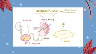
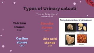
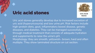
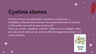
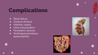
Urolithiasis/
Nephrolithiasis
presentation by:
Dhanik mk
Nephrolithiasis, also known as kidney
stones or renal calculi, refers to the
presence of stones within the kidneys.
So, word nephrolithiasis comes from
“nephro,” which is the latin word for
kidneys, and “lithiasis,” which is the
medical term used to refer to stones.
What is Nephrolithiasis?
Is nephrolithiasis the
same as kidney stones?
Nephrolithiasis refers specifically to kidney
stones, although it is broadly used to refer to
stones in the urinary tract.
Ureterolithiasis, on the other hand, refers to
stones within the ureter, and
urolithiasis refers to stones in any part of the
urinary tract (kidneys, ureter, bladder and
urethra).
Calcium
stones
(75%)
Struvite
stones
(15%)
There are 4 main types of
Urinary calculi:
Cystine
stones
(2%)
Uric acid
stones
(2%)
Types of Urinary calculi
Calcium stones
● They are most common
● The stones maybe in the form of calcium oxalate
which is likely to be the most common
form(50% chance)
● Or it may be in the form of calcium phosphate
(5%)
● Or it may in the composition of both calcium
oxalate and calcium phosphate (45%)
● Almost half the patients with calcium stones have idiopathic
hypercalciuria without hypercalcaemia
● 10% cases are associated with hypercalcaemia and
hypercalciuria, it is most commonly due to hyperparathyroidism,
Or a defect in the bowel or in the kidney.
● 15% of patients with calcium stones have hyperuricosuria with a
normal blood uric acid level and without abnormalities in calcium
metabolism.
● About 25% of patients with calcium stones, the cause is
unknown as there is no abnormalities in urinary excretion of
calcium,uric acid or oxalate and is referred to as ‘idiopathic
calcium stone disease’
Etiology
Pathogenesis
● The mechanism of calcium stone formation is on the
basis of imbalance between the degree of
supersaturation of the ions forming the stone and the
concentration of inhibitors in the urine.
● The likely site of stone formation are tubular lining or
around some fragments of derbis in the tubule acting
as nidus of the stone.
● The stones grows bigger and bigger as more crystals
get deposit around the nidus.
● There are a number of predisposing factor
contributing to formation of calcium stone ( like
alkaline urinary pH, decreased urinary volume and
increased excretion of oxalate & uric acid.
Morphology
● They are usually small, ovoid,
hard, with granular rough surface.
● Their surface may turn to dark
brown due to old blood pigment
deposition.
Struvite stones
They are made of magnesium- ammonium -
calcium phosphate
They are sometimes referred to as infectious stones, because they can be
associated with infections of the urinary tract, especially those caused by
urea-splitting organisms (Proteus mirabilis, Klebsiella, Staphylococcus,
Xanthomonas, Pseudomonas, and Mycoplasma). These bacteria split urea
molecules into ammonium and CO2, thereby raising the urine’s pH to neutral
or alkaline values, and ultimately leading to the precipitation of solutes, to
which the bacteria can adhere. .
Moreover, other types of stones can become infected by
urea-splitting organisms, leading to secondary struvite stone formation.
Struvite stones are more common in biologically-female individuals; they tend
to grow large, filling part of the renal pelvis and calyces, and adopting a
staghorn shape. They are yellow- white or grey in colour and tend to be soft
and friable and irregular in shape. In the urine, they can be seen as “coffin-lid”
shaped crystals. After their removal, if infected fragments of the stone are left
behind in the kidney, they can often grow back, causing a persistent urinary
tract infection and decreased renal function.
Uric acid stones
Uric acid stones generally develop due to increased excretion of
uric acid (hyperuricosuria) and low urine pH. Risk factors include
high-protein diets, gout, inflammatory bowel disease, genetic
diseases, and diabetes. They can be managed successfully
through medical treatment that consists of adequate hydration
and supplements to raise the urine’s pH.
Morphology: they are smooth, yellowish– brown, hard and often
multiple. They show laminated structure on cut section.
Cystine stones are generally caused by cystinuria, a
hereditary disease that causes increased excretion of cystine
in the urine, as well as low urinary pH.
They are small, rounded, smooth , often occurs multiple ,they
are very hard and can be seen as thin hexagonal crystals in a
urine analysis
Cystine stones
Other types
● Occurs due to inherited abnormality of enzyme
metabolism. eg: hereditary xanthinuria
developing xanthine stones.
● drug-induced stones can develop by two
mechanisms. In some cases, excessive use of
laxatives or diuretics can contribute to metabolic
abnormalities that ultimately lead to stone
formation. On the other hand, certain
medications, such as indinavir or ciprofloxacin,
can crystallize in the urine and create stones.
● Renal failure
● Ureteral stricture
● Infection, sepsis
● Urine extravasation
● Perinephric abscess
● Xanthogranulomatous
pyelonephritis
Complications
● NSAIDs
● IV fluids
● Medical expulsive therapy
● Lithotrypsy
● Ureteral endoscopy
Treatment
THANK YOU

Urolithiasis.pdf

  • 1.
  • 3.
    Nephrolithiasis, also knownas kidney stones or renal calculi, refers to the presence of stones within the kidneys. So, word nephrolithiasis comes from “nephro,” which is the latin word for kidneys, and “lithiasis,” which is the medical term used to refer to stones. What is Nephrolithiasis?
  • 4.
    Is nephrolithiasis the sameas kidney stones? Nephrolithiasis refers specifically to kidney stones, although it is broadly used to refer to stones in the urinary tract. Ureterolithiasis, on the other hand, refers to stones within the ureter, and urolithiasis refers to stones in any part of the urinary tract (kidneys, ureter, bladder and urethra).
  • 5.
    Calcium stones (75%) Struvite stones (15%) There are 4main types of Urinary calculi: Cystine stones (2%) Uric acid stones (2%) Types of Urinary calculi
  • 6.
    Calcium stones ● Theyare most common ● The stones maybe in the form of calcium oxalate which is likely to be the most common form(50% chance) ● Or it may be in the form of calcium phosphate (5%) ● Or it may in the composition of both calcium oxalate and calcium phosphate (45%)
  • 7.
    ● Almost halfthe patients with calcium stones have idiopathic hypercalciuria without hypercalcaemia ● 10% cases are associated with hypercalcaemia and hypercalciuria, it is most commonly due to hyperparathyroidism, Or a defect in the bowel or in the kidney. ● 15% of patients with calcium stones have hyperuricosuria with a normal blood uric acid level and without abnormalities in calcium metabolism. ● About 25% of patients with calcium stones, the cause is unknown as there is no abnormalities in urinary excretion of calcium,uric acid or oxalate and is referred to as ‘idiopathic calcium stone disease’ Etiology
  • 8.
    Pathogenesis ● The mechanismof calcium stone formation is on the basis of imbalance between the degree of supersaturation of the ions forming the stone and the concentration of inhibitors in the urine. ● The likely site of stone formation are tubular lining or around some fragments of derbis in the tubule acting as nidus of the stone. ● The stones grows bigger and bigger as more crystals get deposit around the nidus. ● There are a number of predisposing factor contributing to formation of calcium stone ( like alkaline urinary pH, decreased urinary volume and increased excretion of oxalate & uric acid.
  • 9.
    Morphology ● They areusually small, ovoid, hard, with granular rough surface. ● Their surface may turn to dark brown due to old blood pigment deposition.
  • 10.
    Struvite stones They aremade of magnesium- ammonium - calcium phosphate
  • 11.
    They are sometimesreferred to as infectious stones, because they can be associated with infections of the urinary tract, especially those caused by urea-splitting organisms (Proteus mirabilis, Klebsiella, Staphylococcus, Xanthomonas, Pseudomonas, and Mycoplasma). These bacteria split urea molecules into ammonium and CO2, thereby raising the urine’s pH to neutral or alkaline values, and ultimately leading to the precipitation of solutes, to which the bacteria can adhere. . Moreover, other types of stones can become infected by urea-splitting organisms, leading to secondary struvite stone formation. Struvite stones are more common in biologically-female individuals; they tend to grow large, filling part of the renal pelvis and calyces, and adopting a staghorn shape. They are yellow- white or grey in colour and tend to be soft and friable and irregular in shape. In the urine, they can be seen as “coffin-lid” shaped crystals. After their removal, if infected fragments of the stone are left behind in the kidney, they can often grow back, causing a persistent urinary tract infection and decreased renal function.
  • 12.
    Uric acid stones Uricacid stones generally develop due to increased excretion of uric acid (hyperuricosuria) and low urine pH. Risk factors include high-protein diets, gout, inflammatory bowel disease, genetic diseases, and diabetes. They can be managed successfully through medical treatment that consists of adequate hydration and supplements to raise the urine’s pH. Morphology: they are smooth, yellowish– brown, hard and often multiple. They show laminated structure on cut section.
  • 13.
    Cystine stones aregenerally caused by cystinuria, a hereditary disease that causes increased excretion of cystine in the urine, as well as low urinary pH. They are small, rounded, smooth , often occurs multiple ,they are very hard and can be seen as thin hexagonal crystals in a urine analysis Cystine stones
  • 14.
    Other types ● Occursdue to inherited abnormality of enzyme metabolism. eg: hereditary xanthinuria developing xanthine stones. ● drug-induced stones can develop by two mechanisms. In some cases, excessive use of laxatives or diuretics can contribute to metabolic abnormalities that ultimately lead to stone formation. On the other hand, certain medications, such as indinavir or ciprofloxacin, can crystallize in the urine and create stones.
  • 16.
    ● Renal failure ●Ureteral stricture ● Infection, sepsis ● Urine extravasation ● Perinephric abscess ● Xanthogranulomatous pyelonephritis Complications
  • 17.
    ● NSAIDs ● IVfluids ● Medical expulsive therapy ● Lithotrypsy ● Ureteral endoscopy Treatment
  • 18.