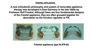
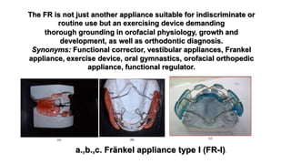
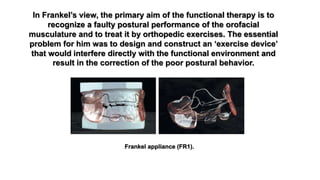
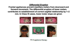
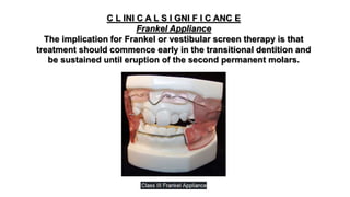
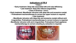
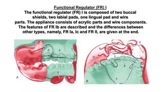
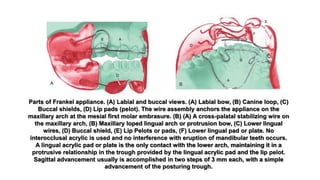
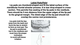
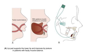
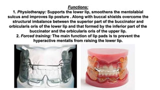
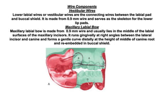
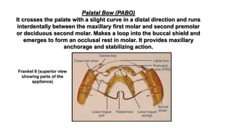
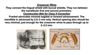
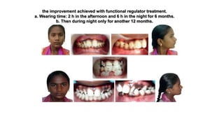
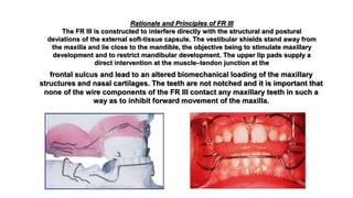
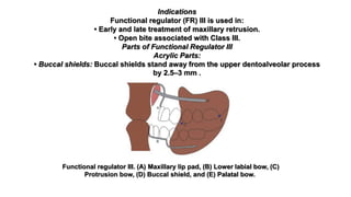
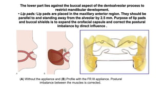
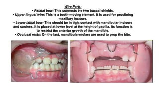
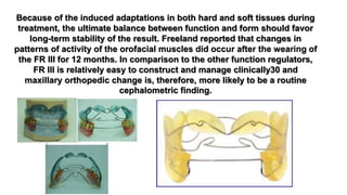
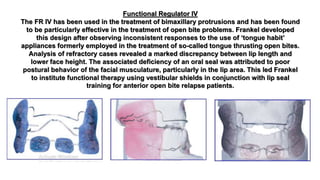
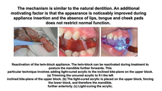
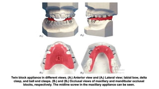
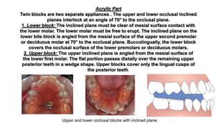
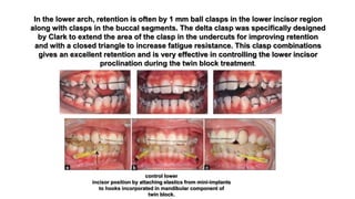
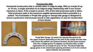
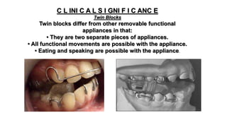
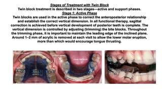
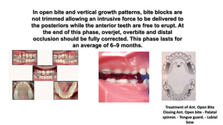
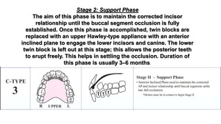
Part 3
dr Maher FOUDA
Faculty of Dentistry
Mansoura Egypt
Professor of orthodontics
Removable functional
appliance
A new orthodontic philosophy and system of removable appliance
therapy was developed in East Germany in the late 1950s by
Professor Rolf Frankel. Although there are four fundamental designs)
of the Frankel appliance, they are often grouped together for
description as the function regulator or FR.
FRANKEL APPLIANCES
Fränkel appliance type III (FR-III))
thorough grounding in orofacial physiology, growth and
development, as well as orthodontic diagnosis.
Synonyms: Functional corrector, vestibular appliances, Frankel
appliance, exercise device, oral gymnastics, orofacial orthopedic
appliance, functional regulator.
The FR is not just another appliance suitable for indiscriminate or
routine use but an exercising device demanding
a.,b.,c. Fränkel appliance type I (FR-I))
T Y P E S O F FUNC T I ONA L R E GUL AT O R S
Functional regulator (FR) I: This is used for correction of Class I and Class II division 1
malocclusion.
FR Ia:
• Used in Class I malocclusion with minor crowding.
• Used in delayed development of the basal bone and dental structure.
FR Ib:
• Used in Class II division 1 malocclusion with deep bite and overjet less than 7 mm.
FR Ic:
• Used in severe Class II division 1 with overjet more than 7 mm.
Functional regulator II: This is used for correction of Class II division 1 and 2 malocclusions.
Functional regulator III: This is used for treatment of Class III due to maxillary deficiency.
Functional regulator IV: This is used for treatment of open bite and bimaxillary protrusion.
Functional regulator V: Functional appliances that incorporate headgear. FR V is used in high
angle cases.
Rationale of Functional Regulator
Frankel’s approach is based on the importance of the form–
function relationship in craniofacial morphogenesis. In general
orthopedics, it has been claimed that of all the functional factors
that play a part in the etiology of skeletal deformities, aberrant
postural performance is the most important. Refer to Box 33.10
for differences between activator and functional regulator.
In Frankel’s view, the primary aim of the functional therapy is to
recognize a faulty postural performance of the orofacial
musculature and to treat it by orthopedic exercises. The essential
problem for him was to design and construct an ‘exercise device’
that would interfere directly with the functional environment and
result in the correction of the poor postural behavior.
Frankel appliance (FR1).
DIFFERENCES BETWE EN F R ANK E L A P P L I ANC E AND A C T I VAT O R
Activator Functional regulator
Activator is a tooth-borne appliance Frankel is a tissue-borne appliance
This is a loose-fitting appliance Firm maxillary anchorage
Activates the muscle, hence called activator. Prevents the aberrant muscle force from acting on the dentition
and arches
Harnesses the muscle force
Does not act as an exercise device Acts as an exercise device
Bulk of the appliance is placed within the teeth Bulk of the appliance is placed outside the teeth in vestibule
Only one wire component Many wire components
Activator is one single acrylic block Three acrylic parts joined by wire components
Mandibular advancement by 6–7 mm Minimum advancement by 2.5–3 mm
Vertical opening is more Minimum vertical opening. Just for the wire to pass
Worn during night-time Worn day and night
Speech is not possible with appliance in mouth Speech is possible with appliance
Faulty muscle posture is seen as having an adverse
environmental effect on:
1. The spatial relationships of the maxilla and/or the
mandible, that is, the sagittal, vertical and transverse basal
arch relationships.
2. The development in space of the dentoalveolar structures.
Frankle 3 appliance.
As the name implies, the actions of the FR are intended to
change or regulate the muscular environment of the face and
teeth, to stretch facial musculature to normal dimensions,
impede abnormal activity of the lips, tongue, and cheeks and
thus allow development of the jaws and teeth in all three
planes.Action of Frankel Appliance
Philosophy of Frankel Appliance/Mechanism of
Action of Frankel Appliance
Frankel applies the functional matrix concept and
terminology of Moss to explain the basis of design of his
appliances. When faulty muscle posture is deemed to have
compromised the spatial relationships of the maxilla and
mandible in a growing child, the appliance is designed to
alter the biomechanical conditions in the periosteal
functional matrices of these bones.
FR 1
1. In the case of a deficient mandible, the cause is considered to be
a postural imbalance between the retractor and protractor muscles.
The FR1 and FR2 appliances are constructed so that the patient is
obliged to posture the mandible forward in order to achieve a
comfortable jaw position. In this way, the periosteal tissues related
to the mandibular condyle are subjected to a biomechanical
stimulus that favors an increased rate of bone deposition until a
position of stability is reached, with the mandible relocated in a
more anterior position.
2. In the case of a deficient maxilla, the FR3 appliance is
constructed so that periosteal tension is produced at the
superior sulci. The vestibular shields and upper lip pads
counteract the restricting effect of aberrant posture
within the labial muscle groups and normal maxillary
growth is restored.
3. In skeletal open bite development, clinical relevance is
placed on the poor postural performance of the muscles
forming the external soft-tissue capsule and of those
suspending the mandible. Accordingly, therapy with the FR4
is directed at restoring a competent anterior oral seal and
establishing a more superior postural position of the tongue
and of the mandible. Subsequent skeletal and dentoalveolar
remodeling mediated via the periosteal matrices will favor
acceptable vertical, dental and facial relationships.8
The second of Moss’s concepts concerns the capsular functional
matrix. Frankel sees the muscular portions of the walls of the
oral and nasopharyngeal spaces as forming a capsular matrix,
the volume of which has an important morphogenetic influence
on the development of the dentoalveolar structures.
Specifically, he claims that an aberrant postural pattern of
the external muscular environment may unfavorably
influence the eruptive path and restrain the normal
physiologic process of decrowding and uprighting of the
teeth during eruption. By means of the vestibular shields of
the functional regulators, the erupting dentition is
protected from the adverse forces generated by the
existing perioral musculature, allowing for spontaneous
decrowding of the teeth.
Vestibular Area of Operation
The major part of Frankel appliance is confined to the oral vestibule. The
buccal shields and lip pads hold the
labial and buccal musculature, the buccinator mechanism, from acting on the dentition.
1. The vestibular shields eliminate perioral soft-tissue pressure that arises from:27
a. Muscular forces associated with aberrant postural patterns in the orofacial muscles.
b. Subatmospheric pressure generated in the oral cavity, which acts to suck the lips
and cheeks against the teeth and into the interocclusal space.
c. Elimination of these restraining muscle pressures allows the inherent bone inducing
potential of the erupting tooth to be expressed in a more buccal direction.
2. The periphery of the vestibular shield is deliberately extended into the vestibular
reflection so that tension is produced in the soft tissues. This pull on the soft
tissue is transferred to the periosteum, with two possible effects:
a. The tension in the periosteum may contribute mechanically to an outward
bending of the thin buccal plate, thereby facilitating outward drift of the teeth.
b. Direct tension in the periosteum is known to stimulate deposition of new
alveolar bone28 and this is claimed to occur on the facial aspect of the alveolus in
response to FR treatment.
3. The outer surfaces of the vestibular shield are presented to the
musculature as a correct configuration of the dentoalveolar process.
The orofacial musculature is trained to function in harmony with the
dental arches as they attain correct width and shape.
4. Bilateral tension on the maxilla at the level of the sulcus is claimed
to stimulate widening of the midpalatal suture.
Tongue Function
Although Frankel appliance gives more importance to buccinator
mechanism, the tongue also plays a significant role in molding the arches.
1. In contrast to most other functional jaw orthopedic appliances, the FR
intervenes very little with the tongue posture and the tongue is free to exert
more force in forward and lateral direction.
2. After removal of the appliance, the perioral soft-tissue pressure will
continue to be reduced while the tongue pressure is increased because it is
elevated into a palatal position. This favors long-term stability of arch
expansion.
Muscle Re-education
Function regulator treatment is claimed to accomplish its skeletal and dental
corrections by modifying behavior aberrations involving the orofacial
musculature. Freeland29 (1979) compared the muscle patterns in a
group of patients before and after treatment with the FR1 to those of a
control group and concluded that the FR did indeed affect muscle activity.
FR1
Exercise Device
Frankel appliance apart from restricting the faulty muscle posture also acts as an exercise
device. That is, it stimulates normal function while eliminating aberrant muscle activity.
Hence, full-time wear of the appliance is recommended along with functional exercises.
Anteroposterior Correction
Increased activity of the superior head of the lateral pterygoid muscle is associated with
functional appliance wear. Skeletal adaptation proceeds until muscle activity is restored.
Maxillary Restraining Effect
Frankel appliances have a restraining effect on the maxillary teeth and arch.30
Decrowding during eruption is a feature of the entire Frankel
appliance. The vestibular screens are deliberately extended into the
vestibular sulcus so that tension is created in the soft tissues. This
causes outward bending of the thin buccal plate, thereby
facilitating outward drift of the teeth.
Decrowding during Eruption
Differential Eruption
Frankel appliances prevent maxillary molars from downward and
forward movement. The differential eruption of lower molars
contributes to establishment of correct sagittal relationship by 1–2
mm. In Class III cases, lower molar stops are given.
C L INI C A L S I GNI F I C ANC E
Frankel Appliance
The implication for Frankel or vestibular screen therapy is that
treatment should commence early in the transitional dentition and
be sustained until eruption of the second permanent molars.
Indications of Functional Regulator
The indications of functional regulator can be studied under each type.
Indications of FR I
• Class I
• Early treatment: Discrepancy between tooth size and arch size in patients with
normal overbite.
• Late treatment: Mild crowding in the presence of adequate apical base.
• Class II division 1
• Early treatment: Mandibular retrusion with normal overbite.
• Late treatment:
- Mandibular retrusion with normal overbite, overjet more than 7 mm.
- Mandibular retrusion with crowding. In this, prefunctional fixed appliance,
treatment is done to correct crowding.
- Mandibular retrusion with open bite.
Indications of FR II
• Class I
• Early treatment: Deep bite associated with arch size deficiency.
• Late treatment: Deep bite without irregularities.
• Class II divisions 1 and 2
• Early treatment: Mandibular retrusion with deep bite and excessive overjet.
• Pretreatment mechanotherapy to correct the upper incisors is required.
• Late treatment:
- Mandibular retrusion with deep bite and excessive overjet without arch
irregularities. Pretreatment mechanotherapy to correct incisors is required.
- Mandibular retrusion with arch irregularities. Pretreatment fixed appliance
mechanotherapy to correct crowding by extraction is required.
Indications of FR III
• Early and late treatment of maxillary retrusion.
• Open bite: Open bite associated with Class III.
Indication of FR IV
• FR IV is used in early treatment of skeletal open bite and bimaxillary
protrusion.
Indications of FR V
• High-angle cases.
• Vertical growth pattern.
Indications of FR as Retainer
• FR type used as the last appliance should be worn as retainer.
• After fixed appliance mechanotherapy.
• After oral surgery, FR is used as exercise device to prevent relapse.
Functional Regulator (FR) I
The functional regulator (FR) I is composed of two buccal
shields, two labial pads, one lingual pad and wire
parts. The appliance consists of acrylic parts and wire components.
The features of FR Ib are described and the differences between
other types, namely, FR Ia, Ic and FR II, are given at the end.
Parts of Frankel appliance. (A) Labial and buccal views. (A) Labial bow, (B) Canine loop, (C)
Buccal shields, (D) Lip pads (pelot). The wire assembly anchors the appliance on the
maxillary arch at the mesial first molar embrasure. (B) (A) A cross-palatal stabilizing wire on
the maxillary arch, (B) Maxillary loped lingual arch or protrusion bow, (C) Lower lingual
wires, (D) Buccal shield, (E) Lip Pelots or pads, (F) Lower lingual pad or plate. No
interocclusal acrylic is used and no interference with eruption of mandibular teeth occurs.
A lingual acrylic pad or plate is the only contact with the lower arch, maintaining it in a
protrusive relationship in the trough provided by the lingual acrylic pad and the lip pelot.
Sagittal advancement usually is accomplished in two steps of 3 mm each, with a simple
advancement of the posturing trough.
Acrylic Parts
Buccal Shields
They extend deep into the sulci in the apical region of the maxillary first premolar and
tuberosity region. In areas where expansion of the dental arch and alveolar process is
required, the shields stand away from the lateral aspects of teeth and alveolus. In the
maxillary teeth and alveolus, the
gap between the shield and teeth surface is double the wax thickness. In the
mandible, only a single layer of wax is added. The thickness of the buccal shield
should be 2.5 mm.
Functions:
1. Physiotherapy: Buccal shields expand the circumoral capsule in
transverse direction causing the soft tissues to adapt to new form.
2. Forced training of the muscles of cheeks to adapt to functional
performance.
3. Correction of spatial disorder: Stimulation of periosteal matrices
corrects spatial disorder.
Labial Pads/Pelot
Lip pads are rhomboid-shaped and fit the labial surface of the
mandibular frontal alveolar process. It is tear drop-shaped in cross-
section. This permits free seating of the lip pads in the vestibule.
There should be 5 mm distance from the upper edges of the lip pad
to the gingival margin. The distal edge of the lip pad should not
overlap the canine root protuberance.
Lip pads hold the lips
away from the teeth and
force the lips to stretch
to form an oral seal. The
lip pad is positioned
low in the vestibule and
must have the proper
inclination
to avoid soft tissue
irritation.
(A) Lip pad supports the lower lip and improves lip posture
in patients with faulty muscle balance.
Functions:
1. Physiotherapy: Supports the lower lip, smoothens the mentolabial
sulcus and improves lip posture . Along with buccal shields overcome the
structural imbalance between the superior part of the buccinator and
orbicularis oris of the lower lip and that formed by the inferior part of the
buccinator and the orbicularis oris of the upper lip.
2. Forced training: The main function of lip pads is to prevent the
hyperactive mentalis from raising the lower lip.
Lingual Shield
The lingual shield is situated or placed below the gingival margin of the mandibular teeth.
This extends up to the distal of the second premolar. It is positioned by the two
connecting wires to the buccal shield.
Functions Forced Training In mandibular retrusion cases, the mandible is kept in the
advanced position by the supporting action of lingual and labial shields. Whenever the
mandible tries to slide back to its original position, the lingual shield elicits a pressure
sensation on the lingual aspect of the alveolar process . This sensory input activates the
proprioceptors in the gingiva and periosteum to stimulate the protractors of mandible.
(B) Lingual shield: Mechanism of action stimulates
the protractor muscles by activating proprioceptors
The lingual pad or flange determines the
anteroposterior and vertical
mandibular posture for most functional
appliances. A, The small lingual pad from a
Frankel appliance;
B, the extensive lingual flange from a
modified activator; C, the lingual components
not only
position the mandible forward but, D, also
exert a protrusive effect on the mandibular
incisors
when the mandible attempts to return to its
original position, especially if some
component of the
appliance contacts these teeth.
Wire Components
Vestibular Wires
Lower labial wires or vestibular wires are the connecting wires between the labial pad
and buccal shield. It is made from 0.9 mm wire and serves as the skeleton for the lower
lip pads.
Maxillary Labial Bow
Maxillary labial bow is made from 0.9 mm wire and usually lies in the middle of the labial
surfaces of the maxillary incisors. It runs gingivally at right angles between the lateral
incisor and canine and forms a gentle curve distally at the height of middle of canine root
and re-embedded in buccal shield.
FIGURE 33.24.
Palatal Bow (PABO)
It crosses the palate with a slight curve in a distal direction and runs
interdentally between the maxillary first molar and second premolar
or deciduous second molar. Makes a loop into the buccal shield and
emerges to form an occlusal rest in molar. It provides maxillary
anchorage and stabilizing action.
Frankel II (superior view
showing parts of the
appliance)
Canine Loop
It starts with its tags in the buccal shield and runs palatally to the lingual
surface of the canine for a distance of about 1 mm. It then crosses the
interproximal contact between canine and lateral incisor. The function of
the canine loop is it keeps the perioral activity away from canine and
provides passive expansion in canine area.
Lower Lingual Springs
These recurved springs, two in number, pass above the cingulum of the
lower incisors. They prevent supraeruption of lower incisors and also
causes bite opening by relative intrusion.
Crossover Wires
They connect the lingual shield with buccal shields. They run between
the mandibular first and second premolars.
Construction Bite for Class II Correction
Frankel advocates minimal sagittal or forward advancement. The
mandible is advanced by 2.5–3 mm only. Vertical opening also should be
very minimal, just enough for the crossover wires to pass through up to
2–2.5 mm.
Differences Between FR Ia and Ic and FR II
• FR Ia: There is lower lingual wire loops instead of lingual shield.
• FR Ic: Buccal shields are split horizontally and vertically into two
parts for incremental advance.
• FR II: Addition of upper palatal protrusion bow behind upper
incisors; modified canine loop.
Clinical Management of Functional Regulators
The treatment with functional regulator can be classified into three stages or
phases: (1) initial treatment phase, (2) active treatment phase, and (3)
retention phase.
1. Initial Treatment Phase
This phase is for the patients to get used to the appliance and handle it as an
orthopedic exercise device combined with lip seal training. The initial phase
consists of the following:
.
FR-2 appliance: Frontal and lateral views.
Delivery of the appliance:
i. All margins are checked for smoothness.
ii. Check for the proper shape of the lip pads.
iii. Check the fit of the appliance in maxilla and mandible separately.
iv. Peripheral portions of shields contact without producing blanching.
v. Instruct the patient the method of appliance wear.
vi. Palpate the face on the outside to make sure there are no sharp edges.
vii. After insertion, ask the patient to speak. During speaking, the vestibular
shields loosen up the tight musculature, which helps in adapting to the
appliance.
C L INI C A L S I GNI F I C ANC E
Features of an Ideal Patient for Treatment with FR I and FR II
Skeletal Features
1. Normal maxillary position in the sagittal and vertical dimensions.
2. Retrognathic mandible.
3. Normal or reduced lower face height.
Dental Features
1. Normal relationship of the maxillary denture to the maxilla in the sagittal and vertical
dimensions.
2. Normal relationship of the mandibular denture to the mandible in the sagittal and vertical
dimensions.
3. Mild crowding in the mandibular arch or in both arches is acceptable, but axial rotations
and bodily displacements of individual teeth require a separate phase of fixed appliance
treatment.
Maturation
Early mixed dentition, 7–9 years.
b. Wearing of the appliance :The objective of the appliance should be well
understood. In traditional orthodontics, we change the form with a
spontaneous adaptation of the neuromuscular pattern. In contrast with
functional approach, the faulty postural function is overcome first, which
results in spontaneous adaptation of form to altered normal function.
i. Worn during daytime also.
ii. Initial wear for 2–3 h for the first 2 weeks.
iii. Lip together exercises to be performed by the patient.
iv. Check-up after 2 weeks—presence of tissue redness and also speech
improvement is a sign of cooperation.
v. Next 3 weeks 4–6 h daytime wear.
vi. Normally 3–4 months for initial phase.
Frankel appliance for correction of Class II division 1
malocclusion
2. Active Treatment Phase
Before advising night-time wear, check for improvement in facial muscle balance.
There must be some change in overcoming the hyperactivity of the muscle. Then
night-time wear is advised.
a. Appliance adjustments like bending of canine loops occlusally, molar rests, labial
bows are carried out, if required.
b. Progress is recorded. After 3 months of full-time wear, expansion of dental arches
will be evident.
c. In 6–8 months, correction from Class II to Class I takes place.
d. Patient will have difficulty in positioning the lower jaw posteriorly.
3. Retention Treatment Phase
A chance for relapse is less with functional appliances; there is
spontaneous adaptation of form to altered function. Therefore, the action
of the retention phase is to stabilize the restraining effect of the exercise
device achieved during active treatment. The last appliance itself is used
as a retention appliance.
the improvement achieved with functional regulator treatment.
a. Wearing time: 2 h in the afternoon and 6 h in the night for 6 months.
b. Then during night only for another 12 months.
Functional Regulator III
In functional orthopedic philosophy, maxillary retrognathia is viewed as
a consequence of failure of expansion of the superior part of the
orofacial capsule. Delaires’ (1978) dissections of normal and cleft lip
cadavers led him to postulate that the nasogenal muscles have a direct
influence on maxillary growth and that functional aberrations of these
muscle groups may play an important role in restricting maxillary
development.
Rationale and Principles of FR III
The FR III is constructed to interfere directly with the structural and postural
deviations of the external soft-tissue capsule. The vestibular shields stand away from
the maxilla and lie close to the mandible, the objective being to stimulate maxillary
development and to restrict mandibular development. The upper lip pads supply a
direct intervention at the muscle–tendon junction at the
frontal sulcus and lead to an altered biomechanical loading of the maxillary
structures and nasal cartilages. The teeth are not notched and it is important that
none of the wire components of the FR III contact any maxillary teeth in such a
way as to inhibit forward movement of the maxilla.
In contrast to the action of the FR II, where the skeletal and dental changes are entirely
functionally induced, the FR III imparts both functional and mechanical effects:
1. The functional effects include stimulation of forward growth of the maxilla,
deposition of new bone on the facial aspect of maxilla33 (about 2 mm in Point A) and
possibly increased growth of the anterior cranial base.34
2. The mechanical aspect of FR III treatment is mediated by contact of the lower labial
connector with the labial surfaces of the mandibular incisors. Lip pressure against the
upper lip pads is transferred to the lower incisors causing retroclination of these teeth
and frequently a posterior rotation of the mandible. This latter effect is particularly
appropriate in Class III malocclusions associated with reduced lower face height.
Diagram illustrating the working principles of FR III
Indications
Functional regulator (FR) III is used in:
• Early and late treatment of maxillary retrusion.
• Open bite associated with Class III.
Parts of Functional Regulator III
Acrylic Parts:
• Buccal shields: Buccal shields stand away from the upper dentoalveolar process
by 2.5–3 mm .
Functional regulator III. (A) Maxillary lip pad, (B) Lower labial bow, (C)
Protrusion bow, (D) Buccal shield, and (E) Palatal bow.
The lower part lies against the buccal aspect of the dentoalveolar process to
restrict mandibular development.
• Lip pads: Lip pads are placed in the maxillary anterior region. They should be
parallel to and standing away from the alveolar by 2.5 mm. Purpose of lip pads
and buccal shields is to expand the orofacial capsule and correct the postural
imbalance by direct influence .
(A) Without the appliance and (B) Profile with the FR III appliance. Postural
imbalance between the muscles is corrected.
Wire Parts:
• Palatal bow: This connects the two buccal shields.
• Upper lingual wire: This is a tooth-moving element. It is used for proclining
maxillary incisors.
• Lower labial bow: This should be in tight contact with mandibular incisors
and canines. It is placed at lower level at the height of papilla. Its function is
to restrict the anterior growth of the mandible.
• Occlusal rests: On the last, mandibular molars are used to prop the bite.
Since the FR III seeks to pre-empt abnormal development and
restore normal growth rather than to correct fully established
malformation, it is important that treatment be initiated as
early as possible. The appliance can be readily used in the
complete deciduous dentition, if necessary.
Because of the induced adaptations in both hard and soft tissues during
treatment, the ultimate balance between function and form should favor
long-term stability of the result. Freeland reported that changes in
patterns of activity of the orofacial muscles did occur after the wearing of
the FR III for 12 months. In comparison to the other function regulators,
FR III is relatively easy to construct and manage clinically30 and
maxillary orthopedic change is, therefore, more likely to be a routine
cephalometric finding.
Functional Regulator IV
The FR IV has been used in the treatment of bimaxillary protrusions and has been found
to be particularly effective in the treatment of open bite problems. Frankel developed
this design after observing inconsistent responses to the use of ‘tongue habit’
appliances formerly employed in the treatment of so-called tongue thrusting open bites.
Analysis of refractory cases revealed a marked discrepancy between lip length and
lower face height. The associated deficiency of an oral seal was attributed to poor
postural behavior of the facial musculature, particularly in the lip area. This led Frankel
to institute functional therapy using vestibular shields in conjunction with lip seal
training for anterior open bite relapse patients.
Frankel reasoned that if alterations in the postural activity of the orofacial
musculature can lead to skeletal open bite, then correction of faulty
postural behavior might help to correct the associated skeletal deformity. A
fundamental aim of his therapy was to overcome the deviant pattern of
mandibular rotation through re-establishment of nose breathing by
correcting the lips-apart condition and faulty tongue posture. In fact, lip
seal training alone, in the absence of any appliance treatment, has been
shown to result in closure of open bites.26 However, voluntary training
exercises without an appliance are difficult to sustain for adequate daily
periods throughout the duration of the growth phase. Furthermore, the FR
IV appliance has additional working
principles that contribute to the desired skeletal remodeling changes .
Illustration of working principles of FR IV.
Correct extension of the posterior margins of the vestibular shields determines
the location of a new center of rotation for the mandible. From receptors in
these areas, sensory feedback is said to cause reflex distraction of the condyle
from the mandibular fossa, while the chin is rotated upward by the
strengthened anterior vertical muscle chain. Compensatory translative growth
may restore the normal condyle–fossa relationship and increase ramus height.
An important question concerning skeletal open bite development is the role
of neuromuscular maturation. Functional disorders in the orofacial area may
be attributed to a failed or incomplete maturation of postural performances,
for which an adverse psychosocial environment may constitute an important
contributory factor.
Treatment with the FR IV is not commenced until the patient has shown
evidence of good cooperation with lip seal exercises during a probationary
period of 3–4 months. As with the other function regulators, therapy should be
initiated in the mixed dentition and lengthy treatment and retention periods
are frequently necessary in the management of these patients.
Twin Block
Twin blocks are bite blocks that effectively modify the occlusal inclined
plane to induce favorably directed occlusal forces by causing a functional
mandibular displacement. Upper and lower bite blocks interlock at a 70°
angle and are designed for full time wear taking advantage of all functional
forces to the dentition including the masticatory forces. Occlusal inclined
planes give more freedom in forward and lateral excursion and cause less
interference with normal function.
The mechanism is similar to the natural dentition. An additional
motivating factor is that the appearance is noticeably improved during
appliance insertion and the absence of lips, tongue and cheek pads
does not restrict normal function..
Reactivation of the twin-block appliance. The twin-block can be reactivated during treatment to
posture the mandible further forwards. This
particular technique involves adding light-cured acrylic to the inclined bite-plane on the upper block.
(a) Trimming the uncured acrylic to fi t the left
inclined bite-plane of the upper block. (b) The light-cured acrylic is placed on the upper block, forcing
the lower block, and therefore the mandible,
further anteriorly. (c) Light-curing the acrylic.
T Y P E S O F TWI N B L O C K S
1. Standard twin block
2. Sagittal twin block
3. Reverse twin block for Class III correction
4. Magnetic twin block
(b) Twin-block appliance
modifi ed for treatment of a
Class II division 2
malocclusion. Note the
additional palatal double-
cantilever spring
(highlighted in red circle) in
the upper arch, which is used
to procline the central
incisors. (c) End of functional
stage. The anteroposterior
discrepancy
has been corrected and the
retroclined upper central
incisors proclined to normal
inclinations. Note the
posterior lateral open bites,
which were
closed in the second phase of
fixed appliances.
Standard Twin Blocks-Class II Div I
(with good arch form)
• Adams or Delta Clasps, and Anterior Ball clasps for
good retention
•Twin Blocks, at a 70 ‫؛‬angle, to advance mandible
• Upper Midline Screw: so upper arch can accomodate
lower arch in advanced position
Twin blocks appliance design. A, Anterior
and lateral views show the following
components.
Upper appliance: (1) labial bow (0.8 mm)
from mesial 6&, (2) clasps (0.8 mm)
incorporating coils to
accommodate the Concorde face-bow, and
(3) occlusal inclined planes occlude at a 45”
angle in @
region. Lower appliance: (1) ball-ended
interdental clasps (1 .O mm) in 21112 region,
(2) delta clasps
(0.8 mm) on q (the delta clasp, designed by
the author, gives excellent retention on
lower premolars
and requires minimal adjustment), and (3)
inclined planes in @ region. 8, Occlusal
views. The upper
appliance has a midline screw for
compensatory lateral expansion. Where
necessary, a midline screw
or recurved lingual bow (as in a Jackson
appliance) can be included in the lower
appliance.
Effectiveness of reverse twin block with lip
pads-RME and face mask with RME in the
early treatment of class III malocclusion
Standard Appliance Design
The parts of the appliance are: (1) acrylic part, which consists of base
plate and occlusal inclined plane and (2) wire components, which
include labial bow, delta clasp and ball end clasp.
Twin block appliance in different views. (A1) Anterior view and (A2) Lateral view; labial bow, delta
clasp, and ball end clasps. (B1) and (B2) Occlusal views of maxillary and mandibular occlusal
blocks, respectively. The midline screw in the maxillary appliance can be seen.
Acrylic Part
Twin blocks are two separate appliances . The upper and lower occlusal inclined
planes interlock at an angle of 70° to the occlusal plane.
1. Lower block: The inclined plane must be clear of mesial surface contact with
the lower molar. The lower molar must be free to erupt. The inclined plane on the
lower bite block is angled from the mesial surface of the upper second premolar
or deciduous molar at 70° to the occlusal plane. Buccolingually, the lower block
covers the occlusal surface of the lower premolars or deciduous molars.
2. Upper block: The upper inclined plane is angled from the mesial surface of
the lower first molar. The flat portion passes distally over the remaining upper
posterior teeth in a wedge shape. Upper blocks cover only the lingual cusps of
the posterior teeth.
Upper and lower occlusal blocks with inclined plane.
Wire Components :
1. Clasps used: Initially, Adams clasps were used . Now delta clasps are used in
lower premolar and upper first molar. In the lower anterior region, ball end clasps
are used.
2. Labial bow: Use of labial bow is optional and it is used for retention purposes.
It is also used when there is severe proclination and if they have to be uprighted.
the acrylic and wire
components of twin block.
1- Headgear tube. 2- Area where the inclined planes meet. 3-
Anterior lower bite block. 4- Posterior upper bite block.
The delta clasps are used for retention in upper appliance.
The clasps incorporate a coiled tube for face bow
attachment, if traction is planned to be applied. Retention may
be increased by placing ball end clasps in the labial or buccal
segments.
An expansion screw in the midline provides compensatory
lateral expansion of the upper arch to accommodate a functional
lower arch protrusion from its retruded position. The labial and
lingual bows, if needed, are included to control upper incisor
angulations.
In the lower arch, retention is often by 1 mm ball clasps in the lower incisor region
along with clasps in the buccal segments. The delta clasp was specifically designed
by Clark to extend the area of the clasp in the undercuts for improving retention
and with a closed triangle to increase fatigue resistance. This clasp combinations
gives an excellent retention and is very effective in controlling the lower incisor
proclination during the twin block treatment.
control lower
incisor position by attaching elastics from mini-implants
to hooks incorporated in mandibular component of
twin block.
The upper bite block covers the lingual cusps of the upper posteriors,
extending to the mesial ridge of the upper second premolar. This improves
retention by allowing the clasp to be flexible. To compensate for the arch
width discrepancy and to allow the inclined planes to interlock in
occlusion, it is necessary to have full occlusal coverage. The lower bite
block extends to the distal marginal ridge of the mandibular second
premolar.
Construction Bite
Horizontal construction bite is usually taken in edge-to-edge. With an overjet of up
to 10 mm, a single activation to an edge-to-edge relationship with 2 mm incisor
clearance is done. If the overjet is severe, 70% of the total protrusive path is taken.
Total protrusive path is the maximum sagittal advancement that is possible in a
patient. The Exactobite or Projet bite gauge or George bite gauge is designed to
record a protrusive interocclusal record or bite registration in wax for construction
of twin blocks.
Projet Bite Gauge: (a) select the appropriate groove for
the upper incisors depending on the size of the overjet and the ease
with which the patient can posture forward; (b) the lower incisors
bite into a single groove to register a protrusive bite; (c) bite
registration for an overjet of up to 10 mm. The blue Projet bite
gauge gives 2 mm interincisal opening and there is 5–6 mm vertical
space between the premolars
The most common fault in twin block construction is to
make the block too thin. In such conditions, patients can
posture out of the appliance reducing the effectiveness of
the treatment. An important principle is that the block should be thick enough
to open the bite slightly beyond the freeway space. On average, blocks are not
less than 5 mm thick in first premolars or first deciduous molar region. This
thickness is normally achieved in Class II division 1 deep bite cases by
registering 2 mm vertical interincisal clearance.
C L INI C A L S I GNI F I C ANC E
Twin Blocks
Twin blocks differ from other removable functional
appliances in that:
• They are two separate pieces of appliances.
• All functional movements are possible with the appliance.
• Eating and speaking are possible with the appliance.
Stages of Treatment with Twin Block
Twin block treatment is described in two stages—active and support phases.
Stage 1: Active Phase
Twin blocks are used in the active phase to correct the anteroposterior relationship
and establish the correct vertical dimension. In all functional therapy, sagittal
correction is achieved before vertical development of posterior teeth is complete. The
vertical dimension is controlled by adjusting (trimming) the bite blocks. Throughout
the trimming phase, it is important to maintain the leading edge of the inclined plane.
Around 1–2 mm of acrylic is removed at each visit to allow the lower molar eruption,
more than which would encourage tongue thrusting.
In open bite and vertical growth patterns, bite blocks are
not trimmed allowing an intrusive force to be delivered to
the posteriors while the anterior teeth are free to erupt. At
the end of this phase, overjet, overbite and distal
occlusion should be fully corrected. This phase lasts for
an average of 6–9 months.
Treatment of Ant. Open Bite
Closing Ant. Open bite - Palatal
spinner. - Tongue guard. - Labial
bow
Stage 2: Support Phase
The aim of this phase is to maintain the corrected incisor
relationship until the buccal segment occlusion is fully
established. Once this phase is accomplished, twin blocks are
replaced with an upper Hawley-type appliance with an anterior
inclined plane to engage the lower incisors and canine. The lower
twin block is left out at this stage; this allows the posterior teeth
to erupt freely. This helps in settling the occlusion. Duration of
this phase is usually 3–6 months.
Retention
Treatment is followed by retention with an upper anterior inclined plane appliance.
A good buccal segment occlusion is cornerstone of stability. Average time taken is
9 months. Twin blocks are the most popular removable functional appliance today
that produce good results . Patient compliance is also better when compared to
other removable functional appliances.
upper anterior inclined plane appliance
Pretreatment and post-treatment photographs of a patient
treated with twin block appliance.
Reference
Removable functional appliance 3

Removable functional appliance 3

  • 1.
    Part 3 dr MaherFOUDA Faculty of Dentistry Mansoura Egypt Professor of orthodontics Removable functional appliance
  • 2.
    A new orthodonticphilosophy and system of removable appliance therapy was developed in East Germany in the late 1950s by Professor Rolf Frankel. Although there are four fundamental designs) of the Frankel appliance, they are often grouped together for description as the function regulator or FR. FRANKEL APPLIANCES Fränkel appliance type III (FR-III))
  • 3.
    thorough grounding inorofacial physiology, growth and development, as well as orthodontic diagnosis. Synonyms: Functional corrector, vestibular appliances, Frankel appliance, exercise device, oral gymnastics, orofacial orthopedic appliance, functional regulator. The FR is not just another appliance suitable for indiscriminate or routine use but an exercising device demanding a.,b.,c. Fränkel appliance type I (FR-I))
  • 4.
    T Y PE S O F FUNC T I ONA L R E GUL AT O R S Functional regulator (FR) I: This is used for correction of Class I and Class II division 1 malocclusion. FR Ia: • Used in Class I malocclusion with minor crowding. • Used in delayed development of the basal bone and dental structure. FR Ib: • Used in Class II division 1 malocclusion with deep bite and overjet less than 7 mm. FR Ic: • Used in severe Class II division 1 with overjet more than 7 mm. Functional regulator II: This is used for correction of Class II division 1 and 2 malocclusions. Functional regulator III: This is used for treatment of Class III due to maxillary deficiency. Functional regulator IV: This is used for treatment of open bite and bimaxillary protrusion. Functional regulator V: Functional appliances that incorporate headgear. FR V is used in high angle cases.
  • 5.
    Rationale of FunctionalRegulator Frankel’s approach is based on the importance of the form– function relationship in craniofacial morphogenesis. In general orthopedics, it has been claimed that of all the functional factors that play a part in the etiology of skeletal deformities, aberrant postural performance is the most important. Refer to Box 33.10 for differences between activator and functional regulator.
  • 6.
    In Frankel’s view,the primary aim of the functional therapy is to recognize a faulty postural performance of the orofacial musculature and to treat it by orthopedic exercises. The essential problem for him was to design and construct an ‘exercise device’ that would interfere directly with the functional environment and result in the correction of the poor postural behavior. Frankel appliance (FR1).
  • 7.
    DIFFERENCES BETWE ENF R ANK E L A P P L I ANC E AND A C T I VAT O R Activator Functional regulator Activator is a tooth-borne appliance Frankel is a tissue-borne appliance This is a loose-fitting appliance Firm maxillary anchorage Activates the muscle, hence called activator. Prevents the aberrant muscle force from acting on the dentition and arches Harnesses the muscle force Does not act as an exercise device Acts as an exercise device Bulk of the appliance is placed within the teeth Bulk of the appliance is placed outside the teeth in vestibule Only one wire component Many wire components Activator is one single acrylic block Three acrylic parts joined by wire components Mandibular advancement by 6–7 mm Minimum advancement by 2.5–3 mm Vertical opening is more Minimum vertical opening. Just for the wire to pass Worn during night-time Worn day and night Speech is not possible with appliance in mouth Speech is possible with appliance
  • 8.
    Faulty muscle postureis seen as having an adverse environmental effect on: 1. The spatial relationships of the maxilla and/or the mandible, that is, the sagittal, vertical and transverse basal arch relationships. 2. The development in space of the dentoalveolar structures. Frankle 3 appliance.
  • 9.
    As the nameimplies, the actions of the FR are intended to change or regulate the muscular environment of the face and teeth, to stretch facial musculature to normal dimensions, impede abnormal activity of the lips, tongue, and cheeks and thus allow development of the jaws and teeth in all three planes.Action of Frankel Appliance Philosophy of Frankel Appliance/Mechanism of Action of Frankel Appliance
  • 10.
    Frankel applies thefunctional matrix concept and terminology of Moss to explain the basis of design of his appliances. When faulty muscle posture is deemed to have compromised the spatial relationships of the maxilla and mandible in a growing child, the appliance is designed to alter the biomechanical conditions in the periosteal functional matrices of these bones. FR 1
  • 11.
    1. In thecase of a deficient mandible, the cause is considered to be a postural imbalance between the retractor and protractor muscles. The FR1 and FR2 appliances are constructed so that the patient is obliged to posture the mandible forward in order to achieve a comfortable jaw position. In this way, the periosteal tissues related to the mandibular condyle are subjected to a biomechanical stimulus that favors an increased rate of bone deposition until a position of stability is reached, with the mandible relocated in a more anterior position.
  • 12.
    2. In thecase of a deficient maxilla, the FR3 appliance is constructed so that periosteal tension is produced at the superior sulci. The vestibular shields and upper lip pads counteract the restricting effect of aberrant posture within the labial muscle groups and normal maxillary growth is restored.
  • 13.
    3. In skeletalopen bite development, clinical relevance is placed on the poor postural performance of the muscles forming the external soft-tissue capsule and of those suspending the mandible. Accordingly, therapy with the FR4 is directed at restoring a competent anterior oral seal and establishing a more superior postural position of the tongue and of the mandible. Subsequent skeletal and dentoalveolar remodeling mediated via the periosteal matrices will favor acceptable vertical, dental and facial relationships.8
  • 14.
    The second ofMoss’s concepts concerns the capsular functional matrix. Frankel sees the muscular portions of the walls of the oral and nasopharyngeal spaces as forming a capsular matrix, the volume of which has an important morphogenetic influence on the development of the dentoalveolar structures.
  • 15.
    Specifically, he claimsthat an aberrant postural pattern of the external muscular environment may unfavorably influence the eruptive path and restrain the normal physiologic process of decrowding and uprighting of the teeth during eruption. By means of the vestibular shields of the functional regulators, the erupting dentition is protected from the adverse forces generated by the existing perioral musculature, allowing for spontaneous decrowding of the teeth.
  • 16.
    Vestibular Area ofOperation The major part of Frankel appliance is confined to the oral vestibule. The buccal shields and lip pads hold the labial and buccal musculature, the buccinator mechanism, from acting on the dentition. 1. The vestibular shields eliminate perioral soft-tissue pressure that arises from:27 a. Muscular forces associated with aberrant postural patterns in the orofacial muscles. b. Subatmospheric pressure generated in the oral cavity, which acts to suck the lips and cheeks against the teeth and into the interocclusal space. c. Elimination of these restraining muscle pressures allows the inherent bone inducing potential of the erupting tooth to be expressed in a more buccal direction.
  • 17.
    2. The peripheryof the vestibular shield is deliberately extended into the vestibular reflection so that tension is produced in the soft tissues. This pull on the soft tissue is transferred to the periosteum, with two possible effects: a. The tension in the periosteum may contribute mechanically to an outward bending of the thin buccal plate, thereby facilitating outward drift of the teeth. b. Direct tension in the periosteum is known to stimulate deposition of new alveolar bone28 and this is claimed to occur on the facial aspect of the alveolus in response to FR treatment.
  • 18.
    3. The outersurfaces of the vestibular shield are presented to the musculature as a correct configuration of the dentoalveolar process. The orofacial musculature is trained to function in harmony with the dental arches as they attain correct width and shape. 4. Bilateral tension on the maxilla at the level of the sulcus is claimed to stimulate widening of the midpalatal suture.
  • 19.
    Tongue Function Although Frankelappliance gives more importance to buccinator mechanism, the tongue also plays a significant role in molding the arches. 1. In contrast to most other functional jaw orthopedic appliances, the FR intervenes very little with the tongue posture and the tongue is free to exert more force in forward and lateral direction. 2. After removal of the appliance, the perioral soft-tissue pressure will continue to be reduced while the tongue pressure is increased because it is elevated into a palatal position. This favors long-term stability of arch expansion.
  • 20.
    Muscle Re-education Function regulatortreatment is claimed to accomplish its skeletal and dental corrections by modifying behavior aberrations involving the orofacial musculature. Freeland29 (1979) compared the muscle patterns in a group of patients before and after treatment with the FR1 to those of a control group and concluded that the FR did indeed affect muscle activity. FR1
  • 21.
    Exercise Device Frankel applianceapart from restricting the faulty muscle posture also acts as an exercise device. That is, it stimulates normal function while eliminating aberrant muscle activity. Hence, full-time wear of the appliance is recommended along with functional exercises. Anteroposterior Correction Increased activity of the superior head of the lateral pterygoid muscle is associated with functional appliance wear. Skeletal adaptation proceeds until muscle activity is restored. Maxillary Restraining Effect Frankel appliances have a restraining effect on the maxillary teeth and arch.30
  • 22.
    Decrowding during eruptionis a feature of the entire Frankel appliance. The vestibular screens are deliberately extended into the vestibular sulcus so that tension is created in the soft tissues. This causes outward bending of the thin buccal plate, thereby facilitating outward drift of the teeth. Decrowding during Eruption
  • 23.
    Differential Eruption Frankel appliancesprevent maxillary molars from downward and forward movement. The differential eruption of lower molars contributes to establishment of correct sagittal relationship by 1–2 mm. In Class III cases, lower molar stops are given.
  • 24.
    C L INIC A L S I GNI F I C ANC E Frankel Appliance The implication for Frankel or vestibular screen therapy is that treatment should commence early in the transitional dentition and be sustained until eruption of the second permanent molars.
  • 25.
    Indications of FunctionalRegulator The indications of functional regulator can be studied under each type. Indications of FR I • Class I • Early treatment: Discrepancy between tooth size and arch size in patients with normal overbite. • Late treatment: Mild crowding in the presence of adequate apical base. • Class II division 1 • Early treatment: Mandibular retrusion with normal overbite. • Late treatment: - Mandibular retrusion with normal overbite, overjet more than 7 mm. - Mandibular retrusion with crowding. In this, prefunctional fixed appliance, treatment is done to correct crowding. - Mandibular retrusion with open bite.
  • 26.
    Indications of FRII • Class I • Early treatment: Deep bite associated with arch size deficiency. • Late treatment: Deep bite without irregularities. • Class II divisions 1 and 2 • Early treatment: Mandibular retrusion with deep bite and excessive overjet. • Pretreatment mechanotherapy to correct the upper incisors is required. • Late treatment: - Mandibular retrusion with deep bite and excessive overjet without arch irregularities. Pretreatment mechanotherapy to correct incisors is required. - Mandibular retrusion with arch irregularities. Pretreatment fixed appliance mechanotherapy to correct crowding by extraction is required.
  • 27.
    Indications of FRIII • Early and late treatment of maxillary retrusion. • Open bite: Open bite associated with Class III. Indication of FR IV • FR IV is used in early treatment of skeletal open bite and bimaxillary protrusion. Indications of FR V • High-angle cases. • Vertical growth pattern. Indications of FR as Retainer • FR type used as the last appliance should be worn as retainer. • After fixed appliance mechanotherapy. • After oral surgery, FR is used as exercise device to prevent relapse.
  • 28.
    Functional Regulator (FR)I The functional regulator (FR) I is composed of two buccal shields, two labial pads, one lingual pad and wire parts. The appliance consists of acrylic parts and wire components. The features of FR Ib are described and the differences between other types, namely, FR Ia, Ic and FR II, are given at the end.
  • 29.
    Parts of Frankelappliance. (A) Labial and buccal views. (A) Labial bow, (B) Canine loop, (C) Buccal shields, (D) Lip pads (pelot). The wire assembly anchors the appliance on the maxillary arch at the mesial first molar embrasure. (B) (A) A cross-palatal stabilizing wire on the maxillary arch, (B) Maxillary loped lingual arch or protrusion bow, (C) Lower lingual wires, (D) Buccal shield, (E) Lip Pelots or pads, (F) Lower lingual pad or plate. No interocclusal acrylic is used and no interference with eruption of mandibular teeth occurs. A lingual acrylic pad or plate is the only contact with the lower arch, maintaining it in a protrusive relationship in the trough provided by the lingual acrylic pad and the lip pelot. Sagittal advancement usually is accomplished in two steps of 3 mm each, with a simple advancement of the posturing trough.
  • 30.
    Acrylic Parts Buccal Shields Theyextend deep into the sulci in the apical region of the maxillary first premolar and tuberosity region. In areas where expansion of the dental arch and alveolar process is required, the shields stand away from the lateral aspects of teeth and alveolus. In the maxillary teeth and alveolus, the gap between the shield and teeth surface is double the wax thickness. In the mandible, only a single layer of wax is added. The thickness of the buccal shield should be 2.5 mm.
  • 31.
    Functions: 1. Physiotherapy: Buccalshields expand the circumoral capsule in transverse direction causing the soft tissues to adapt to new form. 2. Forced training of the muscles of cheeks to adapt to functional performance. 3. Correction of spatial disorder: Stimulation of periosteal matrices corrects spatial disorder.
  • 32.
    Labial Pads/Pelot Lip padsare rhomboid-shaped and fit the labial surface of the mandibular frontal alveolar process. It is tear drop-shaped in cross- section. This permits free seating of the lip pads in the vestibule. There should be 5 mm distance from the upper edges of the lip pad to the gingival margin. The distal edge of the lip pad should not overlap the canine root protuberance. Lip pads hold the lips away from the teeth and force the lips to stretch to form an oral seal. The lip pad is positioned low in the vestibule and must have the proper inclination to avoid soft tissue irritation.
  • 33.
    (A) Lip padsupports the lower lip and improves lip posture in patients with faulty muscle balance.
  • 34.
    Functions: 1. Physiotherapy: Supportsthe lower lip, smoothens the mentolabial sulcus and improves lip posture . Along with buccal shields overcome the structural imbalance between the superior part of the buccinator and orbicularis oris of the lower lip and that formed by the inferior part of the buccinator and the orbicularis oris of the upper lip. 2. Forced training: The main function of lip pads is to prevent the hyperactive mentalis from raising the lower lip.
  • 35.
    Lingual Shield The lingualshield is situated or placed below the gingival margin of the mandibular teeth. This extends up to the distal of the second premolar. It is positioned by the two connecting wires to the buccal shield. Functions Forced Training In mandibular retrusion cases, the mandible is kept in the advanced position by the supporting action of lingual and labial shields. Whenever the mandible tries to slide back to its original position, the lingual shield elicits a pressure sensation on the lingual aspect of the alveolar process . This sensory input activates the proprioceptors in the gingiva and periosteum to stimulate the protractors of mandible. (B) Lingual shield: Mechanism of action stimulates the protractor muscles by activating proprioceptors The lingual pad or flange determines the anteroposterior and vertical mandibular posture for most functional appliances. A, The small lingual pad from a Frankel appliance; B, the extensive lingual flange from a modified activator; C, the lingual components not only position the mandible forward but, D, also exert a protrusive effect on the mandibular incisors when the mandible attempts to return to its original position, especially if some component of the appliance contacts these teeth.
  • 36.
    Wire Components Vestibular Wires Lowerlabial wires or vestibular wires are the connecting wires between the labial pad and buccal shield. It is made from 0.9 mm wire and serves as the skeleton for the lower lip pads. Maxillary Labial Bow Maxillary labial bow is made from 0.9 mm wire and usually lies in the middle of the labial surfaces of the maxillary incisors. It runs gingivally at right angles between the lateral incisor and canine and forms a gentle curve distally at the height of middle of canine root and re-embedded in buccal shield.
  • 37.
  • 38.
    Palatal Bow (PABO) Itcrosses the palate with a slight curve in a distal direction and runs interdentally between the maxillary first molar and second premolar or deciduous second molar. Makes a loop into the buccal shield and emerges to form an occlusal rest in molar. It provides maxillary anchorage and stabilizing action. Frankel II (superior view showing parts of the appliance)
  • 39.
    Canine Loop It startswith its tags in the buccal shield and runs palatally to the lingual surface of the canine for a distance of about 1 mm. It then crosses the interproximal contact between canine and lateral incisor. The function of the canine loop is it keeps the perioral activity away from canine and provides passive expansion in canine area. Lower Lingual Springs These recurved springs, two in number, pass above the cingulum of the lower incisors. They prevent supraeruption of lower incisors and also causes bite opening by relative intrusion.
  • 40.
    Crossover Wires They connectthe lingual shield with buccal shields. They run between the mandibular first and second premolars. Construction Bite for Class II Correction Frankel advocates minimal sagittal or forward advancement. The mandible is advanced by 2.5–3 mm only. Vertical opening also should be very minimal, just enough for the crossover wires to pass through up to 2–2.5 mm.
  • 41.
    Differences Between FRIa and Ic and FR II • FR Ia: There is lower lingual wire loops instead of lingual shield. • FR Ic: Buccal shields are split horizontally and vertically into two parts for incremental advance. • FR II: Addition of upper palatal protrusion bow behind upper incisors; modified canine loop.
  • 42.
    Clinical Management ofFunctional Regulators The treatment with functional regulator can be classified into three stages or phases: (1) initial treatment phase, (2) active treatment phase, and (3) retention phase. 1. Initial Treatment Phase This phase is for the patients to get used to the appliance and handle it as an orthopedic exercise device combined with lip seal training. The initial phase consists of the following: . FR-2 appliance: Frontal and lateral views. Delivery of the appliance: i. All margins are checked for smoothness. ii. Check for the proper shape of the lip pads.
  • 43.
    iii. Check thefit of the appliance in maxilla and mandible separately. iv. Peripheral portions of shields contact without producing blanching. v. Instruct the patient the method of appliance wear. vi. Palpate the face on the outside to make sure there are no sharp edges. vii. After insertion, ask the patient to speak. During speaking, the vestibular shields loosen up the tight musculature, which helps in adapting to the appliance.
  • 44.
    C L INIC A L S I GNI F I C ANC E Features of an Ideal Patient for Treatment with FR I and FR II Skeletal Features 1. Normal maxillary position in the sagittal and vertical dimensions. 2. Retrognathic mandible. 3. Normal or reduced lower face height. Dental Features 1. Normal relationship of the maxillary denture to the maxilla in the sagittal and vertical dimensions. 2. Normal relationship of the mandibular denture to the mandible in the sagittal and vertical dimensions. 3. Mild crowding in the mandibular arch or in both arches is acceptable, but axial rotations and bodily displacements of individual teeth require a separate phase of fixed appliance treatment. Maturation Early mixed dentition, 7–9 years.
  • 45.
    b. Wearing ofthe appliance :The objective of the appliance should be well understood. In traditional orthodontics, we change the form with a spontaneous adaptation of the neuromuscular pattern. In contrast with functional approach, the faulty postural function is overcome first, which results in spontaneous adaptation of form to altered normal function. i. Worn during daytime also. ii. Initial wear for 2–3 h for the first 2 weeks. iii. Lip together exercises to be performed by the patient. iv. Check-up after 2 weeks—presence of tissue redness and also speech improvement is a sign of cooperation. v. Next 3 weeks 4–6 h daytime wear. vi. Normally 3–4 months for initial phase. Frankel appliance for correction of Class II division 1 malocclusion
  • 46.
    2. Active TreatmentPhase Before advising night-time wear, check for improvement in facial muscle balance. There must be some change in overcoming the hyperactivity of the muscle. Then night-time wear is advised. a. Appliance adjustments like bending of canine loops occlusally, molar rests, labial bows are carried out, if required. b. Progress is recorded. After 3 months of full-time wear, expansion of dental arches will be evident. c. In 6–8 months, correction from Class II to Class I takes place. d. Patient will have difficulty in positioning the lower jaw posteriorly.
  • 47.
    3. Retention TreatmentPhase A chance for relapse is less with functional appliances; there is spontaneous adaptation of form to altered function. Therefore, the action of the retention phase is to stabilize the restraining effect of the exercise device achieved during active treatment. The last appliance itself is used as a retention appliance.
  • 48.
    the improvement achievedwith functional regulator treatment. a. Wearing time: 2 h in the afternoon and 6 h in the night for 6 months. b. Then during night only for another 12 months.
  • 49.
    Functional Regulator III Infunctional orthopedic philosophy, maxillary retrognathia is viewed as a consequence of failure of expansion of the superior part of the orofacial capsule. Delaires’ (1978) dissections of normal and cleft lip cadavers led him to postulate that the nasogenal muscles have a direct influence on maxillary growth and that functional aberrations of these muscle groups may play an important role in restricting maxillary development.
  • 50.
    Rationale and Principlesof FR III The FR III is constructed to interfere directly with the structural and postural deviations of the external soft-tissue capsule. The vestibular shields stand away from the maxilla and lie close to the mandible, the objective being to stimulate maxillary development and to restrict mandibular development. The upper lip pads supply a direct intervention at the muscle–tendon junction at the frontal sulcus and lead to an altered biomechanical loading of the maxillary structures and nasal cartilages. The teeth are not notched and it is important that none of the wire components of the FR III contact any maxillary teeth in such a way as to inhibit forward movement of the maxilla.
  • 51.
    In contrast tothe action of the FR II, where the skeletal and dental changes are entirely functionally induced, the FR III imparts both functional and mechanical effects: 1. The functional effects include stimulation of forward growth of the maxilla, deposition of new bone on the facial aspect of maxilla33 (about 2 mm in Point A) and possibly increased growth of the anterior cranial base.34 2. The mechanical aspect of FR III treatment is mediated by contact of the lower labial connector with the labial surfaces of the mandibular incisors. Lip pressure against the upper lip pads is transferred to the lower incisors causing retroclination of these teeth and frequently a posterior rotation of the mandible. This latter effect is particularly appropriate in Class III malocclusions associated with reduced lower face height. Diagram illustrating the working principles of FR III
  • 52.
    Indications Functional regulator (FR)III is used in: • Early and late treatment of maxillary retrusion. • Open bite associated with Class III. Parts of Functional Regulator III Acrylic Parts: • Buccal shields: Buccal shields stand away from the upper dentoalveolar process by 2.5–3 mm . Functional regulator III. (A) Maxillary lip pad, (B) Lower labial bow, (C) Protrusion bow, (D) Buccal shield, and (E) Palatal bow.
  • 53.
    The lower partlies against the buccal aspect of the dentoalveolar process to restrict mandibular development. • Lip pads: Lip pads are placed in the maxillary anterior region. They should be parallel to and standing away from the alveolar by 2.5 mm. Purpose of lip pads and buccal shields is to expand the orofacial capsule and correct the postural imbalance by direct influence . (A) Without the appliance and (B) Profile with the FR III appliance. Postural imbalance between the muscles is corrected.
  • 54.
    Wire Parts: • Palatalbow: This connects the two buccal shields. • Upper lingual wire: This is a tooth-moving element. It is used for proclining maxillary incisors. • Lower labial bow: This should be in tight contact with mandibular incisors and canines. It is placed at lower level at the height of papilla. Its function is to restrict the anterior growth of the mandible. • Occlusal rests: On the last, mandibular molars are used to prop the bite.
  • 55.
    Since the FRIII seeks to pre-empt abnormal development and restore normal growth rather than to correct fully established malformation, it is important that treatment be initiated as early as possible. The appliance can be readily used in the complete deciduous dentition, if necessary.
  • 56.
    Because of theinduced adaptations in both hard and soft tissues during treatment, the ultimate balance between function and form should favor long-term stability of the result. Freeland reported that changes in patterns of activity of the orofacial muscles did occur after the wearing of the FR III for 12 months. In comparison to the other function regulators, FR III is relatively easy to construct and manage clinically30 and maxillary orthopedic change is, therefore, more likely to be a routine cephalometric finding.
  • 57.
    Functional Regulator IV TheFR IV has been used in the treatment of bimaxillary protrusions and has been found to be particularly effective in the treatment of open bite problems. Frankel developed this design after observing inconsistent responses to the use of ‘tongue habit’ appliances formerly employed in the treatment of so-called tongue thrusting open bites. Analysis of refractory cases revealed a marked discrepancy between lip length and lower face height. The associated deficiency of an oral seal was attributed to poor postural behavior of the facial musculature, particularly in the lip area. This led Frankel to institute functional therapy using vestibular shields in conjunction with lip seal training for anterior open bite relapse patients.
  • 58.
    Frankel reasoned thatif alterations in the postural activity of the orofacial musculature can lead to skeletal open bite, then correction of faulty postural behavior might help to correct the associated skeletal deformity. A fundamental aim of his therapy was to overcome the deviant pattern of mandibular rotation through re-establishment of nose breathing by correcting the lips-apart condition and faulty tongue posture. In fact, lip seal training alone, in the absence of any appliance treatment, has been shown to result in closure of open bites.26 However, voluntary training exercises without an appliance are difficult to sustain for adequate daily periods throughout the duration of the growth phase. Furthermore, the FR IV appliance has additional working principles that contribute to the desired skeletal remodeling changes .
  • 59.
    Illustration of workingprinciples of FR IV. Correct extension of the posterior margins of the vestibular shields determines the location of a new center of rotation for the mandible. From receptors in these areas, sensory feedback is said to cause reflex distraction of the condyle from the mandibular fossa, while the chin is rotated upward by the strengthened anterior vertical muscle chain. Compensatory translative growth may restore the normal condyle–fossa relationship and increase ramus height.
  • 60.
    An important questionconcerning skeletal open bite development is the role of neuromuscular maturation. Functional disorders in the orofacial area may be attributed to a failed or incomplete maturation of postural performances, for which an adverse psychosocial environment may constitute an important contributory factor. Treatment with the FR IV is not commenced until the patient has shown evidence of good cooperation with lip seal exercises during a probationary period of 3–4 months. As with the other function regulators, therapy should be initiated in the mixed dentition and lengthy treatment and retention periods are frequently necessary in the management of these patients.
  • 61.
    Twin Block Twin blocksare bite blocks that effectively modify the occlusal inclined plane to induce favorably directed occlusal forces by causing a functional mandibular displacement. Upper and lower bite blocks interlock at a 70° angle and are designed for full time wear taking advantage of all functional forces to the dentition including the masticatory forces. Occlusal inclined planes give more freedom in forward and lateral excursion and cause less interference with normal function.
  • 62.
    The mechanism issimilar to the natural dentition. An additional motivating factor is that the appearance is noticeably improved during appliance insertion and the absence of lips, tongue and cheek pads does not restrict normal function.. Reactivation of the twin-block appliance. The twin-block can be reactivated during treatment to posture the mandible further forwards. This particular technique involves adding light-cured acrylic to the inclined bite-plane on the upper block. (a) Trimming the uncured acrylic to fi t the left inclined bite-plane of the upper block. (b) The light-cured acrylic is placed on the upper block, forcing the lower block, and therefore the mandible, further anteriorly. (c) Light-curing the acrylic.
  • 63.
    T Y PE S O F TWI N B L O C K S 1. Standard twin block 2. Sagittal twin block 3. Reverse twin block for Class III correction 4. Magnetic twin block (b) Twin-block appliance modifi ed for treatment of a Class II division 2 malocclusion. Note the additional palatal double- cantilever spring (highlighted in red circle) in the upper arch, which is used to procline the central incisors. (c) End of functional stage. The anteroposterior discrepancy has been corrected and the retroclined upper central incisors proclined to normal inclinations. Note the posterior lateral open bites, which were closed in the second phase of fixed appliances.
  • 64.
    Standard Twin Blocks-ClassII Div I (with good arch form) • Adams or Delta Clasps, and Anterior Ball clasps for good retention •Twin Blocks, at a 70 ‫؛‬angle, to advance mandible • Upper Midline Screw: so upper arch can accomodate lower arch in advanced position
  • 65.
    Twin blocks appliancedesign. A, Anterior and lateral views show the following components. Upper appliance: (1) labial bow (0.8 mm) from mesial 6&, (2) clasps (0.8 mm) incorporating coils to accommodate the Concorde face-bow, and (3) occlusal inclined planes occlude at a 45” angle in @ region. Lower appliance: (1) ball-ended interdental clasps (1 .O mm) in 21112 region, (2) delta clasps (0.8 mm) on q (the delta clasp, designed by the author, gives excellent retention on lower premolars and requires minimal adjustment), and (3) inclined planes in @ region. 8, Occlusal views. The upper appliance has a midline screw for compensatory lateral expansion. Where necessary, a midline screw or recurved lingual bow (as in a Jackson appliance) can be included in the lower appliance.
  • 66.
    Effectiveness of reversetwin block with lip pads-RME and face mask with RME in the early treatment of class III malocclusion
  • 67.
    Standard Appliance Design Theparts of the appliance are: (1) acrylic part, which consists of base plate and occlusal inclined plane and (2) wire components, which include labial bow, delta clasp and ball end clasp.
  • 68.
    Twin block appliancein different views. (A1) Anterior view and (A2) Lateral view; labial bow, delta clasp, and ball end clasps. (B1) and (B2) Occlusal views of maxillary and mandibular occlusal blocks, respectively. The midline screw in the maxillary appliance can be seen.
  • 69.
    Acrylic Part Twin blocksare two separate appliances . The upper and lower occlusal inclined planes interlock at an angle of 70° to the occlusal plane. 1. Lower block: The inclined plane must be clear of mesial surface contact with the lower molar. The lower molar must be free to erupt. The inclined plane on the lower bite block is angled from the mesial surface of the upper second premolar or deciduous molar at 70° to the occlusal plane. Buccolingually, the lower block covers the occlusal surface of the lower premolars or deciduous molars. 2. Upper block: The upper inclined plane is angled from the mesial surface of the lower first molar. The flat portion passes distally over the remaining upper posterior teeth in a wedge shape. Upper blocks cover only the lingual cusps of the posterior teeth. Upper and lower occlusal blocks with inclined plane.
  • 70.
    Wire Components : 1.Clasps used: Initially, Adams clasps were used . Now delta clasps are used in lower premolar and upper first molar. In the lower anterior region, ball end clasps are used. 2. Labial bow: Use of labial bow is optional and it is used for retention purposes. It is also used when there is severe proclination and if they have to be uprighted. the acrylic and wire components of twin block. 1- Headgear tube. 2- Area where the inclined planes meet. 3- Anterior lower bite block. 4- Posterior upper bite block.
  • 71.
    The delta claspsare used for retention in upper appliance. The clasps incorporate a coiled tube for face bow attachment, if traction is planned to be applied. Retention may be increased by placing ball end clasps in the labial or buccal segments. An expansion screw in the midline provides compensatory lateral expansion of the upper arch to accommodate a functional lower arch protrusion from its retruded position. The labial and lingual bows, if needed, are included to control upper incisor angulations.
  • 72.
    In the lowerarch, retention is often by 1 mm ball clasps in the lower incisor region along with clasps in the buccal segments. The delta clasp was specifically designed by Clark to extend the area of the clasp in the undercuts for improving retention and with a closed triangle to increase fatigue resistance. This clasp combinations gives an excellent retention and is very effective in controlling the lower incisor proclination during the twin block treatment. control lower incisor position by attaching elastics from mini-implants to hooks incorporated in mandibular component of twin block.
  • 73.
    The upper biteblock covers the lingual cusps of the upper posteriors, extending to the mesial ridge of the upper second premolar. This improves retention by allowing the clasp to be flexible. To compensate for the arch width discrepancy and to allow the inclined planes to interlock in occlusion, it is necessary to have full occlusal coverage. The lower bite block extends to the distal marginal ridge of the mandibular second premolar.
  • 74.
    Construction Bite Horizontal constructionbite is usually taken in edge-to-edge. With an overjet of up to 10 mm, a single activation to an edge-to-edge relationship with 2 mm incisor clearance is done. If the overjet is severe, 70% of the total protrusive path is taken. Total protrusive path is the maximum sagittal advancement that is possible in a patient. The Exactobite or Projet bite gauge or George bite gauge is designed to record a protrusive interocclusal record or bite registration in wax for construction of twin blocks. Projet Bite Gauge: (a) select the appropriate groove for the upper incisors depending on the size of the overjet and the ease with which the patient can posture forward; (b) the lower incisors bite into a single groove to register a protrusive bite; (c) bite registration for an overjet of up to 10 mm. The blue Projet bite gauge gives 2 mm interincisal opening and there is 5–6 mm vertical space between the premolars
  • 75.
    The most commonfault in twin block construction is to make the block too thin. In such conditions, patients can posture out of the appliance reducing the effectiveness of the treatment. An important principle is that the block should be thick enough to open the bite slightly beyond the freeway space. On average, blocks are not less than 5 mm thick in first premolars or first deciduous molar region. This thickness is normally achieved in Class II division 1 deep bite cases by registering 2 mm vertical interincisal clearance.
  • 76.
    C L INIC A L S I GNI F I C ANC E Twin Blocks Twin blocks differ from other removable functional appliances in that: • They are two separate pieces of appliances. • All functional movements are possible with the appliance. • Eating and speaking are possible with the appliance.
  • 77.
    Stages of Treatmentwith Twin Block Twin block treatment is described in two stages—active and support phases. Stage 1: Active Phase Twin blocks are used in the active phase to correct the anteroposterior relationship and establish the correct vertical dimension. In all functional therapy, sagittal correction is achieved before vertical development of posterior teeth is complete. The vertical dimension is controlled by adjusting (trimming) the bite blocks. Throughout the trimming phase, it is important to maintain the leading edge of the inclined plane. Around 1–2 mm of acrylic is removed at each visit to allow the lower molar eruption, more than which would encourage tongue thrusting.
  • 78.
    In open biteand vertical growth patterns, bite blocks are not trimmed allowing an intrusive force to be delivered to the posteriors while the anterior teeth are free to erupt. At the end of this phase, overjet, overbite and distal occlusion should be fully corrected. This phase lasts for an average of 6–9 months. Treatment of Ant. Open Bite Closing Ant. Open bite - Palatal spinner. - Tongue guard. - Labial bow
  • 79.
    Stage 2: SupportPhase The aim of this phase is to maintain the corrected incisor relationship until the buccal segment occlusion is fully established. Once this phase is accomplished, twin blocks are replaced with an upper Hawley-type appliance with an anterior inclined plane to engage the lower incisors and canine. The lower twin block is left out at this stage; this allows the posterior teeth to erupt freely. This helps in settling the occlusion. Duration of this phase is usually 3–6 months.
  • 80.
    Retention Treatment is followedby retention with an upper anterior inclined plane appliance. A good buccal segment occlusion is cornerstone of stability. Average time taken is 9 months. Twin blocks are the most popular removable functional appliance today that produce good results . Patient compliance is also better when compared to other removable functional appliances. upper anterior inclined plane appliance
  • 81.
    Pretreatment and post-treatmentphotographs of a patient treated with twin block appliance.
  • 82.