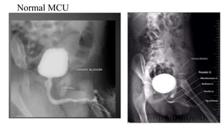
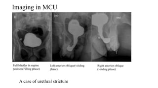
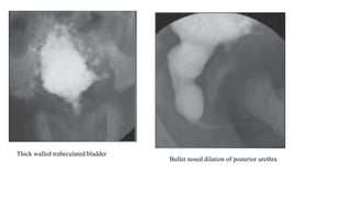
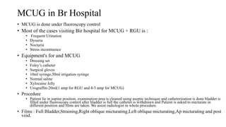
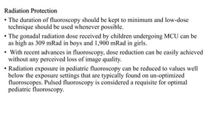
Radiographic round on
MCUG
By Sanju Timilsina
B.Sc,MIT 3rd year
What is MCUG?
Anatomy
Indications
Urinary tract infection Dysuria
Urethral Diverticulum Vesico uretric reflux
Stress Incontinence
Contrast Media
HOCM or LOCM 200 to 500 ml diluted with normal saline in ratio of 1:10/1:20
Equipments
 Fluoroscopy unit with spot film device and tilting table
 Video recorder
 Jaques or foleycatheter
 In small infants a fine (5-7F) feeding tube is adequate
 Syringes
 Xylocaine jelly 1-2%
 Aseptic tray
Procedure
• Under aseptic conditions, catheterize the urinary bladder with patient in
supine position.
• Push the diluted contrast medium slowly under fluoroscopic guidance
• Ask the patient to inform you when he has urge to micturate
• Ask the patient to micturate in a urine receiver in an erect oblique
position
• Spot images are taken during micturition in right and left oblique
projections and any reflux is recorded
• Finally,a full length view of abdomen is taken to demonstrate any
undetected reflux of the contrast medium that might have occurred in
kidneys and to record the post micturationresidue
Complications
• Contrast reaction
• Contrast induced cystitis
• UTI
• Catheter Trauma
• Bladder perforation-due to overfilling of contrast
• Retention of Foley’s catheter
After Care
• Patient should be warned of rare dysuria and retention of urine
• In case of reflux- antibiotics are to be prescribed
Normal MCU
Imaging in MCU
A case of urethral stricture
Full bladder in supine
position(Filling phase)
Left anterior oblique(voiding
phase)
Right anterior oblique
(voiding phase)
Thick walled trabeculated bladder
Bullet nosed dilation of posterior urethra
Vesico uretric reflux
• Refers to retrograde passage of urine from bladder into the ureter and
often into the cycles
• Most significant risk factor for childhood renal scarring and its sequalae
• VUR in most cases is the result of primary maturation abnormality of
the vesicoureteral junction resulting in short distal ureteric submucosal
tunnel
• Imaging of VUR
• MCUG
• Radionuclide cystography
• MR voiding cystography
Grading of VUR in radiographic image
Grade 1 Grade 2 Grade 3 Grade 4
Grade 5
Strenghts
• MCUG is superior method for demonstrating the anatomical detail of male
urethra,the bladder and vesicoureteric junction.Calyceal anatomy may be seen if
VUR is present; sometimes this is only opportunity to visualize calyces if child is
in renal failure
Weakness
• An MCUG requires bladder catheterization and entails high radiation burden to
gonads,especially in girls.with increasing availability of digital
equipment,particularly when combined with pulsed
fluoroscopy,however,absorbed doses from conventional MCUG can be reduced
provided screening times can be kept to minimum
MCUG in Br Hospital
• MCUG is done under fluoroscopy control
• Most of the cases visiting Bir hospital for MCUG + RGU is :
• Frequent Urination
• Dysuria
• Nocturia
• Stress incontinence
• Equipment's for and MCUG
• Dressing set
• Foley’s catheter
• Surgical gloves
• 10ml syringe,50ml irrigation syringe
• Normal saline
• Xylocaine Jelly
• Urograffin-20ml(1 amp for RGU and 4-5 amp for MCUG)
• Procedure
• Patient lie in supine position, examination area is cleaned using aseptic technique and catheterization is done bladder is
filled under fluoroscopy control after bladder is full the catheter is withdrawn and Patient is asked to micturate in
different position and films are taken. We assist radiologist in whole procedure.
• Films : Full Bladder,Straining,Right oblique micturating,Left oblique micturating,Ap micturating and post
void.
Radiation Protection
• The duration of fluoroscopy should be kept to minimum and low-dose
technique should be used whenever possible.
• The gonadal radiation dose received by children undergoing MCU can be
as high as 309 mRad in boys and 1,900 mRad in girls.
• With recent advances in fluoroscopy, dose reduction can be easily achieved
without any perceived loss of image quality.
• Radiation exposure in pediatric fluoroscopy can be reduced to values well
below the exposure settings that are typically found on un-optimized
fluoroscopes. Pulsed fluoroscopy is considered a requisite for optimal
pediatric fluoroscopy.
References
• Diagnostic radiology text book of medical imaging by Grainger &
Allison’s fourth edition volume 2
• A Guide to Radiological Procedures, Chapman & Nakielny - 4th
Edition.
• Clark’s Special Procedure.
• http://www.sjkdt.org/article.asp?issn=1319-
2442;year=2008;volume=19;issue=1;spage=20;epage=25;aulast=Al-
Imam
• https://www.slideshare.net/shubhamxsinghal/rgu-mcu?qid=27fbe1f9-
28de-4219-aa20-3e08ba555e61&v=&b=&from_search=1
Radiographic round on mcug

Radiographic round on mcug

  • 1.
    Radiographic round on MCUG BySanju Timilsina B.Sc,MIT 3rd year
  • 2.
  • 3.
  • 5.
    Indications Urinary tract infectionDysuria Urethral Diverticulum Vesico uretric reflux Stress Incontinence
  • 6.
    Contrast Media HOCM orLOCM 200 to 500 ml diluted with normal saline in ratio of 1:10/1:20
  • 7.
    Equipments  Fluoroscopy unitwith spot film device and tilting table  Video recorder  Jaques or foleycatheter  In small infants a fine (5-7F) feeding tube is adequate  Syringes  Xylocaine jelly 1-2%  Aseptic tray
  • 8.
    Procedure • Under asepticconditions, catheterize the urinary bladder with patient in supine position. • Push the diluted contrast medium slowly under fluoroscopic guidance • Ask the patient to inform you when he has urge to micturate • Ask the patient to micturate in a urine receiver in an erect oblique position • Spot images are taken during micturition in right and left oblique projections and any reflux is recorded • Finally,a full length view of abdomen is taken to demonstrate any undetected reflux of the contrast medium that might have occurred in kidneys and to record the post micturationresidue
  • 9.
    Complications • Contrast reaction •Contrast induced cystitis • UTI • Catheter Trauma • Bladder perforation-due to overfilling of contrast • Retention of Foley’s catheter After Care • Patient should be warned of rare dysuria and retention of urine • In case of reflux- antibiotics are to be prescribed
  • 10.
  • 11.
    Imaging in MCU Acase of urethral stricture Full bladder in supine position(Filling phase) Left anterior oblique(voiding phase) Right anterior oblique (voiding phase)
  • 12.
    Thick walled trabeculatedbladder Bullet nosed dilation of posterior urethra
  • 13.
    Vesico uretric reflux •Refers to retrograde passage of urine from bladder into the ureter and often into the cycles • Most significant risk factor for childhood renal scarring and its sequalae • VUR in most cases is the result of primary maturation abnormality of the vesicoureteral junction resulting in short distal ureteric submucosal tunnel • Imaging of VUR • MCUG • Radionuclide cystography • MR voiding cystography
  • 14.
    Grading of VURin radiographic image Grade 1 Grade 2 Grade 3 Grade 4 Grade 5
  • 15.
    Strenghts • MCUG issuperior method for demonstrating the anatomical detail of male urethra,the bladder and vesicoureteric junction.Calyceal anatomy may be seen if VUR is present; sometimes this is only opportunity to visualize calyces if child is in renal failure Weakness • An MCUG requires bladder catheterization and entails high radiation burden to gonads,especially in girls.with increasing availability of digital equipment,particularly when combined with pulsed fluoroscopy,however,absorbed doses from conventional MCUG can be reduced provided screening times can be kept to minimum
  • 16.
    MCUG in BrHospital • MCUG is done under fluoroscopy control • Most of the cases visiting Bir hospital for MCUG + RGU is : • Frequent Urination • Dysuria • Nocturia • Stress incontinence • Equipment's for and MCUG • Dressing set • Foley’s catheter • Surgical gloves • 10ml syringe,50ml irrigation syringe • Normal saline • Xylocaine Jelly • Urograffin-20ml(1 amp for RGU and 4-5 amp for MCUG) • Procedure • Patient lie in supine position, examination area is cleaned using aseptic technique and catheterization is done bladder is filled under fluoroscopy control after bladder is full the catheter is withdrawn and Patient is asked to micturate in different position and films are taken. We assist radiologist in whole procedure. • Films : Full Bladder,Straining,Right oblique micturating,Left oblique micturating,Ap micturating and post void.
  • 17.
    Radiation Protection • Theduration of fluoroscopy should be kept to minimum and low-dose technique should be used whenever possible. • The gonadal radiation dose received by children undergoing MCU can be as high as 309 mRad in boys and 1,900 mRad in girls. • With recent advances in fluoroscopy, dose reduction can be easily achieved without any perceived loss of image quality. • Radiation exposure in pediatric fluoroscopy can be reduced to values well below the exposure settings that are typically found on un-optimized fluoroscopes. Pulsed fluoroscopy is considered a requisite for optimal pediatric fluoroscopy.
  • 18.
    References • Diagnostic radiologytext book of medical imaging by Grainger & Allison’s fourth edition volume 2 • A Guide to Radiological Procedures, Chapman & Nakielny - 4th Edition. • Clark’s Special Procedure. • http://www.sjkdt.org/article.asp?issn=1319- 2442;year=2008;volume=19;issue=1;spage=20;epage=25;aulast=Al- Imam • https://www.slideshare.net/shubhamxsinghal/rgu-mcu?qid=27fbe1f9- 28de-4219-aa20-3e08ba555e61&v=&b=&from_search=1

Editor's Notes

  • #6 Contraindication say myself
  • #8 Patient preparation- patient is asked to micturate prior to examination
  • #13 Case of posterior urethral valves
  • #14 Primary diagnosticprocedure for evaluation of VUJ is MCUG However radionuclidecystography is better as a screening tool as the radiation dose is lower
  • #17 Fluoroscopy unit here is of Shimadzu with rotating anode over couch tube with maximum Kvp of 150 and maximum mA of 800 but working range of mA is 500 and mA used during fluoroscopy is 2-5mA