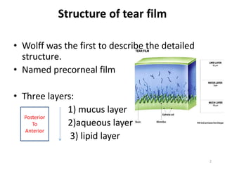
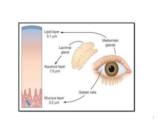
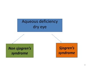
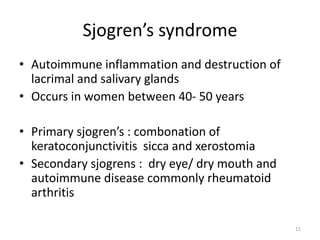
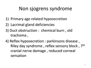
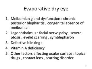
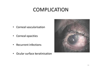
TEAR FILM
AND
DRY EYEPRESENTER: SURAJKCMBBS
1
Structure of tear film
• Wolff was the first to describe the detailed
structure.
• Named precorneal film
• Three layers:
1) mucus layer
2)aqueous layer
3) lipid layer
Posterior
To
Anterior
2
3
Layers of tear film
1. Mucus layer.
• innermost and thinnest stratum of the tear
film.
• consists of mucin secreted by conjunctival
goblet cells and glands of Manz.
• converts the hydrophobic corneal surface into
hydrophilic .
• Helps in lubrication
4
Layers of tear film
2 . Aqueous layer:
• intermediate layer which consists of tears
secreted by the main and accessory lacrimal
glands.
• The tears mainly comprise of water and small
quantities of solutes such as sodium chloride,
sugar,urea and proteins.
• It also contains antibacterial substances like
lysozyme, betalysin and lactoferrin.
5
Layers of tear film
3 Lipid or oily layer:
• This is the outermost layer of tear film formed
at air-tear interface
• Secreted by : Meibomian, Zeis, and Moll
glands.
• prevents the overflow of tears, retards their
evaporation and lubricates the eyelids
6
Functions of tear film
• 1. Keeps the cornea and conjunctiva moist.
• 2. It provides oxygen to the corneal
epithelium.
• 3. Washes away debris and noxious irritants.
• 4. Prevents infection due to presence of
antibacterialsubstances.
• 5. Facilitates movements of the lids over the
globe.
7
Dry eye
Dry eye occurs when there is inadequate tear
volume or function resulting in an unstable tear
film and ocular surface disease.
1 Keratoconjunctivitis sicca (KCS) refers to any eye
with some degree of dryness
2 Xerophthalmia describes a dry eye associated
with vitamin A deficiency.
3 Xerosis refers to the extreme ocular dryness
and keratinization that occurs in eyes with severe
conjunctival cicatrization.
8
ETIOLOGY
 Accroding to International Dry Eye Workshop
report 2007 , causes can be divided into :
1) Aqueous deficiency dry eye
2) Evaporative dry eye
9
Aqueous deficiency
dry eye
Sjogren’s
syndrome
Non sjogren’s
syndrome
10
Sjogren’s syndrome
• Autoimmune inflammation and destruction of
lacrimal and salivary glands
• Occurs in women between 40- 50 years
• Primary sjogren’s : combonation of
keratoconjunctivitis sicca and xerostomia
• Secondary sjogrens : dry eye/ dry mouth and
autoimmune disease commonly rheumatoid
arthritis
11
Non sjogrens syndrome
1) Primary age related hyposecretion
2) Lacrimal gland deficiencies
3) Duct obstruction : chemical burn , old
trachoma ,
4) Reflex hyposecretion : parkinsons disease ,
Riley day syndrome , reflex sensory block , 7th
cranial nerve damage , reduced corneal
sensation
12
Evaporative dry eye
1. Meibomian gland dysfunction : chronic
posterior blepharitis , congenital absence of
meibomian
2. Lagophthalmus : facial nerve palsy , severe
ptosis , eyelid scarring , symblepharon
3. Defective blinking :
4. Vitamin A deficiency
5. Other factors affecting ocular surface : topical
drugs , contact lens , scarring disorder
13
Clinical features
 Irritation
 foreign body sensation
Itching
Non specific ocular discomfort
Feeling of dryness
14
Clinical features
Signs :
Tear film signs : stingy mucous , marginal tear
strip is reduced, froth in the tear
Conjunctival sign : lustureless , mild
congestion , xerosis and keratinisation may
occur
Corneal signs : punctuate epithelial erosion ,
filaments or mucus plaques ,
Signs of causative disease :
15
Conjunctival
Signs
Hyperemia Bitot’s spots
Symblepheron
16
CORNEAL
SIGNS
17
Approach to dry eye
1 ) HISTORY
2) PHYSICAL EXAMINATION
3) INVESTIGATIONS
18
1. Tear film break-up (BUT).
 Interval between a complete blink and appearance
of first randomly distributed dry spot on the cornea.
 Noted after instilling a drop of fluorescein and
examining in acobalt-blue light of a slit-lamp.
 Indicator of adequacy of mucin component of tears.
 Normal values : 15 to 35 seconds.
 Values less than 10 seconds imply an unstable tear
film
19
TEAR BREAK UP TIME
15-34 sec Normal
<10 sec Abnormal
5-9 sec Borderline Dry eye
<5 sec Severe Dry Eye
20
Schirmer-I test
 measures total tear secretions.
 performed with the help of a 5 × 35 mm strip of
Whatman-41 filter paper which is folded 5 mm
from one end and kept in the lower fornix at the
junction of lateral one-third and medial two-
third.
Normal values of Schirmer-I test are more than
15 mm.
 Values of 5-10 mm are suggestive of moderate to
mild keratoconjunctivitis sicca (KCS) and less than
5 mm of severe KCS.
21
23
3. Rose Bengal staining
PATTERN INDICATION
C mild or
early cases with fine punctate stains
B moderate cases with
extensive staining
A severe cases with
confluent staining of conjunctiva and
cornea
24
25
Treatment
1) Supplementation with tear substitute :
 available in drop , ointment
slow release inserts
 most available tear drop contains
either cellulose derivatives
( 0.25 – 0.7 methy cellulose )
or polyvinyl alcohol
26
2) Topical cyclosporine (0.05% , 0.1% ) :
reduces cell mediated inflammation of lacrimal
tissues
3) Mucolytics : 5% acetyl cystine
disperses the mucus thread and decreases the
tear viscosity
27
4) Preserving the existing tears :
reducing the
evaporation
decreasing the drainage
( punctal occlusion )
Decreasing room temperature Collagen implants
Use of moist chamber and protective
glass
Cyanoacrylate tissue adhesives ,
electrocauterisation
Argon laser occlusion
Surgical occlusion
28
29
5) Treatment of causative disease :
30
COMPLICATION
• Corneal vascularisation
• Corneal opacities
• Recurrent infections
• Ocular surface keratinisation
31
REFERENCES
• A K khurana comprehensive ophthalmology
6TH
• Kanski clinical ophthalmology 7th edition
• Color atlas of ophthalmology
32
THANKYOU
33

Dry eye final

  • 1.
  • 2.
    Structure of tearfilm • Wolff was the first to describe the detailed structure. • Named precorneal film • Three layers: 1) mucus layer 2)aqueous layer 3) lipid layer Posterior To Anterior 2
  • 3.
  • 4.
    Layers of tearfilm 1. Mucus layer. • innermost and thinnest stratum of the tear film. • consists of mucin secreted by conjunctival goblet cells and glands of Manz. • converts the hydrophobic corneal surface into hydrophilic . • Helps in lubrication 4
  • 5.
    Layers of tearfilm 2 . Aqueous layer: • intermediate layer which consists of tears secreted by the main and accessory lacrimal glands. • The tears mainly comprise of water and small quantities of solutes such as sodium chloride, sugar,urea and proteins. • It also contains antibacterial substances like lysozyme, betalysin and lactoferrin. 5
  • 6.
    Layers of tearfilm 3 Lipid or oily layer: • This is the outermost layer of tear film formed at air-tear interface • Secreted by : Meibomian, Zeis, and Moll glands. • prevents the overflow of tears, retards their evaporation and lubricates the eyelids 6
  • 7.
    Functions of tearfilm • 1. Keeps the cornea and conjunctiva moist. • 2. It provides oxygen to the corneal epithelium. • 3. Washes away debris and noxious irritants. • 4. Prevents infection due to presence of antibacterialsubstances. • 5. Facilitates movements of the lids over the globe. 7
  • 8.
    Dry eye Dry eyeoccurs when there is inadequate tear volume or function resulting in an unstable tear film and ocular surface disease. 1 Keratoconjunctivitis sicca (KCS) refers to any eye with some degree of dryness 2 Xerophthalmia describes a dry eye associated with vitamin A deficiency. 3 Xerosis refers to the extreme ocular dryness and keratinization that occurs in eyes with severe conjunctival cicatrization. 8
  • 9.
    ETIOLOGY  Accroding toInternational Dry Eye Workshop report 2007 , causes can be divided into : 1) Aqueous deficiency dry eye 2) Evaporative dry eye 9
  • 10.
  • 11.
    Sjogren’s syndrome • Autoimmuneinflammation and destruction of lacrimal and salivary glands • Occurs in women between 40- 50 years • Primary sjogren’s : combonation of keratoconjunctivitis sicca and xerostomia • Secondary sjogrens : dry eye/ dry mouth and autoimmune disease commonly rheumatoid arthritis 11
  • 12.
    Non sjogrens syndrome 1)Primary age related hyposecretion 2) Lacrimal gland deficiencies 3) Duct obstruction : chemical burn , old trachoma , 4) Reflex hyposecretion : parkinsons disease , Riley day syndrome , reflex sensory block , 7th cranial nerve damage , reduced corneal sensation 12
  • 13.
    Evaporative dry eye 1.Meibomian gland dysfunction : chronic posterior blepharitis , congenital absence of meibomian 2. Lagophthalmus : facial nerve palsy , severe ptosis , eyelid scarring , symblepharon 3. Defective blinking : 4. Vitamin A deficiency 5. Other factors affecting ocular surface : topical drugs , contact lens , scarring disorder 13
  • 14.
    Clinical features  Irritation foreign body sensation Itching Non specific ocular discomfort Feeling of dryness 14
  • 15.
    Clinical features Signs : Tearfilm signs : stingy mucous , marginal tear strip is reduced, froth in the tear Conjunctival sign : lustureless , mild congestion , xerosis and keratinisation may occur Corneal signs : punctuate epithelial erosion , filaments or mucus plaques , Signs of causative disease : 15
  • 16.
  • 17.
  • 18.
    Approach to dryeye 1 ) HISTORY 2) PHYSICAL EXAMINATION 3) INVESTIGATIONS 18
  • 19.
    1. Tear filmbreak-up (BUT).  Interval between a complete blink and appearance of first randomly distributed dry spot on the cornea.  Noted after instilling a drop of fluorescein and examining in acobalt-blue light of a slit-lamp.  Indicator of adequacy of mucin component of tears.  Normal values : 15 to 35 seconds.  Values less than 10 seconds imply an unstable tear film 19
  • 20.
    TEAR BREAK UPTIME 15-34 sec Normal <10 sec Abnormal 5-9 sec Borderline Dry eye <5 sec Severe Dry Eye 20
  • 21.
    Schirmer-I test  measurestotal tear secretions.  performed with the help of a 5 × 35 mm strip of Whatman-41 filter paper which is folded 5 mm from one end and kept in the lower fornix at the junction of lateral one-third and medial two- third. Normal values of Schirmer-I test are more than 15 mm.  Values of 5-10 mm are suggestive of moderate to mild keratoconjunctivitis sicca (KCS) and less than 5 mm of severe KCS. 21
  • 22.
  • 23.
    3. Rose Bengalstaining PATTERN INDICATION C mild or early cases with fine punctate stains B moderate cases with extensive staining A severe cases with confluent staining of conjunctiva and cornea 24
  • 24.
  • 25.
    Treatment 1) Supplementation withtear substitute :  available in drop , ointment slow release inserts  most available tear drop contains either cellulose derivatives ( 0.25 – 0.7 methy cellulose ) or polyvinyl alcohol 26
  • 26.
    2) Topical cyclosporine(0.05% , 0.1% ) : reduces cell mediated inflammation of lacrimal tissues 3) Mucolytics : 5% acetyl cystine disperses the mucus thread and decreases the tear viscosity 27
  • 27.
    4) Preserving theexisting tears : reducing the evaporation decreasing the drainage ( punctal occlusion ) Decreasing room temperature Collagen implants Use of moist chamber and protective glass Cyanoacrylate tissue adhesives , electrocauterisation Argon laser occlusion Surgical occlusion 28
  • 28.
  • 29.
    5) Treatment ofcausative disease : 30
  • 30.
    COMPLICATION • Corneal vascularisation •Corneal opacities • Recurrent infections • Ocular surface keratinisation 31
  • 31.
    REFERENCES • A Kkhurana comprehensive ophthalmology 6TH • Kanski clinical ophthalmology 7th edition • Color atlas of ophthalmology 32
  • 32.