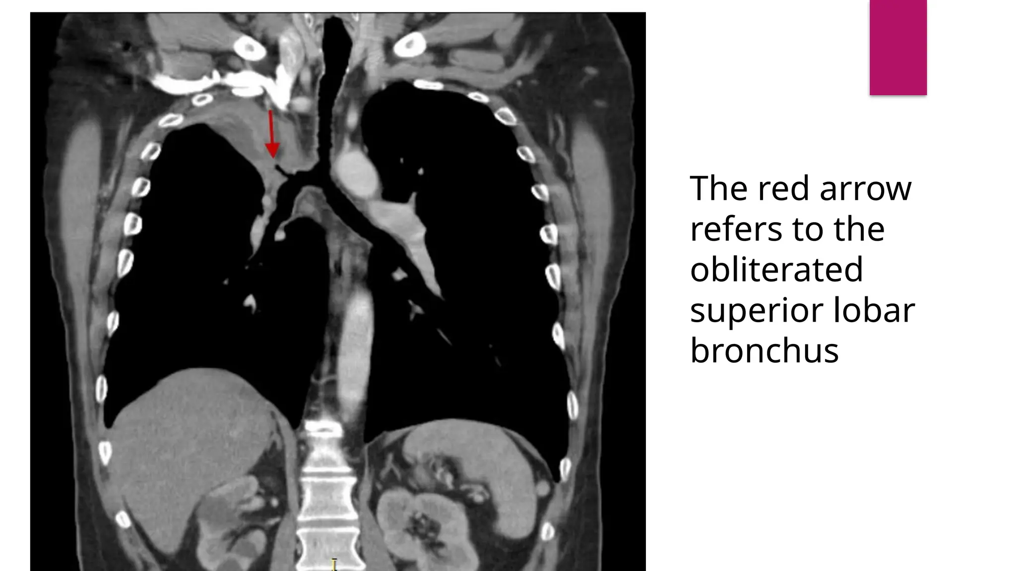
TUMORS OF LUNGS
PREPARED BY. DR USMAN JAVED
PGR DIAGNOSTIC RADIOLOGY
DIAGNOSTIC IMAGING AND
CARCINOMA OF LUNGS
 Imaging makes an important contribution to three aspects of the
managementt of lung cancer. These are
1. Making the diagnosis
2. Staging the tumou
3. Assessingg treatment.
Classification of tumors of lungs
 I . Squamous cell (or epidermoid ) carcinoma,
30-35% of cases of primary lung cancer
 2. Adenocarcinoma (including alveolar cell carcinoma)
30-35%; of cases
 3. Large cell undifferentiated
15-20% of cases
 4. Small (oat) cell carcinoma,
20-25% of cases.
 Some lung cancers do not fall neatly into one of these categories, eg, adenosquamous
carcinoma.
RADIOLOGICAL FEATURES
 The radiological features oh lung cancer are a reflection of the pathologyy, and depends
upon the size and site of the tumour.
1. Hilar enlargement
2. Airway obstruction
3. Peripheral mass
4. Mediastinal involvement
5. Pleural involvement
6. Bone involvement
Hilar enlargement
 common radiographic manifestation of lung cancer.
 if the primary tumour is central, this represents the tumour itself.
 If the tumour is peripheral, it represents metastasis to bronchopulmonary
lymph nodes and the primary tumor may or may not be visible.
 Some times hilar involvement Is minor And presents as increased density of
the hilum rather than as enlargement.
Airway obstruction
 Bronchial narrowing due to tumour growth eventually causes collapse of
the lung distal to the tumour. Which may be
1. Segmental collapse
2. Lobar Collapse
3. Entire lung (less often)
 Prior to collapse of a lobe or segment, infection may deyelop distal to the bronchial obstruction.
Consequently, segmental or lobar consolidation may be a manifestation of lung cancer,
 Diffuse or central Calcification in a peripheral Mass is very suggestive of a benign lesion.
Peripheral mass
 peripheral pulmonary mass in the chest X-ray is a common presentation of
lung cancer
 There are no radiological features that can reliably differentiate between a
benign and a malignant pulmonary nodule or mass. However , malignant
tumours are usually larger than benign lesions at the time of presentation.
 peripheral Iung cancers tend to have poorly defined lobulated or
umbilieated margins.
Mediastinal involvement
 Enlargement of mediastinal lymph nodes is a typical feature of smalll cell
tumours, but also occurs with other bronchial carcinomas. The
mediastinum appears widened and may have a lobulated outline.
 Enlarged mediastinal lymph nodes or central tumours may distort the
oesophagus, Barium swallow may therefore be used to assess the
mediastinum- and is essential in patients with dysphagia.
 Mediastinal involvement can also cause Superior vena cava obstruction
that can be assessed by Vanacavography and dynamically enhanced CT
or MRI.
 Invasion of the pericardium by metastatic lymph nodes
or the primary tumour itself may result in pericarditis and
pericardial effusion.
Pleural involvement
 Pleural effusion may be due to
1. Direct spread of tumor
2. Lymphatic obstruction
3. Secondary to obstructive pneumonitis
4. sympathetic response to the tumour,
 Rarely a cavitating subpleural tu.mour will cause a spontaneous pneumothorax
Bone involvement
 Peripheral carcinomas may invade the ribs or spine directly.
 Haematogenous metastases from lung to bone are usually osteolytic.
 They are often painful, and are identified earliest by isotope bone scan.
 On plain films the affected bones show well defined periosteal new bone
formation.
 Isotope bone scan may be Positivee before radiographic changes are
visible.
Making diagnosis
 Sputum cytology an bronchoscopic biopsies or washings usually provide the cell type of
central tumours, but peripheral tumours may require percutaneous biopsy. This may be done
with fluoroscopic, CT or ultrasound guidance.
 Comparison with previous imaging is invaluable, as a nodule that has not changed on the
chest radiograph over 2 years is likely to be benign.
 Recentl developed strategics to differentiate benign from malignant tumours include CT
densitometry and positron emission tomography (PET using18F-fluorodeoxyglucosee ( FDG ).
 Compared to benign nodules, malignantt nodules show a greater degree of enhancement
following intravenous injection of iodinated contrast medium, such that an increase in
attenuation on CT scanning of greater than 20 Hounsfield units is very suggestive of
malignancy
Staging the tumour
 The main purposes of accurate staging of lung cancer are:
1. To identify those patients with non-small cell tumours who wil Benefit from surger
2. To avoid surgery in those who will not benefit, and
3. To Provide accurate data for assessing and comparing differentMethods of treatment.
Adenocarcinoma
 Adenocarcinoma of the lung is the most common
histologic type of lung cancer. Grouped under the
non-small cell carcinomas of the lung, it is a malignant
tumour with glandular differentiation or mucin
production expressing in different patterns and
degrees of differentiation.
 Often it is impossible to radiographically distinguish between other
histological lung cancer types.
 lung nodule is a rounded or irregular region of increased attenuation. The
amount of attenuation can further classify the nodules as either
ground glass, subsolid or solid.
Ill-defined
opacity in the
medial right
upper zone.
Cardiomediastin
al contour is
unremarkable
Squamous cell carcinoma
 0-35% of all lung cancers
 In most instances are due to heavy smoking.
 may be the commonest type accounting for Pancoast tumours.
 traditionally known to arise centrally (66-90%), the incidence of peripherally located SCC is
increasing.
Radiographic features
 Chest radiograph
 The appearance depends on the location of the lesion.
 The more central lesions may merely appear as a bulky hilum, representing the tumour and local
nodal involvement.
 Lobar collapse may be seen due to obstruction of a bronchus. When the right upper lobe is collapsed
and a hilar mass is present, this is known as the Golden S sign.
 A more peripheral location may appear as a rounded or spiculated mass.
 Cavitation may be seen as an air-fluid level.
 Chest wall invasion is difficult to identify on plain films unless there is the destruction of the adjacent
rib or evidence of soft tissue growing into the soft tissues superficial to the ribs.
 Pleural effusion may also be seen.
CT
 CT is the modality of choice for the evaluation of possible lung cancer.
 Central SCC often results in intraluminal obstruction and causes lung
collapse and/or obstructive pneumonitis.
 Peripheral SCC may be seen as a solid nodule/mass with or without an
irregular border. The irregular margin can be attributed to infiltrative
growth.
 Similar to central lung cancer, peripheral cancer can also result in
obstructive changes such as a mucocele.
 Cavitation is a frequent finding in primary lung SCC but can also be
encountered in metastatic SCC.
Chest x-ray
demonstrates
increased
density in the
right upper
medial
hemithorax with
loss of volume,
and shift of the
trachea to the
"Golden S sign" or
"reverse S sign of
Golden
 There is collapse of the right lower lobe, the
remainder of the lungs appear
unremarkable. There is mild deviation of the
distal trachea to the right and the right
paratracheal stripe appears enlarged. No
pneumothorax or subphrenic free Fluid.gas.
The red arrow
refers to the
obliterated
superior lobar
bronchus
Tumors of Lungs (Respiratory System) ppt

Tumors of Lungs (Respiratory System) ppt

  • 1.
    TUMORS OF LUNGS PREPAREDBY. DR USMAN JAVED PGR DIAGNOSTIC RADIOLOGY
  • 2.
    DIAGNOSTIC IMAGING AND CARCINOMAOF LUNGS  Imaging makes an important contribution to three aspects of the managementt of lung cancer. These are 1. Making the diagnosis 2. Staging the tumou 3. Assessingg treatment.
  • 3.
    Classification of tumorsof lungs  I . Squamous cell (or epidermoid ) carcinoma, 30-35% of cases of primary lung cancer  2. Adenocarcinoma (including alveolar cell carcinoma) 30-35%; of cases  3. Large cell undifferentiated 15-20% of cases  4. Small (oat) cell carcinoma, 20-25% of cases.  Some lung cancers do not fall neatly into one of these categories, eg, adenosquamous carcinoma.
  • 4.
    RADIOLOGICAL FEATURES  Theradiological features oh lung cancer are a reflection of the pathologyy, and depends upon the size and site of the tumour. 1. Hilar enlargement 2. Airway obstruction 3. Peripheral mass 4. Mediastinal involvement 5. Pleural involvement 6. Bone involvement
  • 5.
    Hilar enlargement  commonradiographic manifestation of lung cancer.  if the primary tumour is central, this represents the tumour itself.  If the tumour is peripheral, it represents metastasis to bronchopulmonary lymph nodes and the primary tumor may or may not be visible.  Some times hilar involvement Is minor And presents as increased density of the hilum rather than as enlargement.
  • 6.
    Airway obstruction  Bronchialnarrowing due to tumour growth eventually causes collapse of the lung distal to the tumour. Which may be 1. Segmental collapse 2. Lobar Collapse 3. Entire lung (less often)  Prior to collapse of a lobe or segment, infection may deyelop distal to the bronchial obstruction. Consequently, segmental or lobar consolidation may be a manifestation of lung cancer,  Diffuse or central Calcification in a peripheral Mass is very suggestive of a benign lesion.
  • 7.
    Peripheral mass  peripheralpulmonary mass in the chest X-ray is a common presentation of lung cancer  There are no radiological features that can reliably differentiate between a benign and a malignant pulmonary nodule or mass. However , malignant tumours are usually larger than benign lesions at the time of presentation.  peripheral Iung cancers tend to have poorly defined lobulated or umbilieated margins.
  • 8.
    Mediastinal involvement  Enlargementof mediastinal lymph nodes is a typical feature of smalll cell tumours, but also occurs with other bronchial carcinomas. The mediastinum appears widened and may have a lobulated outline.  Enlarged mediastinal lymph nodes or central tumours may distort the oesophagus, Barium swallow may therefore be used to assess the mediastinum- and is essential in patients with dysphagia.  Mediastinal involvement can also cause Superior vena cava obstruction that can be assessed by Vanacavography and dynamically enhanced CT or MRI.
  • 9.
     Invasion ofthe pericardium by metastatic lymph nodes or the primary tumour itself may result in pericarditis and pericardial effusion.
  • 10.
    Pleural involvement  Pleuraleffusion may be due to 1. Direct spread of tumor 2. Lymphatic obstruction 3. Secondary to obstructive pneumonitis 4. sympathetic response to the tumour,  Rarely a cavitating subpleural tu.mour will cause a spontaneous pneumothorax
  • 11.
    Bone involvement  Peripheralcarcinomas may invade the ribs or spine directly.  Haematogenous metastases from lung to bone are usually osteolytic.  They are often painful, and are identified earliest by isotope bone scan.  On plain films the affected bones show well defined periosteal new bone formation.  Isotope bone scan may be Positivee before radiographic changes are visible.
  • 12.
    Making diagnosis  Sputumcytology an bronchoscopic biopsies or washings usually provide the cell type of central tumours, but peripheral tumours may require percutaneous biopsy. This may be done with fluoroscopic, CT or ultrasound guidance.  Comparison with previous imaging is invaluable, as a nodule that has not changed on the chest radiograph over 2 years is likely to be benign.  Recentl developed strategics to differentiate benign from malignant tumours include CT densitometry and positron emission tomography (PET using18F-fluorodeoxyglucosee ( FDG ).  Compared to benign nodules, malignantt nodules show a greater degree of enhancement following intravenous injection of iodinated contrast medium, such that an increase in attenuation on CT scanning of greater than 20 Hounsfield units is very suggestive of malignancy
  • 13.
    Staging the tumour The main purposes of accurate staging of lung cancer are: 1. To identify those patients with non-small cell tumours who wil Benefit from surger 2. To avoid surgery in those who will not benefit, and 3. To Provide accurate data for assessing and comparing differentMethods of treatment.
  • 16.
    Adenocarcinoma  Adenocarcinoma ofthe lung is the most common histologic type of lung cancer. Grouped under the non-small cell carcinomas of the lung, it is a malignant tumour with glandular differentiation or mucin production expressing in different patterns and degrees of differentiation.
  • 17.
     Often itis impossible to radiographically distinguish between other histological lung cancer types.  lung nodule is a rounded or irregular region of increased attenuation. The amount of attenuation can further classify the nodules as either ground glass, subsolid or solid.
  • 18.
    Ill-defined opacity in the medialright upper zone. Cardiomediastin al contour is unremarkable
  • 19.
    Squamous cell carcinoma 0-35% of all lung cancers  In most instances are due to heavy smoking.  may be the commonest type accounting for Pancoast tumours.  traditionally known to arise centrally (66-90%), the incidence of peripherally located SCC is increasing.
  • 20.
    Radiographic features  Chestradiograph  The appearance depends on the location of the lesion.  The more central lesions may merely appear as a bulky hilum, representing the tumour and local nodal involvement.  Lobar collapse may be seen due to obstruction of a bronchus. When the right upper lobe is collapsed and a hilar mass is present, this is known as the Golden S sign.  A more peripheral location may appear as a rounded or spiculated mass.  Cavitation may be seen as an air-fluid level.  Chest wall invasion is difficult to identify on plain films unless there is the destruction of the adjacent rib or evidence of soft tissue growing into the soft tissues superficial to the ribs.  Pleural effusion may also be seen.
  • 21.
    CT  CT isthe modality of choice for the evaluation of possible lung cancer.  Central SCC often results in intraluminal obstruction and causes lung collapse and/or obstructive pneumonitis.  Peripheral SCC may be seen as a solid nodule/mass with or without an irregular border. The irregular margin can be attributed to infiltrative growth.  Similar to central lung cancer, peripheral cancer can also result in obstructive changes such as a mucocele.  Cavitation is a frequent finding in primary lung SCC but can also be encountered in metastatic SCC.
  • 22.
    Chest x-ray demonstrates increased density inthe right upper medial hemithorax with loss of volume, and shift of the trachea to the "Golden S sign" or "reverse S sign of Golden
  • 24.
     There iscollapse of the right lower lobe, the remainder of the lungs appear unremarkable. There is mild deviation of the distal trachea to the right and the right paratracheal stripe appears enlarged. No pneumothorax or subphrenic free Fluid.gas.
  • 25.
    The red arrow refersto the obliterated superior lobar bronchus