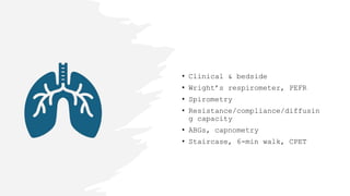
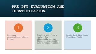
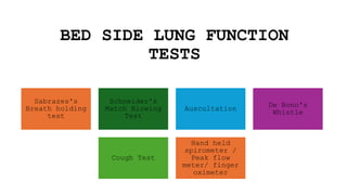
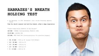
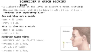
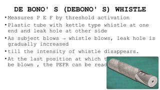
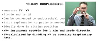
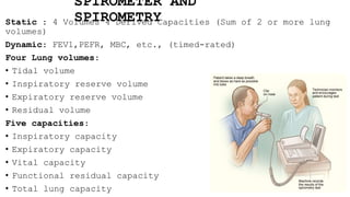
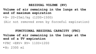
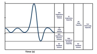
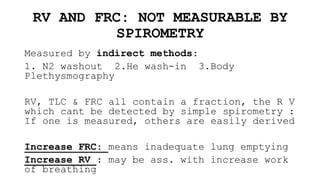
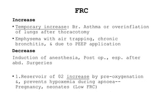
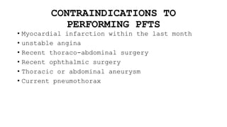
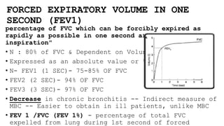
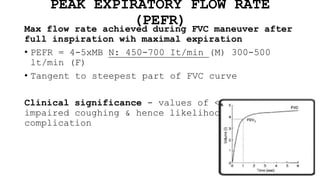
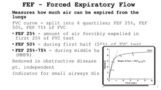
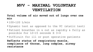
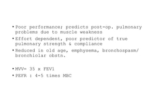
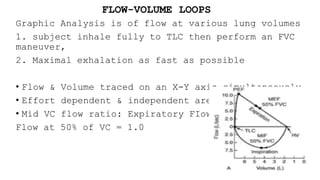
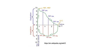
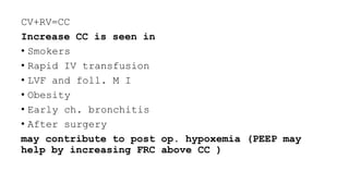
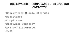
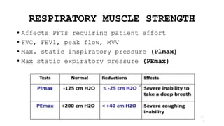
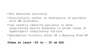
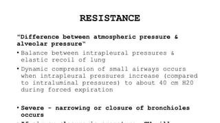
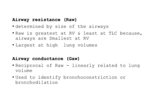
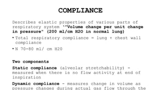
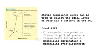
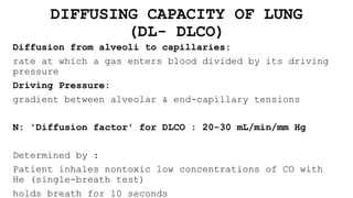
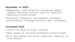
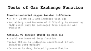
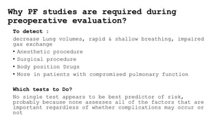
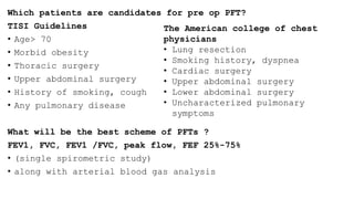
Pulmonary
Function Test
INDICATIONS
DETECT DISEASE :
IDENTIFICATION
OF TYPE OF LUNG
DISEASE -
OBSTRUCTIVE VS.
RESTRICTIVE
QUANTIFICATION
OF THE EXTENT OF
LUNG DISEASE
MONITORING RATE
OF PROGRESSION
DETERMINATION OF
THE RESPONSE TO
THERAPY
ASSESS RISK FOR
SURGICAL
PROCEDURES
EVALUATION FOR
WEANING FROM A
VENTILATOR
• Clinical & bedside
• Wright’s respirometer, PEFR
• Spirometry
• Resistance/compliance/diffusin
g capacity
• ABGs, capnometry
• Staircase, 6-min walk, CPET
PRE PFT EVALUATION AND
IDENTIFICATION
History,
examination, chest
X ray
1
Chest x-ray film :
Marker for
clinically severe
disease, especially
lung hyperinflation
2
Basic Bed Side Lung
Function Tests
3
BED SIDE LUNG FUNCTION
TESTS
Sabrazes's
Breath holding
test
Schneider's
Match Blowing
Test
Auscultation
De Bono's
Whistle
Cough Test
Hand held
spirometer /
Peak flow
meter/ finger
oximeter
SABRAZES'S BREATH
HOLDING TEST
• By Sabrazes, in 1902 (Bordeaux): also called Voluntary Apnoeic
Pause
Time for which a person can hold his breath, after a deep inspiration
"Cardio-respiratory Reserve of the Patient“
>25 SEC.- NORMAL Cardiopulmonary Reserve (CPR)
15-25 SEC- LIMITED CPR
<15 SEC- VERY POOR CPR (Contraindication for elective surgery)
• 25- 30 SEC - 3500 ml VC (normal-3100-4800ml)
• 20 – 25 SEC - 3000 ml VC
• 15 - 20 SEC - 2500 ml VC
• 10 - 15 SEC - 2000 ml VC
• 5 - 10 SEC - 1500 ml VC
SCHNEIDER'S MATCH BLOWING
TEST
• A lighted candle at the level of patient's mouth (sitting)
• Minimum Distance, ability to blow it off: 25 cm. <15 cm -
"Reduced Peak Expiratory Flow"
Can not blow out a match
• MBC < 60 L/min
• FEV1 < 1.6L
Able to blow out a match
• MBC > 60 L/min
• FEV1 > 1.6L
MODIFIED MATCH TEST:
• DISTANCE MBC (N-150-175 L/min)
• 9”inch >150 L/MIN.
• 6”inch >60 L/MIN.
• 3”inch > 40 L/MIN.
DE BONO' S (DEBONO' S) WHISTLE
• Measures P E F by threshold activation
• Plastic tube with kettle type whistle at one
end and leak hole at other side
• As subject blows → whistle blows, leak hole is
gradually increased
• till the intensity of whistle disappears.
• At the last position at which the whistle can
be blown , the PEFR can be read off the scale
COUGH TEST: DEEP BREATH F/BY COUGH
• ABILITY TO COUGH
• STRENGTH
• EFFECTIVENESS
-VC ~ 3 TIMES TV FOR EFFECTIVE COUGH.
• A wet productive cough / self propagated paroxysms of
coughing – patient susceptible for pulmonary Complication.
AUSCULATION (WHEEZE TEST)
• Patient asked to take 5 deep breaths, then auscultated to
check presence or absence of wheeze.
WRIGHT RESPIROMETER
• measures TV, MV
• Simple and rapid
• Can be connected to endotracheal tube or face mask
• Prior explanation to patients needed.
• Ideally done in sitting position
• MV- instrument records for 1 min and reads directly.
• TV-calculated by dividing MV by counting Respiratory
Rate.
SPIROMETER AND
SPIROMETRY
Static : 4 Volumes 4 Derived Capacities (Sum of 2 or more lung
volumes)
Dynamic: FEV1,PEFR, MBC, etc., (timed-rated)
Four Lung volumes:
• Tidal volume
• Inspiratory reserve volume
• Expiratory reserve volume
• Residual volume
Five capacities:
• Inspiratory capacity
• Expiratory capacity
• Vital capacity
• Functional residual capacity
• Total lung capacity
TIDAL VOLUME (TV) AND MINUTE
VENTILATION
"Vol of air inspired expired during normal quiet
breathing“
• N : 500 ml (6-8ml /kg)
Minute Ventilation
TV X RR= Min. Ventilation = 3.7 – 20 L/min
(TV-VD) x RR= Alv.Ventilation= 2- 3.5 L/min/cm2
ALVEOLAR VENTILATION (VA)
That part of MV that takes part in gas exchange
• Affected by TV, RR, Vd and controls the excretion
of CO2
• VD : VD Anatomical / VD Physiological / VD
Aparatures
• Deep anesthesia, respiratory dep. drugs decrease
VA
• Hence, among other factors, this mandates an FiO2
of atleast 0.33
INSPIRATORY RESERVE VOLUME (IRV)
Maximum amount of air that can be inhaled
after a normal tidal volume inspiration (N:
1900-3300 ml)
EXPIRATORY RESERVE VOLUME (ERV)
Maximum amount of air that can be exhaled from
the resting expiratory level (N : 700- 1100
ml)
INSPIRATORY CAPACITY (IC)
Maximum amount of air that can be inhaled from
the end of a tidal volume (IRV+ TV) ( N: 2400-
RESIDUAL VOLUME (RV)
Volume of air remaining in the lungs at the
end of maximum expiration
• N- 20-25ml/kg (1200-1500)
(Air not removed even by forceful expiration)
FUNCTIONAL RESIDUAL CAPACITY (FRC)
Volume of air remaining in the lungs at the
end of a TV expiration
• FRC =ERV+ RV= 1100+1200
• N: 2300 ml
RV AND FRC: NOT MEASURABLE BY
SPIROMETRY
Measured by indirect methods:
1. N2 washout 2.He wash-in 3.Body
Plethysmography
RV, TLC & FRC all contain a fraction, the R V
which cant be detected by simple spirometry :
If one is measured, others are easily derived
Increase FRC: means inadequate lung emptying
Increase RV : may be ass. with increase work
of breathing
FRC
Increase
• Temporary increase: Br. Asthma or overinflation
of lungs after thoracotomy
• Emphysema with air trapping, chronic
bronchitis, & due to PEEP application
Decrease
Induction of anesthesia, Post op., esp. after
abd. Surgeries
• 1.Reservoir of 02 increase by pre-oxygenation
&, prevents hypoxemia during apnoea--
Pregnancy, neonates (Low FRC)
TOTAL LUNG CAPACITY (TLC)
Volume of air in the lungs after a maximum
inspiration
• TLC = IRV + TV + ERV + RV
• 3000 + 500 + 1100 + 1200 = 5800 ml (80-100
ml/kg)
VITAL CAPACITY VC
Maximal volume of gas that can be expelled
from the lungs by a forceful effort
following a maximal inspiration
VC =TV+IRV+ERV= 500 + 3000 + 1100= 4600 ml
(60-70 ml/kg)
Preoxygenation : 2-3 VC breaths = 2-3
minutes of Tidal breathing
VC IS
AFFECTED BY
• Physical dimensions- directly proportional to height
• SEX – More in males : large chest size, more muscle
power.
• AGE – decreases with increasing age
• STRENGTH OF RESPIRATORY MUSCLES
• POSTURE – decreases in supine position
• PREGNANCY- unchanged or increases by 10%( increase in AP
diameter in pregnancy)
• PuL Disease and Pul. Congestion
• Space occupying lesions in the chest
CLINICAL SIGNIFICANCE OF VC
• VC correlates with capability for deep breathing
and effective cough.
• So in Post Operative period if VC falls below 3
times TV– Artificial Respiration is needed to
maintain airway clear of secretions.
CONTRAINDICATIONS TO
PERFORMING PFTS
• Myocardial infarction within the last month
• unstable angina
• Recent thoraco-abdominal surgery
• Recent ophthalmic surgery
• Thoracic or abdominal aneurysm
• Current pneumothorax
FORCED EXPIRATORY VOLUME IN ONE
SECOND (FEV1)
percentage of FVC which can be forcibly expired as
rapidly as possible in one second after maximal
inspiration"
• N : 80% of FVC & Dependent on Voluntary Effort
• Expressed as an absolute value or % of FVC
• N- FEV1 (1 SEC)- 75-85% OF FVC
• FEV2 (2 SEC)- 94% OF FVC
• FEV3 (3 SEC)- 97% OF FVC
• Decrease in chronic bronchitis -- Indirect measure of
MBC -- Easier to obtain in ill patients, unlike MBC
• FEV 1 /FVC (FEV 1%) - percentage of total FVC
expelled from lung during 1st second of forced
PEAK EXPIRATORY FLOW RATE
(PEFR)
Max flow rate achieved during FVC maneuver after
full inspiration wih maximal expiration
• PEFR = 4-5xMB N: 450-700 It/min (M) 300-500
lt/min (F)
• Tangent to steepest part of FVC curve
Clinical significance - values of <200L/min-
impaired coughing & hence likelihood of post-op
complication
FEF - Forced Expiratory Flow
Measures how much air can be expired from the
lungs
FVC curve - split into 4 quartiles; FEF 25%, FEF
50%, FEF 75% of FVC
• FEF 25% - amount of air forcibly expelled in
first 25% of FVC test
• FEF 50% - during first half (50%) of FVC test
• FEF 25%-75% - during middle half of FVC test
(MMFR)
Reduved in obstructive disease
pt. independent
Indicator for small airways disease
MVV - MAXIMAL VOLUNTARY
VENTILATION
Total volume of air moved out of lungs over one
min
• 100-120 L/min
• Dynamic test as opposed to the VC (static test)
• Patient breathes in & out as rapidly & fully as
possible for 12-15 seconds X 3-4
• Difficult for ill or post operative patients
Reflects status of respiratory muscle,
compliance of thorax, lung complex, airway
resistance
• Poor performance; predicts post-op. pulmonary
problems due to muscle weakness
• Effort dependent, poor predictor of true
pulmonary strength & compliance
• Reduced in old age, emphysema, bronchospasm/
bronchiolar obstn.
• MVV= 35 x FEV1
• PEFR : 4-5 times MBC
FLOW-VOLUME LOOPS
Graphic Analysis is of flow at various lung volumes
1. subject inhale fully to TLC then perform an FVC
maneuver,
2. Maximal exhalation as fast as possible
• Flow & Volume traced on an X-Y axis simultaneously
• Effort dependent & independent areas
• Mid VC flow ratio: Expiratory FIow to Inspiratory
Flow at 50% of VC = 1.0
OBSTRUCTIVE
Limitation of expiratory airflow as airways cannot
empty as rapidly compared to normal (e.g., narrowed
airways from bronchospasm, inflammation, etc.)
Examples:
Asthma/ Emphysema/ Cystic Fibrosis
RESTRICTIVE
Characterized by reduced lung volumes/decreased lung
compliance
Examples:
Interstitial Fibrosis/ Scoliosis/ Obesity/ Lung
Resection/ Neuromuscular diseases/ Cystic Fibrosis
CLOSING CAPACITY
Lung vol at which airways in dependent area of
lung begin to close/ stop contributing to expired
gas⇒ Air Trapping occurs
• Airway narrowing due to gravitational forces,
tested by Single breath N2 washout technique
Relationship of CC to FRC:
• If CC rises > FRC during part or later perhaps
the whole of normal range of ventilation, blood
passing through the closed areas of lung will not
be fully oxygenated, and PaO2 will fall
CV+RV=CC
Increase CC is seen in
• Smokers
• Rapid IV transfusion
• LVF and foll. M I
• Obesity
• Early ch. bronchitis
• After surgery
may contribute to post op. hypoxemia (PEEP may
help by increasing FRC above CC )
RESISTANCE, COMPLIANCE, DIFFUSING
CAPACITY
• Respiratory Muscle Strength
• Resistance
• Compliance
• Diffusing Capacity
• A-a P02 Difference
• Pa02
RESPIRATORY MUSCLE STRENGTH
• Affects PFTs requiring patient effort
• FVC, FEV1, peak flow, MVV
• Max. static inspiratory pressure (Plmax)
• Max static expiratory pressure (PEmax)
• Not measured routinely
• Particularly useful in evaluation of patients
with NM disorders
• Can readily identify patients in whom
respiratory muscle weakness is prime Cause of
hypercapnic respiratory failure
• Extubation Criteria after GA & Weaning From MV
:
PImax at least -20 to - 30 cm H2O
RESISTANCE
"Difference between atmospheric pressure &
alveolar pressure“
• Balance between intrapleural pressures &
elastic recoil of lung
• Dynamic compression of small airways occurs
when intrapleural pressures increase (compared
to intraluminal pressures) to about 40 cm H20
during forced expiration
• Severe - narrowing or closure of bronchioles
occurs
Airway resistance (Raw)
• determined by size of the airways
• Raw is greatest at RV & least at TLC because,
airways are Smallest at RV
• Largest at high lung volumes
Airway conductance (Gaw)
• Reciprocal of Raw - linearly related to lung
volume
• Used to identify bronchoconstriction or
bronchodilation
COMPLIANCE
Describes elastic properties of various parts of
respiratory system ‘”Volume change per unit change
in pressure" (200 ml/cm H2O in normal lung)
• Total respiratory compliance = lung + chest wall
compliance
• N 70-80 ml/ cm H20
Two components
Static compliance (alveolar stretchability) -
measured when there is no flow activity at end of
inspiration
Dynamic compliance - measures change in volume as
pressure changes during actual gas flow through the
Static compliance curve can be
used to select the ideal level
of PEEP for a patient in the ICU
Ideal PEEP:
• Corresponds to a point on
favorable part of pressure
volume curve for alveoli,
maximizing oxygenation &
minimizing over-distension
DIFFUSING CAPACITY OF LUNG
(DL- DLCO)
Diffusion from alveoli to capillaries:
rate at which a gas enters blood divided by its driving
pressure
Driving Pressure:
gradient between alveolar & end-capillary tensions
N: 'Diffusion factor' for DLCO : 20-30 mL/min/mm Hg
Determined by :
Patient inhales nontoxic low concentrations of CO with
He (single-breath test)
holds breath for 10 seconds
Decreases in DLCO:
• Emphysema, lung resection pulmonary emboli
anemia, decrease surface area by reducing
capillary blood volume
• Pulmonary fibrosis, sarcoidosis, alveolar
proteinosis, increase alveolar wall thickness
Increased DLCO
• rarely of much clinical concern
• when there is increase pulmonary blood volume
• E.g. the supine position, exercise, obesity, L-
to-R shunts
Tests of Gas Exchange Function
Alveolar-arterial oxygen tension difference
• N: 8 - 25 mm Hg & and increase with age
• Not widely used because of difficulty in measuring
PAO2 which must be estimated from alveolar air
equation
Arterial 02 tension (Pa02) in room air
• Useful estimate of lung function
• Value <60 mm hg indicates significant if not
advanced lung disease
• Decrease in drug induced hypoventilation
Why PF studies are required during
preoperative evaluation?
To detect :
decrease Lung volumes, rapid & shallow breathing, impaired
gas exchange
• Anesthetic procedure
• Surgical procedure
• Body position Drugs
• More in patients with compromised pulmonary function
Which tests to Do?
No single test appears to be best predictor of risk,
probably because none assesses all of the factors that are
important regardless of whether complications may occur or
not
Which patients are candidates for pre op PFT?
TISI Guidelines
• Age> 70
• Morbid obesity
• Thoracic surgery
• Upper abdominal surgery
• History of smoking, cough
• Any pulmonary disease
What will be the best scheme of PFTs ?
FEV1, FVC, FEV1 /FVC, peak flow, FEF 25%-75%
• (single spirometric study)
• along with arterial blood gas analysis
The American college of chest
physicians
• Lung resection
• Smoking history, dyspnea
• Cardiac surgery
• Upper abdominal surgery
• Lower abdominal surgery
• Uncharacterized pulmonary
symptoms
P F CRITERIA INDICATING INCREASED RISK
FOR POST OP COMPLICATIONS FROM
RESPIRATORY FAILURE
pulmonary%20function%20test%20-%20Copy.pptx

pulmonary%20function%20test%20-%20Copy.pptx

  • 1.
  • 2.
    INDICATIONS DETECT DISEASE : IDENTIFICATION OFTYPE OF LUNG DISEASE - OBSTRUCTIVE VS. RESTRICTIVE QUANTIFICATION OF THE EXTENT OF LUNG DISEASE MONITORING RATE OF PROGRESSION DETERMINATION OF THE RESPONSE TO THERAPY ASSESS RISK FOR SURGICAL PROCEDURES EVALUATION FOR WEANING FROM A VENTILATOR
  • 3.
    • Clinical &bedside • Wright’s respirometer, PEFR • Spirometry • Resistance/compliance/diffusin g capacity • ABGs, capnometry • Staircase, 6-min walk, CPET
  • 4.
    PRE PFT EVALUATIONAND IDENTIFICATION History, examination, chest X ray 1 Chest x-ray film : Marker for clinically severe disease, especially lung hyperinflation 2 Basic Bed Side Lung Function Tests 3
  • 5.
    BED SIDE LUNGFUNCTION TESTS Sabrazes's Breath holding test Schneider's Match Blowing Test Auscultation De Bono's Whistle Cough Test Hand held spirometer / Peak flow meter/ finger oximeter
  • 6.
    SABRAZES'S BREATH HOLDING TEST •By Sabrazes, in 1902 (Bordeaux): also called Voluntary Apnoeic Pause Time for which a person can hold his breath, after a deep inspiration "Cardio-respiratory Reserve of the Patient“ >25 SEC.- NORMAL Cardiopulmonary Reserve (CPR) 15-25 SEC- LIMITED CPR <15 SEC- VERY POOR CPR (Contraindication for elective surgery) • 25- 30 SEC - 3500 ml VC (normal-3100-4800ml) • 20 – 25 SEC - 3000 ml VC • 15 - 20 SEC - 2500 ml VC • 10 - 15 SEC - 2000 ml VC • 5 - 10 SEC - 1500 ml VC
  • 7.
    SCHNEIDER'S MATCH BLOWING TEST •A lighted candle at the level of patient's mouth (sitting) • Minimum Distance, ability to blow it off: 25 cm. <15 cm - "Reduced Peak Expiratory Flow" Can not blow out a match • MBC < 60 L/min • FEV1 < 1.6L Able to blow out a match • MBC > 60 L/min • FEV1 > 1.6L MODIFIED MATCH TEST: • DISTANCE MBC (N-150-175 L/min) • 9”inch >150 L/MIN. • 6”inch >60 L/MIN. • 3”inch > 40 L/MIN.
  • 8.
    DE BONO' S(DEBONO' S) WHISTLE • Measures P E F by threshold activation • Plastic tube with kettle type whistle at one end and leak hole at other side • As subject blows → whistle blows, leak hole is gradually increased • till the intensity of whistle disappears. • At the last position at which the whistle can be blown , the PEFR can be read off the scale
  • 9.
    COUGH TEST: DEEPBREATH F/BY COUGH • ABILITY TO COUGH • STRENGTH • EFFECTIVENESS -VC ~ 3 TIMES TV FOR EFFECTIVE COUGH. • A wet productive cough / self propagated paroxysms of coughing – patient susceptible for pulmonary Complication. AUSCULATION (WHEEZE TEST) • Patient asked to take 5 deep breaths, then auscultated to check presence or absence of wheeze.
  • 10.
    WRIGHT RESPIROMETER • measuresTV, MV • Simple and rapid • Can be connected to endotracheal tube or face mask • Prior explanation to patients needed. • Ideally done in sitting position • MV- instrument records for 1 min and reads directly. • TV-calculated by dividing MV by counting Respiratory Rate.
  • 11.
    SPIROMETER AND SPIROMETRY Static :4 Volumes 4 Derived Capacities (Sum of 2 or more lung volumes) Dynamic: FEV1,PEFR, MBC, etc., (timed-rated) Four Lung volumes: • Tidal volume • Inspiratory reserve volume • Expiratory reserve volume • Residual volume Five capacities: • Inspiratory capacity • Expiratory capacity • Vital capacity • Functional residual capacity • Total lung capacity
  • 12.
    TIDAL VOLUME (TV)AND MINUTE VENTILATION "Vol of air inspired expired during normal quiet breathing“ • N : 500 ml (6-8ml /kg) Minute Ventilation TV X RR= Min. Ventilation = 3.7 – 20 L/min (TV-VD) x RR= Alv.Ventilation= 2- 3.5 L/min/cm2
  • 13.
    ALVEOLAR VENTILATION (VA) Thatpart of MV that takes part in gas exchange • Affected by TV, RR, Vd and controls the excretion of CO2 • VD : VD Anatomical / VD Physiological / VD Aparatures • Deep anesthesia, respiratory dep. drugs decrease VA • Hence, among other factors, this mandates an FiO2 of atleast 0.33
  • 14.
    INSPIRATORY RESERVE VOLUME(IRV) Maximum amount of air that can be inhaled after a normal tidal volume inspiration (N: 1900-3300 ml) EXPIRATORY RESERVE VOLUME (ERV) Maximum amount of air that can be exhaled from the resting expiratory level (N : 700- 1100 ml) INSPIRATORY CAPACITY (IC) Maximum amount of air that can be inhaled from the end of a tidal volume (IRV+ TV) ( N: 2400-
  • 15.
    RESIDUAL VOLUME (RV) Volumeof air remaining in the lungs at the end of maximum expiration • N- 20-25ml/kg (1200-1500) (Air not removed even by forceful expiration) FUNCTIONAL RESIDUAL CAPACITY (FRC) Volume of air remaining in the lungs at the end of a TV expiration • FRC =ERV+ RV= 1100+1200 • N: 2300 ml
  • 17.
    RV AND FRC:NOT MEASURABLE BY SPIROMETRY Measured by indirect methods: 1. N2 washout 2.He wash-in 3.Body Plethysmography RV, TLC & FRC all contain a fraction, the R V which cant be detected by simple spirometry : If one is measured, others are easily derived Increase FRC: means inadequate lung emptying Increase RV : may be ass. with increase work of breathing
  • 18.
    FRC Increase • Temporary increase:Br. Asthma or overinflation of lungs after thoracotomy • Emphysema with air trapping, chronic bronchitis, & due to PEEP application Decrease Induction of anesthesia, Post op., esp. after abd. Surgeries • 1.Reservoir of 02 increase by pre-oxygenation &, prevents hypoxemia during apnoea-- Pregnancy, neonates (Low FRC)
  • 19.
    TOTAL LUNG CAPACITY(TLC) Volume of air in the lungs after a maximum inspiration • TLC = IRV + TV + ERV + RV • 3000 + 500 + 1100 + 1200 = 5800 ml (80-100 ml/kg)
  • 20.
    VITAL CAPACITY VC Maximalvolume of gas that can be expelled from the lungs by a forceful effort following a maximal inspiration VC =TV+IRV+ERV= 500 + 3000 + 1100= 4600 ml (60-70 ml/kg) Preoxygenation : 2-3 VC breaths = 2-3 minutes of Tidal breathing
  • 21.
    VC IS AFFECTED BY •Physical dimensions- directly proportional to height • SEX – More in males : large chest size, more muscle power. • AGE – decreases with increasing age • STRENGTH OF RESPIRATORY MUSCLES • POSTURE – decreases in supine position • PREGNANCY- unchanged or increases by 10%( increase in AP diameter in pregnancy) • PuL Disease and Pul. Congestion • Space occupying lesions in the chest
  • 22.
    CLINICAL SIGNIFICANCE OFVC • VC correlates with capability for deep breathing and effective cough. • So in Post Operative period if VC falls below 3 times TV– Artificial Respiration is needed to maintain airway clear of secretions.
  • 23.
    CONTRAINDICATIONS TO PERFORMING PFTS •Myocardial infarction within the last month • unstable angina • Recent thoraco-abdominal surgery • Recent ophthalmic surgery • Thoracic or abdominal aneurysm • Current pneumothorax
  • 24.
    FORCED EXPIRATORY VOLUMEIN ONE SECOND (FEV1) percentage of FVC which can be forcibly expired as rapidly as possible in one second after maximal inspiration" • N : 80% of FVC & Dependent on Voluntary Effort • Expressed as an absolute value or % of FVC • N- FEV1 (1 SEC)- 75-85% OF FVC • FEV2 (2 SEC)- 94% OF FVC • FEV3 (3 SEC)- 97% OF FVC • Decrease in chronic bronchitis -- Indirect measure of MBC -- Easier to obtain in ill patients, unlike MBC • FEV 1 /FVC (FEV 1%) - percentage of total FVC expelled from lung during 1st second of forced
  • 25.
    PEAK EXPIRATORY FLOWRATE (PEFR) Max flow rate achieved during FVC maneuver after full inspiration wih maximal expiration • PEFR = 4-5xMB N: 450-700 It/min (M) 300-500 lt/min (F) • Tangent to steepest part of FVC curve Clinical significance - values of <200L/min- impaired coughing & hence likelihood of post-op complication
  • 26.
    FEF - ForcedExpiratory Flow Measures how much air can be expired from the lungs FVC curve - split into 4 quartiles; FEF 25%, FEF 50%, FEF 75% of FVC • FEF 25% - amount of air forcibly expelled in first 25% of FVC test • FEF 50% - during first half (50%) of FVC test • FEF 25%-75% - during middle half of FVC test (MMFR) Reduved in obstructive disease pt. independent Indicator for small airways disease
  • 27.
    MVV - MAXIMALVOLUNTARY VENTILATION Total volume of air moved out of lungs over one min • 100-120 L/min • Dynamic test as opposed to the VC (static test) • Patient breathes in & out as rapidly & fully as possible for 12-15 seconds X 3-4 • Difficult for ill or post operative patients Reflects status of respiratory muscle, compliance of thorax, lung complex, airway resistance
  • 28.
    • Poor performance;predicts post-op. pulmonary problems due to muscle weakness • Effort dependent, poor predictor of true pulmonary strength & compliance • Reduced in old age, emphysema, bronchospasm/ bronchiolar obstn. • MVV= 35 x FEV1 • PEFR : 4-5 times MBC
  • 29.
    FLOW-VOLUME LOOPS Graphic Analysisis of flow at various lung volumes 1. subject inhale fully to TLC then perform an FVC maneuver, 2. Maximal exhalation as fast as possible • Flow & Volume traced on an X-Y axis simultaneously • Effort dependent & independent areas • Mid VC flow ratio: Expiratory FIow to Inspiratory Flow at 50% of VC = 1.0
  • 31.
    OBSTRUCTIVE Limitation of expiratoryairflow as airways cannot empty as rapidly compared to normal (e.g., narrowed airways from bronchospasm, inflammation, etc.) Examples: Asthma/ Emphysema/ Cystic Fibrosis RESTRICTIVE Characterized by reduced lung volumes/decreased lung compliance Examples: Interstitial Fibrosis/ Scoliosis/ Obesity/ Lung Resection/ Neuromuscular diseases/ Cystic Fibrosis
  • 34.
    CLOSING CAPACITY Lung volat which airways in dependent area of lung begin to close/ stop contributing to expired gas⇒ Air Trapping occurs • Airway narrowing due to gravitational forces, tested by Single breath N2 washout technique Relationship of CC to FRC: • If CC rises > FRC during part or later perhaps the whole of normal range of ventilation, blood passing through the closed areas of lung will not be fully oxygenated, and PaO2 will fall
  • 35.
    CV+RV=CC Increase CC isseen in • Smokers • Rapid IV transfusion • LVF and foll. M I • Obesity • Early ch. bronchitis • After surgery may contribute to post op. hypoxemia (PEEP may help by increasing FRC above CC )
  • 36.
    RESISTANCE, COMPLIANCE, DIFFUSING CAPACITY •Respiratory Muscle Strength • Resistance • Compliance • Diffusing Capacity • A-a P02 Difference • Pa02
  • 37.
    RESPIRATORY MUSCLE STRENGTH •Affects PFTs requiring patient effort • FVC, FEV1, peak flow, MVV • Max. static inspiratory pressure (Plmax) • Max static expiratory pressure (PEmax)
  • 38.
    • Not measuredroutinely • Particularly useful in evaluation of patients with NM disorders • Can readily identify patients in whom respiratory muscle weakness is prime Cause of hypercapnic respiratory failure • Extubation Criteria after GA & Weaning From MV : PImax at least -20 to - 30 cm H2O
  • 39.
    RESISTANCE "Difference between atmosphericpressure & alveolar pressure“ • Balance between intrapleural pressures & elastic recoil of lung • Dynamic compression of small airways occurs when intrapleural pressures increase (compared to intraluminal pressures) to about 40 cm H20 during forced expiration • Severe - narrowing or closure of bronchioles occurs
  • 40.
    Airway resistance (Raw) •determined by size of the airways • Raw is greatest at RV & least at TLC because, airways are Smallest at RV • Largest at high lung volumes Airway conductance (Gaw) • Reciprocal of Raw - linearly related to lung volume • Used to identify bronchoconstriction or bronchodilation
  • 41.
    COMPLIANCE Describes elastic propertiesof various parts of respiratory system ‘”Volume change per unit change in pressure" (200 ml/cm H2O in normal lung) • Total respiratory compliance = lung + chest wall compliance • N 70-80 ml/ cm H20 Two components Static compliance (alveolar stretchability) - measured when there is no flow activity at end of inspiration Dynamic compliance - measures change in volume as pressure changes during actual gas flow through the
  • 42.
    Static compliance curvecan be used to select the ideal level of PEEP for a patient in the ICU Ideal PEEP: • Corresponds to a point on favorable part of pressure volume curve for alveoli, maximizing oxygenation & minimizing over-distension
  • 43.
    DIFFUSING CAPACITY OFLUNG (DL- DLCO) Diffusion from alveoli to capillaries: rate at which a gas enters blood divided by its driving pressure Driving Pressure: gradient between alveolar & end-capillary tensions N: 'Diffusion factor' for DLCO : 20-30 mL/min/mm Hg Determined by : Patient inhales nontoxic low concentrations of CO with He (single-breath test) holds breath for 10 seconds
  • 44.
    Decreases in DLCO: •Emphysema, lung resection pulmonary emboli anemia, decrease surface area by reducing capillary blood volume • Pulmonary fibrosis, sarcoidosis, alveolar proteinosis, increase alveolar wall thickness Increased DLCO • rarely of much clinical concern • when there is increase pulmonary blood volume • E.g. the supine position, exercise, obesity, L- to-R shunts
  • 45.
    Tests of GasExchange Function Alveolar-arterial oxygen tension difference • N: 8 - 25 mm Hg & and increase with age • Not widely used because of difficulty in measuring PAO2 which must be estimated from alveolar air equation Arterial 02 tension (Pa02) in room air • Useful estimate of lung function • Value <60 mm hg indicates significant if not advanced lung disease • Decrease in drug induced hypoventilation
  • 46.
    Why PF studiesare required during preoperative evaluation? To detect : decrease Lung volumes, rapid & shallow breathing, impaired gas exchange • Anesthetic procedure • Surgical procedure • Body position Drugs • More in patients with compromised pulmonary function Which tests to Do? No single test appears to be best predictor of risk, probably because none assesses all of the factors that are important regardless of whether complications may occur or not
  • 47.
    Which patients arecandidates for pre op PFT? TISI Guidelines • Age> 70 • Morbid obesity • Thoracic surgery • Upper abdominal surgery • History of smoking, cough • Any pulmonary disease What will be the best scheme of PFTs ? FEV1, FVC, FEV1 /FVC, peak flow, FEF 25%-75% • (single spirometric study) • along with arterial blood gas analysis The American college of chest physicians • Lung resection • Smoking history, dyspnea • Cardiac surgery • Upper abdominal surgery • Lower abdominal surgery • Uncharacterized pulmonary symptoms
  • 48.
    P F CRITERIAINDICATING INCREASED RISK FOR POST OP COMPLICATIONS FROM RESPIRATORY FAILURE

Editor's Notes

  • #4 Perf – peak exp flow rate Cpet – cardio pul exercise test
  • #7 To access – CPR IT is indirectly linked to vital capacity
  • #8 To detect Ability of the patient to cough and bring out secretions Maximum Breathing Capacity - MBC= 35x FEVl PEFR= 3-4 x MBC
  • #9 Blowing will produce sound which increases at high airflow rate
  • #10 Vc- vital capacity Tv – tidal volume
  • #11 Tv- tidal volume Mv- minute ventilation
  • #12 Dynaic are time rated Pefr – peak expirartory flow rate Mbc- minimum breathing capacity
  • #13 Alv vent – removed the dead space(vd) from minut ventilation
  • #14 Vd – dead space Vd app – during gen anesth aparatures
  • #15 Irv- isp reserve volume
  • #16 Erv-Exp reserve vol Rv- residual vol
  • #18 N2 – nitrogen He – helium Frc – func residual capacity
  • #19 Decrease during anes due to central and peripheral depressions. 20% reduced in preg Low frc fast induction
  • #20 Irc- insp reserve vol Tv – tidal vol Erv – ecp reserve vol Rv- residulvol
  • #22 because of increase in pul blood volume and tendency for abd contents to push diaphragm upwards Alteration in muscle power- d/t drugs, n-m diseases. Pulmonary diseases – pneumonia, chronic bronchitis, asthma, fibrosis, emphysema, pulmonary edema,. Space occupying lesions in chest- tumours, pleural/pericardial effusion, kyphoscoliosis Abdominal tumours, ascites. Depression of respiration : Opioids/ Volatile agents Abdominal Splinting – Abdominal binders, tight bandages, hip spica. Abdominal pain – decreases by 50% & 75% in lower & upper abdominal Surgeries respectively. Posture
  • #23 < 3 time – approx. 1.5 L
  • #25 It’s a dynamic lung test. It has fvc tracing. Whole curce took 5-6 sec. to reach fvc of around 3.5 to 4 L. at one sec 80% value reached. Fvc – forced vital capacity
  • #26 It is measured at the steepest part. In this curve around 3 is steep before the curve.
  • #27 Mmfr – maximum mid expiratory flow rate.
  • #29 Mvv – maximal volantary ventilation Pefr – peak exp flow rate
  • #30 Lower inspiration. Upper expiration. Join n show tht 50% fr mid vc
  • #31 0= rv - residual vol Inspiratory – saddle shape. Exp – initial rapid rise(effort dependent). Peak- peak exp flow rate. Then linear fall.(effort independent)
  • #33 Obs – insp curv normal, change in exp part. Effort independent part bcmes concave as it depends on elastic recoil. Rest –decrease in compliance of lung bt recoil normal. Pt breath only less vol of air, insp curv normal but reduced in isze.exp curve tall with steep limb. Overall size small. Variable upper airway –e..g. laryngomalacia/ vpcal cord abnormal/ tracheomalacia – during insp neg intra luminal pressure. More ext atmosp pressure. So insp flow rate decrease,. Exp normal. Obs intra pul – eg,tracheomalcia, tumors.- during insp plueral pressure negative than intra luminal pressure. So insp normal. Exp more plural preesure. Airway narrow accrows n reduced exp flow rates Fixed airway – both insp and exp flow rate reduced.
  • #34 Obst – resi vol increase due to air traping Rest – coz resd vol fall – fev1 remains normal
  • #35 Frc – funct residual capacity
  • #36 Cv – closing volume
  • #37 Last 2 abg. A-a – arveloar arteria; o2 partcial presure
  • #40 Cv- closing volume
  • #44 Co- carbon monoxide He - helium
  • #49 Fvc-forced vital capacity Fev1 –forced exp vol n 1 sec Mvv –maximal voluntary ventilation Dlco – difusing capacity for carbon monoxide Rc- residual vol Tlc – total lung caopacity