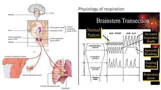
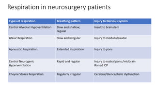
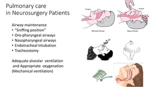
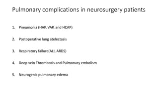
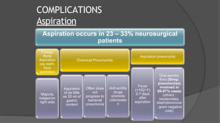
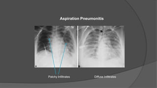
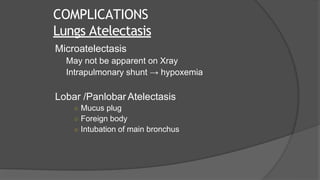
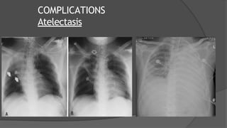
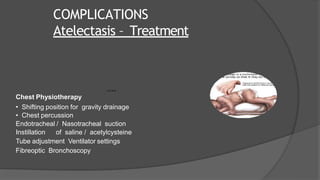
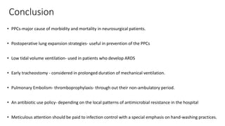
Pulmonary consideration
and complication in
neurosurgery
Dr Fakir Mohan Sahu
MCh Neurosurgery
AIIMS Bhubaneswar
14/03/2020
Learning objectives
• Anatomy
• Physiology
• Pulmonary care in Neurosurgery Patients
• Endotracheal intubation
• Tracheostomy
• Mechanical ventilation
• Post operative Pulmonary complications(PPCs)
• Pathogenesis of PPCs
• Management of PPCs
Anatomy
Tracheobronchial tree
Dependent parts
• Posterior and Superior Segments of
Right and Left Lower lobes
• Posterior Segment of Right Upper Lobe
• Right main bronchus in direct alignment
with trachea
Anatomy
• Pulmonary circulation
• Low pressure (25/8 mm Hg)
• Filter for micro emboli
• Bronchial circulation
• Small normal physiological shunt
• Innervation
• Afferent - Myelinated A fibres provide stretch feedback
Unmyelinated C fibers respond to chemical stimulation (e.g. bradykinnin)
• Efferent- Sympathetic – vasoconstriction and mucus secretion
Parasympathetic-- bronchoconstriction and mucus secretion
• Afferent
Physiology of respiration
Respiration in neurosurgery patients
Types of respiration Breathing pattern Injury to Nervous system
Central Alveolar Hypoventilation Slow and shallow;
regular
Insult to brainstem
Ataxic Respiration Slow and irregular Injury to medulla/caudal
Apneustic Respiration: Extended inspiration Injury to pons
Central Neurogenic
Hyperventilation
Rapid and regular Injury to rostral pons /midbrain
Raised ICP
Cheyne Stokes Respiration Regularly irregular Cerebral/diencephalic dysfunction
Pulmonary care
in Neurosurgery Patients
Airway maintenance
• “Sniffing position”
• Oro-pharyngeal airways
• Nasopharyngeal airways
• Endotracheal Intubation
• Tracheostomy
Adequate alveolar ventilation
and Appropriate oxygenation
(Mechanical ventilation)
Pulmonary complications in neurosurgery patients
1. Pneumonia (HAP, VAP, and HCAP)
2. Postoperative lung atelectasis
3. Respiratory failure(ALI, ARDS)
4. Deep vein Thrombosis and Pulmonary embolism
5. Neurogenic pulmonary edema
Risk factor of PPCs in neurosurgery patient
• Infratentorial surgery
• Mechanical ventilation ≥48 hours
• Time spent in the ICU >3 days
• Decrease in level of consciousness
• Duration of surgery ≥300 minutes
• Previous chronic lung disease
Sogame LC, Vidotto MC, Jardim JR, Faresin SM. Incidence and risk factors for
postoperative pulmonary complications in elective intracranial surgery. J Neurosurg
2008;109:222-7.
• 2 years of study, 236 patients,
• PPCs occurred in 58 patients (24.6%) and 23 patients (10%) died.
• Most frequent PPCs - purulent tracheobronchitis,
followed by pneumonia, bronchospasm, and atelectasis.
Pathogenesis of pulmonary complications in neurosurgical patients
Type of injury pathogenesis
Respiratory depression central (TBI, CNS disease, sedative use) or
peripheral (muscle ds./NM blockade)
High Spinal cord injury impairment of diaphragmatic function,
Injury at thoracic cord levels weakness of thoracoabdominal muscles leading to
paradoxical respiration, reduction in FRC
Associated traumatic airway injuries tracheobronchial disruptions, tracheoesophageal fistula, or
diaphragmatic rupture
Neurogenic pulmonary edema Impaired consciousness
Nasotracheal or orotracheal intubation bacterial colonization and nosocomial pneumonia
Prolonged immobility DVT and pulmonary embolism
Definitions of HAP, VAP, and HCAP
• Hospital-acquired pneumonia (HAP) - 48 h or more after admission
(not incubating at the time of admission)
• Health-care-associated pneumonia (HCAP)
• in hospitalized pt. in an acute care hospital for 2 or more days within 90 days of the infection,
• received recent iv antibiotic therapy, chemotherapy, or wound care within the past 30 days of the current
infection
• attended a haemodialysis clinic
• Ventilator-associated pneumonia (VAP)- more than 48–72 h after endotracheal intubation
• Early VAP/HAP -pneumonia occurring within first 4 days of hospitalization
• Late VAP/HAP - pneumonia occurring after 5 days of hospitalization
(American Thoracic Society Documents. Am J Respir Crit Care Med 2005. 171; 388–416)
COMPLICATIONS
Mechanical Ventilation
• 86% of all nosocomial pneumonias
• Upto 50% mortality
• Acinetobacter the commonest organism
• Suspicion : a new onset (<48hrs) or progressive
radiographic infiltrate with at least 2 of :
• Temp >38o C or < 36o C
• TLC > 10000 or <5000/ml
• Purulent tracheal secretions
• Gas exchange degradation
Ventilator
Associated
Pneumonia
(VAP)
Clinical pulmonary infection score(CPIS)
Clinical characteristics points
Temperature (°C)
≥36.5 and ≤38.4 0
≥38.5 and ≤38.9 1
≥39 and ≤36.5 2
Blood leukocytes (/mm3)
≥4,000 and ≤11,000 0
< 4,000 or >11,000 1
+ band forms ≥50% Add 1
Tracheal secretions
Absence of tracheal secretions 0
Presence of non-purulent tracheal
secretions
1
Presence of purulent tracheal secretions 2
Clinical characteristics points
Oxygenation: PaO2/FIO2
>240 or ARDS 0
<240 and No ARDS 2
Pulmonary radiography
No infiltrate 0
Diffuse (or patchy) infiltrate 1
Localized infiltrate 2
Progression of pulmonary infiltrate
No radiographic progression 0
Radiographic progression (after CHF and ARDS excluded) 2
Culture of tracheal aspirate
Pathogenic bacteria cultured in rare or light quantity or no
growth
0
Pathogenic bacteria cultured in moderate or heavy quantity 1
Same pathogenic bacteria seen on Gram stain Add 1 points
COMPLICATIONS
Mechanical Ventilation
• Culture : Endotrachaeal Aspirate,BAL
• Management :
• Broad spectrum empirical Antibiotic (eg quinolones)
with an anti pseudomonal drug (ceftazidime, imipenem,
piperacillin)
• Specific antibiotic after culture reports
• Supportive care
Ventilator
Associated
Pneumonia
(VAP)
Koenig SM, Truwit JD.. Ventilator-associated pneumonia: diagnosis, treatment, and prevention. Clin Microbiol Rev.
2006;19:637–57
COMPLICATIONS
Aspiration
Aspiration occurs in 23 – 33% neurosurgical
patients
Foreign
Body
Aspiration
(eg teeth,
food
particles)
Majority
lodged on
right side
Chemical Pneumonitis
Aspiration
of as little
as 25 ml of
gastric
content
Often does
not
progress to
bacterial
pneumonia
Anti acidity
drugs
promote
colonizatio
n
Aspiration pneumonia
Fever
(>102o F)
2-7 days
after
aspiration
Oral aerobic
flora (Strep.
pneumoniae)
involved in
66-87% cases
(others :
bacteroides,
staphylococcus
, gram negative
rods)
Patchy Infiltrates Diffuse Infiltrates
Aspiration Pneumonitis
COMPLICATIONS
Lungs Atelectasis
Microatelectasis
May not be apparent on Xray
Intrapulmonary shunt → hypoxemia
Lobar /Panlobar Atelectasis
○ Mucus plug
○ Foreign body
○ Intubation of main bronchus
COMPLICATIONS
Atelectasis
COMPLICATIONS
Atelectasis – Treatment
A.D.A.M.
Chest Physiotherapy
• Shifting position for gravity drainage
• Chest percussion
Endotracheal / Nasotracheal suction
Instillation of saline / acetylcysteine
Tube adjustment Ventilator settings
Fibreoptic Bronchoscopy
COMPLICATIONS
Neurogenic Pulmonary Edema
NPE is a syndrome characterized by the acute onset of pulmonary edema
following significant central nervous system (CNS) injury
Incidence 11 -71% (SAH, Head Injury)
Cl/F- acute development of dyspnoea, tachypnoea, and hypoxemia within minutes.
Pink, frothy sputum is commonly seen and bilateral crackles are audible
Mechanism :
○ Increased Sympathetic Discharge
○ Inflammatory mediators
Treatment : Supportive
COMPLICATIONS
AIR EMBOLISM
Incidence 1.2
– 60%
Morbidity –
mortality ~
3% Risk factors
Surgery in sitting
position
Dehydration /
blood loss
causing low CVP
Diagnosis
Transesophage
al
Echocardiograp
hy and Doppler
Expired N2
End Tidal
CO2
Treatment
Left lateral
decubitus
position
Aspiration of
through atrial
catheter
Irrigation of
surgical field
and sealing all
portals of entry
Hemodynamic
stabilization
COMPLICATIONS
Deep venous thrombosis
• Incidence of DVT 18-50%
• Thrombo Embolism occurs in 15% cases
Venous Thrombosis
• Prolonged surgery and immobilization,
• Previous DVT
• Malignancy(Astrocytoma)
• Direct lower extremity, spinal trauma
• Limb weakness
• Advanced age, ischemic stroke
• Hypercoagulability
Risk factors
• Ankle, calf swelling, calf tenderness, Homan’s signClinically
• D-dimer, LL compression USG, MDCT
Angiography)Diagnosis
COMPLICATIONS DVT
• Mechanical prophylaxis methods
• early ambulation
• intermittent pneumatic compression
• sequential compression devices
• Pharmacological prophylaxis
• low-dose subcutaneous unfractionated heparin (5000 U every 8–12 h)
• low-molecular-weight heparins (enoxaparin)
IVC Filter- Greenfield filter
Venous
Thrombosis
treatment
COMPLICATIONS
Pulmonary Embolism
Pulmonary Embolism: presentation
• Tachypnea
• Pleuritic chest pain
• Dyspnea
• Cough
• Jugular venous distension
• Fever
• Altered sensorium
Investigations
• Po2 of < 80 mm Hg (85%)
• Right axis deviation on ECG
• Infiltrates on Chest X-ray
• Spiral CT (preferred)
• Angiography (Gold Standard)
• Radio nucleotide perfusion scan (sensitive)
COMPLICATIONS
Pulmonary Embolism
Treatment
• Ventilatory support
• Vasopressors
• Full anticoagulation with heparin infusion
(target partial thromboplastin time of 45 to
60 seconds) despite risk of intracranial
bleed.
• i/v Heparin 5000-10000 IU bolus followed
by 1000 IU/hr infusion
• Pulmonary embolectomy (last resort)
COMPLICATIONS
Central Venous Access
Reported Pulmonary Complications
Catheter
malposition
(3.9%)
Pneumothorax
(4.3%)
Catheter
associated
pleural/pericardial
effusion (few
case reports)
(0.2%)
Vandoni RE, et al .Randomised comparison of complications from three different permanent central venous access systems.
Swiss Med Wkly. 2009 May 30;139(21-22):313-6
Walshe C, et al . Vascular erosion by central venous catheters used for total parenteral nutrition. Intensive Care Med. 2007 Mar;
33(3):534-
COMPLICATIONS
Central Venous Access
Central line Chest Tube
COMPLICATIONS
Barotrauma
Barotrauma : Associated with positive pressure ventilation.
Incidence 5 -10%
• Pneumothorax
• Dyspnea, sharp pleuritic chest pain
• Tachypnea, tachycardia
• Contralateral tracheal deviation , hyperresonant percussion
Subcutaneous and Mediastinal Emphysema
PNEUMOTHORAX
Deep Sulcus Sign
COMPLICATIONS
Pneumothorax
Treatment
○ Emergent : 14 – 16 Gauge i/v needle in 2nd – 3rd IC space (midclavicular
line)
○ Definitive : Tube Thoracostomy
To be kept in place till < 100 ml fluid drains /day
No air leak
Lung fully expanded
Post op pneumothorax
Conclusion
• PPCs-major cause of morbidity and mortality in neurosurgical patients.
• Postoperative lung expansion strategies- useful in prevention of the PPCs
• Low tidal volume ventilation- used in patients who develop ARDS
• Early tracheostomy - considered in prolonged duration of mechanical ventilation.
• Pulmonary Embolism- thromboprophylaxis- through out their non-ambulatory period.
• An antibiotic use policy- depending on the local patterns of antimicrobial resistance in the hospital
• Meticulous attention should be paid to infection control with a special emphasis on hand-washing practices.
THANK YOU

Pulmonary Consideration and Complication in Neurosurgery

  • 1.
    Pulmonary consideration and complicationin neurosurgery Dr Fakir Mohan Sahu MCh Neurosurgery AIIMS Bhubaneswar 14/03/2020
  • 2.
    Learning objectives • Anatomy •Physiology • Pulmonary care in Neurosurgery Patients • Endotracheal intubation • Tracheostomy • Mechanical ventilation • Post operative Pulmonary complications(PPCs) • Pathogenesis of PPCs • Management of PPCs
  • 3.
    Anatomy Tracheobronchial tree Dependent parts •Posterior and Superior Segments of Right and Left Lower lobes • Posterior Segment of Right Upper Lobe • Right main bronchus in direct alignment with trachea
  • 4.
    Anatomy • Pulmonary circulation •Low pressure (25/8 mm Hg) • Filter for micro emboli • Bronchial circulation • Small normal physiological shunt • Innervation • Afferent - Myelinated A fibres provide stretch feedback Unmyelinated C fibers respond to chemical stimulation (e.g. bradykinnin) • Efferent- Sympathetic – vasoconstriction and mucus secretion Parasympathetic-- bronchoconstriction and mucus secretion • Afferent
  • 5.
  • 6.
    Respiration in neurosurgerypatients Types of respiration Breathing pattern Injury to Nervous system Central Alveolar Hypoventilation Slow and shallow; regular Insult to brainstem Ataxic Respiration Slow and irregular Injury to medulla/caudal Apneustic Respiration: Extended inspiration Injury to pons Central Neurogenic Hyperventilation Rapid and regular Injury to rostral pons /midbrain Raised ICP Cheyne Stokes Respiration Regularly irregular Cerebral/diencephalic dysfunction
  • 7.
    Pulmonary care in NeurosurgeryPatients Airway maintenance • “Sniffing position” • Oro-pharyngeal airways • Nasopharyngeal airways • Endotracheal Intubation • Tracheostomy Adequate alveolar ventilation and Appropriate oxygenation (Mechanical ventilation)
  • 8.
    Pulmonary complications inneurosurgery patients 1. Pneumonia (HAP, VAP, and HCAP) 2. Postoperative lung atelectasis 3. Respiratory failure(ALI, ARDS) 4. Deep vein Thrombosis and Pulmonary embolism 5. Neurogenic pulmonary edema
  • 9.
    Risk factor ofPPCs in neurosurgery patient • Infratentorial surgery • Mechanical ventilation ≥48 hours • Time spent in the ICU >3 days • Decrease in level of consciousness • Duration of surgery ≥300 minutes • Previous chronic lung disease Sogame LC, Vidotto MC, Jardim JR, Faresin SM. Incidence and risk factors for postoperative pulmonary complications in elective intracranial surgery. J Neurosurg 2008;109:222-7. • 2 years of study, 236 patients, • PPCs occurred in 58 patients (24.6%) and 23 patients (10%) died. • Most frequent PPCs - purulent tracheobronchitis, followed by pneumonia, bronchospasm, and atelectasis.
  • 10.
    Pathogenesis of pulmonarycomplications in neurosurgical patients Type of injury pathogenesis Respiratory depression central (TBI, CNS disease, sedative use) or peripheral (muscle ds./NM blockade) High Spinal cord injury impairment of diaphragmatic function, Injury at thoracic cord levels weakness of thoracoabdominal muscles leading to paradoxical respiration, reduction in FRC Associated traumatic airway injuries tracheobronchial disruptions, tracheoesophageal fistula, or diaphragmatic rupture Neurogenic pulmonary edema Impaired consciousness Nasotracheal or orotracheal intubation bacterial colonization and nosocomial pneumonia Prolonged immobility DVT and pulmonary embolism
  • 11.
    Definitions of HAP,VAP, and HCAP • Hospital-acquired pneumonia (HAP) - 48 h or more after admission (not incubating at the time of admission) • Health-care-associated pneumonia (HCAP) • in hospitalized pt. in an acute care hospital for 2 or more days within 90 days of the infection, • received recent iv antibiotic therapy, chemotherapy, or wound care within the past 30 days of the current infection • attended a haemodialysis clinic • Ventilator-associated pneumonia (VAP)- more than 48–72 h after endotracheal intubation • Early VAP/HAP -pneumonia occurring within first 4 days of hospitalization • Late VAP/HAP - pneumonia occurring after 5 days of hospitalization (American Thoracic Society Documents. Am J Respir Crit Care Med 2005. 171; 388–416)
  • 12.
    COMPLICATIONS Mechanical Ventilation • 86%of all nosocomial pneumonias • Upto 50% mortality • Acinetobacter the commonest organism • Suspicion : a new onset (<48hrs) or progressive radiographic infiltrate with at least 2 of : • Temp >38o C or < 36o C • TLC > 10000 or <5000/ml • Purulent tracheal secretions • Gas exchange degradation Ventilator Associated Pneumonia (VAP)
  • 13.
    Clinical pulmonary infectionscore(CPIS) Clinical characteristics points Temperature (°C) ≥36.5 and ≤38.4 0 ≥38.5 and ≤38.9 1 ≥39 and ≤36.5 2 Blood leukocytes (/mm3) ≥4,000 and ≤11,000 0 < 4,000 or >11,000 1 + band forms ≥50% Add 1 Tracheal secretions Absence of tracheal secretions 0 Presence of non-purulent tracheal secretions 1 Presence of purulent tracheal secretions 2 Clinical characteristics points Oxygenation: PaO2/FIO2 >240 or ARDS 0 <240 and No ARDS 2 Pulmonary radiography No infiltrate 0 Diffuse (or patchy) infiltrate 1 Localized infiltrate 2 Progression of pulmonary infiltrate No radiographic progression 0 Radiographic progression (after CHF and ARDS excluded) 2 Culture of tracheal aspirate Pathogenic bacteria cultured in rare or light quantity or no growth 0 Pathogenic bacteria cultured in moderate or heavy quantity 1 Same pathogenic bacteria seen on Gram stain Add 1 points
  • 14.
    COMPLICATIONS Mechanical Ventilation • Culture: Endotrachaeal Aspirate,BAL • Management : • Broad spectrum empirical Antibiotic (eg quinolones) with an anti pseudomonal drug (ceftazidime, imipenem, piperacillin) • Specific antibiotic after culture reports • Supportive care Ventilator Associated Pneumonia (VAP) Koenig SM, Truwit JD.. Ventilator-associated pneumonia: diagnosis, treatment, and prevention. Clin Microbiol Rev. 2006;19:637–57
  • 15.
    COMPLICATIONS Aspiration Aspiration occurs in23 – 33% neurosurgical patients Foreign Body Aspiration (eg teeth, food particles) Majority lodged on right side Chemical Pneumonitis Aspiration of as little as 25 ml of gastric content Often does not progress to bacterial pneumonia Anti acidity drugs promote colonizatio n Aspiration pneumonia Fever (>102o F) 2-7 days after aspiration Oral aerobic flora (Strep. pneumoniae) involved in 66-87% cases (others : bacteroides, staphylococcus , gram negative rods)
  • 16.
    Patchy Infiltrates DiffuseInfiltrates Aspiration Pneumonitis
  • 17.
    COMPLICATIONS Lungs Atelectasis Microatelectasis May notbe apparent on Xray Intrapulmonary shunt → hypoxemia Lobar /Panlobar Atelectasis ○ Mucus plug ○ Foreign body ○ Intubation of main bronchus
  • 18.
  • 19.
    COMPLICATIONS Atelectasis – Treatment A.D.A.M. ChestPhysiotherapy • Shifting position for gravity drainage • Chest percussion Endotracheal / Nasotracheal suction Instillation of saline / acetylcysteine Tube adjustment Ventilator settings Fibreoptic Bronchoscopy
  • 20.
    COMPLICATIONS Neurogenic Pulmonary Edema NPEis a syndrome characterized by the acute onset of pulmonary edema following significant central nervous system (CNS) injury Incidence 11 -71% (SAH, Head Injury) Cl/F- acute development of dyspnoea, tachypnoea, and hypoxemia within minutes. Pink, frothy sputum is commonly seen and bilateral crackles are audible Mechanism : ○ Increased Sympathetic Discharge ○ Inflammatory mediators Treatment : Supportive
  • 21.
    COMPLICATIONS AIR EMBOLISM Incidence 1.2 –60% Morbidity – mortality ~ 3% Risk factors Surgery in sitting position Dehydration / blood loss causing low CVP Diagnosis Transesophage al Echocardiograp hy and Doppler Expired N2 End Tidal CO2 Treatment Left lateral decubitus position Aspiration of through atrial catheter Irrigation of surgical field and sealing all portals of entry Hemodynamic stabilization
  • 22.
    COMPLICATIONS Deep venous thrombosis •Incidence of DVT 18-50% • Thrombo Embolism occurs in 15% cases Venous Thrombosis • Prolonged surgery and immobilization, • Previous DVT • Malignancy(Astrocytoma) • Direct lower extremity, spinal trauma • Limb weakness • Advanced age, ischemic stroke • Hypercoagulability Risk factors • Ankle, calf swelling, calf tenderness, Homan’s signClinically • D-dimer, LL compression USG, MDCT Angiography)Diagnosis
  • 23.
    COMPLICATIONS DVT • Mechanicalprophylaxis methods • early ambulation • intermittent pneumatic compression • sequential compression devices • Pharmacological prophylaxis • low-dose subcutaneous unfractionated heparin (5000 U every 8–12 h) • low-molecular-weight heparins (enoxaparin) IVC Filter- Greenfield filter Venous Thrombosis treatment
  • 24.
    COMPLICATIONS Pulmonary Embolism Pulmonary Embolism:presentation • Tachypnea • Pleuritic chest pain • Dyspnea • Cough • Jugular venous distension • Fever • Altered sensorium Investigations • Po2 of < 80 mm Hg (85%) • Right axis deviation on ECG • Infiltrates on Chest X-ray • Spiral CT (preferred) • Angiography (Gold Standard) • Radio nucleotide perfusion scan (sensitive)
  • 25.
    COMPLICATIONS Pulmonary Embolism Treatment • Ventilatorysupport • Vasopressors • Full anticoagulation with heparin infusion (target partial thromboplastin time of 45 to 60 seconds) despite risk of intracranial bleed. • i/v Heparin 5000-10000 IU bolus followed by 1000 IU/hr infusion • Pulmonary embolectomy (last resort)
  • 26.
    COMPLICATIONS Central Venous Access ReportedPulmonary Complications Catheter malposition (3.9%) Pneumothorax (4.3%) Catheter associated pleural/pericardial effusion (few case reports) (0.2%) Vandoni RE, et al .Randomised comparison of complications from three different permanent central venous access systems. Swiss Med Wkly. 2009 May 30;139(21-22):313-6 Walshe C, et al . Vascular erosion by central venous catheters used for total parenteral nutrition. Intensive Care Med. 2007 Mar; 33(3):534-
  • 27.
  • 28.
    COMPLICATIONS Barotrauma Barotrauma : Associatedwith positive pressure ventilation. Incidence 5 -10% • Pneumothorax • Dyspnea, sharp pleuritic chest pain • Tachypnea, tachycardia • Contralateral tracheal deviation , hyperresonant percussion Subcutaneous and Mediastinal Emphysema
  • 29.
  • 30.
    COMPLICATIONS Pneumothorax Treatment ○ Emergent :14 – 16 Gauge i/v needle in 2nd – 3rd IC space (midclavicular line) ○ Definitive : Tube Thoracostomy To be kept in place till < 100 ml fluid drains /day No air leak Lung fully expanded
  • 31.
  • 34.
    Conclusion • PPCs-major causeof morbidity and mortality in neurosurgical patients. • Postoperative lung expansion strategies- useful in prevention of the PPCs • Low tidal volume ventilation- used in patients who develop ARDS • Early tracheostomy - considered in prolonged duration of mechanical ventilation. • Pulmonary Embolism- thromboprophylaxis- through out their non-ambulatory period. • An antibiotic use policy- depending on the local patterns of antimicrobial resistance in the hospital • Meticulous attention should be paid to infection control with a special emphasis on hand-washing practices.
  • 35.

Editor's Notes

  • #5 Dual circulation reduces incidence of infarction Chemoreceptors Central (near medulla) : CSF pH and PaCO2 Peripheral(Carotid and aortic bodies) : O2 delivery Mechanoreceptors (feedback and irritant) j receptor or juxtacapilary receptor
  • #6 Control of Breathing Rhythm and Pattern Generator DRG and VRG located in medulla Pneumotaxic center Pons Inspiratory cut off
  • #8 ( temporizing measure) Airway keeps tongue anterior- May stimulate gag Bypasses tongue mechanism Adequate size to be used (e.g. Size 34 or more for 70 kg adult)
  • #10 This has to priorly kept in mind from preoperative period
  • #11 decreased cough strength , and decreased ability to handle secretions. Impaired consciousness, abnormal swallowing mechanism, delayed gastric emptying, depressed gag reflexes, and decreased gastrointestinal motility can predispose to aspiration
  • #12 These definitions are important because these help in identification multidrug‑resistant (MDR) organisms as a cause of pneumonia and therefore are critically important in deciding the choice of antibiotic therapy major difference is early HAP/VAP is likely to be caused by antibiotic sensitive organisms and associated with a better prognosis. On the other hand, late HAP/VAP is likely caused by antibiotic‑resistant (MDR) organisms and associated with higher morbidity and mortality. Head injury staph aures
  • #14 Clinical pulmonary infection score (CPIS) is a tool that was described to objectify the diagnosis of patients with suspected VAP. CPIS combines clinical, radiographic, physiological (PaO2/FiO2), and microbiologic data into a single numerical result. CPIS >6 has demonstrated a good correlation with the presence of pneumonia if <6 than reassess in 48 hr if < 6 then less likely VAP/HAP
  • #16 —Bronchoscopy for foreign body Empirical antibiotics Penicillin group drugs Piperacillin + aminoglycoside Change as per culture reports Empirical corticosteroids and PEEP may not be beneficial
  • #18 Depression in respiratory drive impaired consciousness GA Hyperventilation in patients with raised ICP—leads to hypocapnia and respiratory alkalosis. associated with increase in the intrapulmonary shunt and shift of the oxyhemoglobin dissociation curve to the left. This can lead to worsening of hypoxemia
  • #19 Collapse Parenchymal density on X ray; rib crowding
  • #21 Diagnosis is made after the exclusion of other plausible causes. Characteristically, the onset occurs within minutes to hours after a neurologic insult and usually resolution occurs within 72 h. Alpha adrenergic blocker -phentolamine
  • #24 Thromboprophylaxis is an extremely important intervention which should be considered in all neurosurgical patients
  • #30 Diagnosis : chest radiograph Erect film ideal Deep sulcus sign on supine film Lateral decubitus film for smaller quantities of air Life threating condition tension pneumothorax
  • #33 Journal of anaesthesiology and clinical pharmoacology
  • #35 incentive spirometry, deep breathing exercises, intermittent positive‑pressure breathing, and continuous positive airway pressure decrease in the anatomical dead space, reduced work of breathing, ease of suctioning, improved patient comfort, and reduced sedation requirements. Pulmonary embolism is a killer. It is imperative that Prevention and timely management of these complications can help to decrease the morbidity and mortality associated with pulmonary complications.