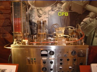
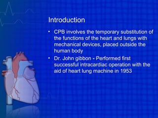
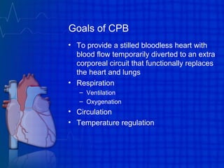
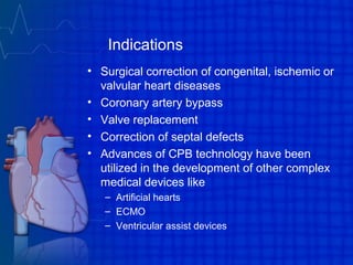
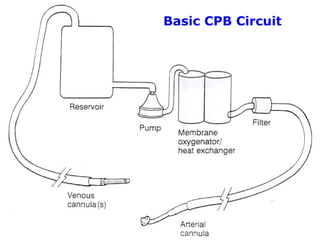
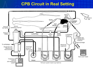
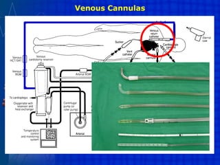
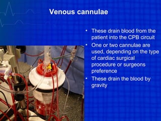
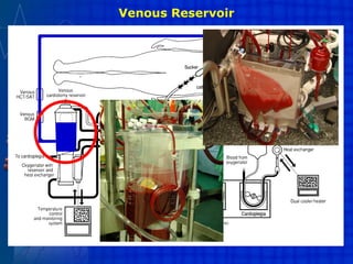
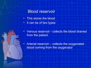
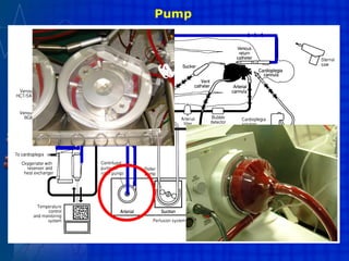
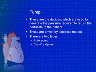
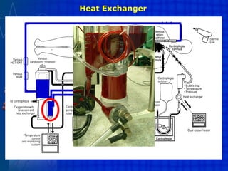
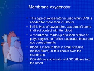
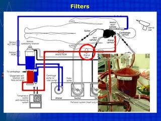
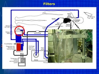
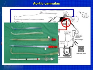
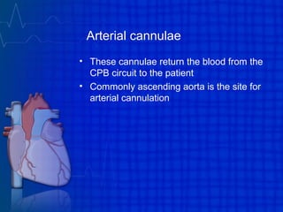
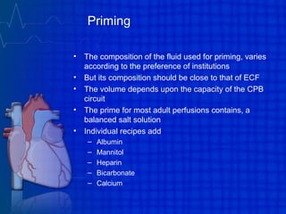
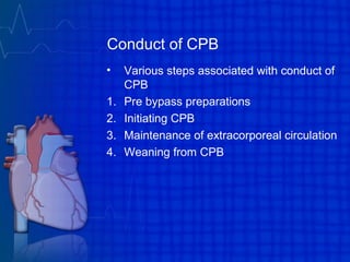
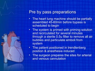
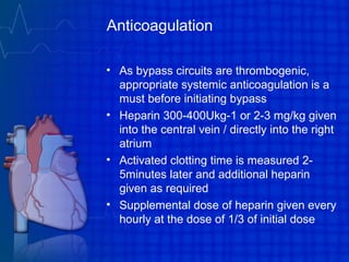
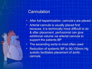
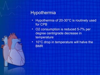
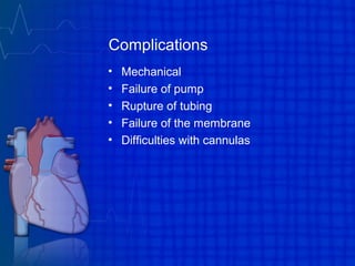
Extra corporeal circulation
Dr. Arati Mohan Badgandi
Introduction
• A procedure in which blood is taken from a
patient's circulation to have a process
applied to it before it is returned to the
circulation
• All of the apparatus carrying the blood
outside the body is termed extracorporeal
circuit
Extra corporeal circulation
• Hemodialysis
• Hemofiltration
• Plasmapheresis
• Extracorporeal membrane oxygenation
(ECMO)
• Cardiopulmonary bypass during open
heart surgery
CPB
CPB
Introduction
• CPB involves the temporary substitution of
the functions of the heart and lungs with
mechanical devices, placed outside the
human body
• Dr. John gibbon - Performed first
successful intracardiac operation with the
aid of heart lung machine in 1953
Goals of CPB
• To provide a stilled bloodless heart with
blood flow temporarily diverted to an extra
corporeal circuit that functionally replaces
the heart and lungs
• Respiration
– Ventilation
– Oxygenation
• Circulation
• Temperature regulation
Indications
• Surgical correction of congenital, ischemic or
valvular heart diseases
• Coronary artery bypass
• Valve replacement
• Correction of septal defects
• Advances of CPB technology have been
utilized in the development of other complex
medical devices like
– Artificial hearts
– ECMO
– Ventricular assist devices
Basic CPB Circuit
CPB Circuit in Real Setting
Venous Cannulas
Venous cannulae
• These drain blood from the
patient into the CPB circuit
• One or two cannulae are
used, depending on the type
of cardiac surgical
procedure or surgeons
preference
• These drain the blood by
gravity
Venous Reservoir
Blood reservoir
• This stores the blood
• It can be of two types
• Venous reservoir - collects the blood drained
from the patient
• Arterial reservoir – collects the oxygenated
blood coming from the oxygenator
Pump
Pump
• These are the devices, which are used to
generate the pressure required to return the
perfusate to the patient
• These are driven by electrical motors
• There are two types
– Roller pump
– Centrifugal pump
Heat Exchanger
Heat exchanger
• It adjusts the temperature of the perfusate to
provide moderate systemic hypothermia
during the period of cardiac repair and
gradually rewarms the blood during the
discontinuation of CPB
• Water circulates within the heat exchanger in
a counter current fashion to the flow of blood
at temperatures between 1-2°C and 42°C
• The temperature gradient between the water
and blood is kept at 10°C or less
Oxygenator
Oxygenators
• It is an apparatus where O2 and Co2
exchange takes place
• There are two types
– Bubble oxygenator
– Membrane oxygenator
Bubble oxygenator
• This type of oxygenator is used when CPB is
needed for less than 2hours
• In this, O2 is directly infused into a column of
systemic venous blood through a diffusion
plate
• The diffusion plate produces thousands of
small O2 bubbles within the blood
• Gas exchange occurs across a thin film at the
blood gas interface around each bubble
• Co2 diffuses into the bubble and O2 diffuses
into the plasma
Membrane oxygenator
• This type of oxygenator is used when CPB is
needed for more than 2-3 hours
• In this type of oxygenator, gas doesn’t come
in direct contact with the blood
• A membrane, made up of silicon rubber or
polypropylene or Teflon, separates blood and
gas compartments
• Blood is made to flow in small streams
(hollow fibers) or thin sheets over the
membrane
• CO2 diffuses outwards and O2 diffuses into
the blood
Filters
Filters
• These are designed to trap particulate
matter and gaseous emboli
• In the suction line - Prevents the debris
from the operating site from reaching the
oxygenator
• Arterial filter - This removes the emboli
from the blood in the arterial tubing prior to
entering the body
• Venous filler - Remove the particulate
matter from the cardiotomy suction
devices
Filters
Filters
Aortic cannulas
Arterial cannulae
• These cannulae return the blood from the
CPB circuit to the patient
• Commonly ascending aorta is the site for
arterial cannulation
Priming
• The composition of the fluid used for priming, varies
according to the preference of institutions
• But its composition should be close to that of ECF
• The volume depends upon the capacity of the CPB
circuit
• The prime for most adult perfusions contains, a
balanced salt solution
• Individual recipes add
– Albumin
– Mannitol
– Heparin
– Bicarbonate
– Calcium
Conduct of CPB
• Various steps associated with conduct of
CPB
1. Pre bypass preparations
2. Initiating CPB
3. Maintenance of extracorporeal circulation
4. Weaning from CPB
Pre by pass preparations
• The heart lung machine should be partially
assembled 45-60min before bypass is
scheduled to begin
• The system is primed with priming solution
and recirculated for several minutes
through a sterile 0.5u filter to remove all air
bubbles and particulate emboli from
system
• The patient positioned in trendlenberg
position & anesthesia induced
• The surgeon prepares the sites for arterial
and venous cannulation
Anticoagulation
• As bypass circuits are thrombogenic,
appropriate systemic anticoagulation is a
must before initiating bypass
• Heparin 300-400Ukg-1 or 2-3 mg/kg given
into the central vein / directly into the right
atrium
• Activated clotting time is measured 2-
5minutes later and additional heparin
given as required
• Supplemental dose of heparin given every
hourly at the dose of 1/3 of initial dose
Cannulation
• After full heparinization, cannula’s are placed
• Arterial cannula is usually placed first
because, it is technically more difficult to do
& after placement, perfusionist can give
additional volume via arterial cannula to
support the patients BP
• The ascending aorta is most often used
• Reduction of systemic BP to 90-100mm Hg
systolic facilitates placement of aortic
cannula
Cannulation
• The anesthesiologist should palpate both
carotids to confirm cannula is not
obstructing right-sided carotid blood flow
• Once the surgeon, perfusionist &
anesthesiologist are satisfied with
functioning & placement of arterial line,
venous cannula is then placed
Initiating CPB
• Bypass begins when surgeon unclamps
venous lines & blood begins to enter
extracorporeal circuit
• As blood starts to fill up venous reservoir of
oxygenator, arterial pump is turned on
• The perfusionist gradually increases bypass
flow rates until all systemic perfusion is
machine generated
• Left ventricular vent introduced through right
superior pulmonary vein & tapes around SVC
& IVC are tightened
Initiating CPB
• Once machine is at full flow, all venous
blood is bypassing cardiopulmonary
circulation
• Ventilation at this point is discontinued
• Once circulation begins, aorta is cross-
clamped & cardioplegic myocardial
protection given before surgical correction
is undertaken
Maintenance of bypass
• The primary goal of this period is to obtain
desired levels of hypothermia, maintain
adequate systemic perfusion, tissue
oxygenation & manage arterial blood
gases
• Ischemic depletion of high energy
phosphate compounds & accumulation of
intracellular calcium, depletes the energy
stores of myocardium
Maintenance of bypass
• To maintain normal myocardial cellular
integrity and function during CPB, the
available high energy phosphate
compounds have to be spared
• This is accomplished by
– Hypothermia
– Cardioplegia
– Intermittent periods of myocardial perfusion
Hypothermia
• Hypothermia of 20-30°C is routinely used
for CPB
• O2 consumption is reduced 5-7% per
degree centrigrade decrease in
temperature
• 10°C drop in temperature will halve the
BMR
Cardioplegia
• A number of different solutions are
described
• Most common compounds are
– Potassium = 15-40 meq/l
– Sodium = 100-120 meq/l
– Chloride = 110-120 meq/l
– Calcium = 0.7 meq/l
– Magnesium = 15 meq/l
– Glucose = 28mmol/l
– Bicarbonate = 27mmol/l
Cardioplegia
• 14G needle is inserted into the aortic root
below cross clamp
• 1 litre of cold (0-40C) cardioplegic solution
is rapidly infused through needle
• In presence of aortic incompetence, the
infusion is done directly through cannulae
inserted into each coronary osteum
Mechanism of action
Increased extracellular potassium
↓
Decrease in transmembrane potential
↓
Impairment of Na+ transport
↓
Abolition of action potential generation
↓
Cardiac arrest in diastole
Intermittent periods of myocardial perfusion
• Intermittent arotic cross clamping
• Intermittent coronary perfusion with
arterialized blood from heart – lung
machine via 2 small cannulae inserted into
coronary ostia
• Alternating ventricular fibrillation with
defibrillation to allow heart to beat
intermittently
Blood gas management
• Acid base management during CPB is
done by two methods
– Alpha stat method
– pH stat method
Alpha stat method
• It considers alkaline pH seen during CPB
is physiological (increased solubility of
carbon dioxide seen during hypothermia
raises pH)
• Hence no additional measures to correct
pH/Pco2 levels is undertaken
• This is more commonly used method & it
appears to preserve cerebral
autoregulation & improve myocardial
preservation
pH stat method
• In this, pH & Pco2 are maintained at
normal values regardless of body
temperature
• In order to maintain Pco2, Co2 is added to
ventilating gas mixture
• This method is not preferred because
patients tend to have higher CBF because
of increase in Co2 content and there will
be loss of cerebral autoregulation
Blood pressure & flow rate management
• A 50 —60 ml/kg of flow rate maintains mean
arterial pressure at 40- 60 mmHg
• To preserve CNS function MAP should be
maintained >50mmHg
• As the patient is rewarmed, MAP should also
be increased accordingly, to ensure adequate
CBF
• Renal function is maintained with pump flows
of more than1.6L/min
Rewarming
• When the surgeon begins last phase of
procedure,perfusionist begins to rewarm
patient
• The rewarming should be gradual & is done
over a 30 minute period
• A gradient of I0°c is maintained between
patient & perfusate to prevent formation of
gas bubbles due to their increased solubility
as blood gets warmed
• Anaesthesiologist should ensure amnesia of
patient by administering additional doses of
benzodiazepines
Rewarming
• Upon completion of the surgical repair, any
residual air in ventricles to be removed
• The anaesthesiologist vigorously inflates
lungs to remove air from pulmonary veins
& aids in filling of cardiac chambers
• TEE assesses the effectiveness of
deairing process
• The aortic clamp removed to resume
myocardial perfusion
• The heart is defibrillated & allowed to beat
empty
Weaning from CPB
• Ensure that patients are rewarmed
adequately & myocardial contractility rhythms
are acceptable
• If necessary, heart rate & rhythm can be
regulated either pharmacologically or
electrically with appropriate pacing
• Ventilation begun with 100%-O2
• Venous drainage lines are gradually
occluded, allowing, arterial return to
progressively raise the circulating volume
• Venous cannulas are removed following a
satisfactory interval of stable hemodynamic
functions
Weaning from CPB
• Reversal of anticoagulation done with
protamine
• Dose - 1 to 1.3ml (10mg/ml) for every
1000U of heparin or dose is calculated
based on the heparin dose response curve
• Arterial cannulas remain in place for
continued transfusion of pump contents
• When this is completed & bleeding is
controlled, arterial cannula is removed &
chest is closed
Post bypass period
• Patient should be shifted to the ICU, kept on
mechanical ventilatory support
• He should be continuously monitored for his
haemodynamic stability & cardiac electrical activity
• SBP should be maintained at 90-110mmHg, HR -
70-80bpm & Hct kept at 25-30%.
• Hypertension may be due to pain & should be
treated with analgesics & sedatives
• Extubation considered when muscle paralysis has
been worn off and patient is hemodynamically
stable
• Most patients can be extubated by following
morning
Complications
Physiologic
• CNS - Intracranial Haemorrhage (14%),
bleeding,seizures
• Respiratory - Pulmonary oedema, pulmonary
haemorrhage
• CVS - hypo/hypervolemia leading to hypo/HTN
• Alteration in R-Angiotensin – aldosterone cycle,
secondary to the non pulsatile perfusion, may lead to
Renal complications
• Haematologic - Anaemia,
leucopenia,thrombocytopenia (because of
consumption in membrane oxygenator)
• Infections
Complications
• Mechanical
• Failure of pump
• Rupture of tubing
• Failure of the membrane
• Difficulties with cannulas
Extracorporeal circulation - CPB, ECMO

Extracorporeal circulation - CPB, ECMO

  • 1.
    Extra corporeal circulation Dr.Arati Mohan Badgandi
  • 2.
    Introduction • A procedurein which blood is taken from a patient's circulation to have a process applied to it before it is returned to the circulation • All of the apparatus carrying the blood outside the body is termed extracorporeal circuit
  • 3.
    Extra corporeal circulation •Hemodialysis • Hemofiltration • Plasmapheresis • Extracorporeal membrane oxygenation (ECMO) • Cardiopulmonary bypass during open heart surgery
  • 4.
  • 5.
    Introduction • CPB involvesthe temporary substitution of the functions of the heart and lungs with mechanical devices, placed outside the human body • Dr. John gibbon - Performed first successful intracardiac operation with the aid of heart lung machine in 1953
  • 6.
    Goals of CPB •To provide a stilled bloodless heart with blood flow temporarily diverted to an extra corporeal circuit that functionally replaces the heart and lungs • Respiration – Ventilation – Oxygenation • Circulation • Temperature regulation
  • 7.
    Indications • Surgical correctionof congenital, ischemic or valvular heart diseases • Coronary artery bypass • Valve replacement • Correction of septal defects • Advances of CPB technology have been utilized in the development of other complex medical devices like – Artificial hearts – ECMO – Ventricular assist devices
  • 8.
  • 9.
    CPB Circuit inReal Setting
  • 10.
  • 11.
    Venous cannulae • Thesedrain blood from the patient into the CPB circuit • One or two cannulae are used, depending on the type of cardiac surgical procedure or surgeons preference • These drain the blood by gravity
  • 12.
  • 13.
    Blood reservoir • Thisstores the blood • It can be of two types • Venous reservoir - collects the blood drained from the patient • Arterial reservoir – collects the oxygenated blood coming from the oxygenator
  • 14.
  • 15.
    Pump • These arethe devices, which are used to generate the pressure required to return the perfusate to the patient • These are driven by electrical motors • There are two types – Roller pump – Centrifugal pump
  • 16.
  • 17.
    Heat exchanger • Itadjusts the temperature of the perfusate to provide moderate systemic hypothermia during the period of cardiac repair and gradually rewarms the blood during the discontinuation of CPB • Water circulates within the heat exchanger in a counter current fashion to the flow of blood at temperatures between 1-2°C and 42°C • The temperature gradient between the water and blood is kept at 10°C or less
  • 18.
  • 19.
    Oxygenators • It isan apparatus where O2 and Co2 exchange takes place • There are two types – Bubble oxygenator – Membrane oxygenator
  • 20.
    Bubble oxygenator • Thistype of oxygenator is used when CPB is needed for less than 2hours • In this, O2 is directly infused into a column of systemic venous blood through a diffusion plate • The diffusion plate produces thousands of small O2 bubbles within the blood • Gas exchange occurs across a thin film at the blood gas interface around each bubble • Co2 diffuses into the bubble and O2 diffuses into the plasma
  • 21.
    Membrane oxygenator • Thistype of oxygenator is used when CPB is needed for more than 2-3 hours • In this type of oxygenator, gas doesn’t come in direct contact with the blood • A membrane, made up of silicon rubber or polypropylene or Teflon, separates blood and gas compartments • Blood is made to flow in small streams (hollow fibers) or thin sheets over the membrane • CO2 diffuses outwards and O2 diffuses into the blood
  • 22.
  • 23.
    Filters • These aredesigned to trap particulate matter and gaseous emboli • In the suction line - Prevents the debris from the operating site from reaching the oxygenator • Arterial filter - This removes the emboli from the blood in the arterial tubing prior to entering the body • Venous filler - Remove the particulate matter from the cardiotomy suction devices
  • 24.
  • 25.
  • 26.
  • 27.
    Arterial cannulae • Thesecannulae return the blood from the CPB circuit to the patient • Commonly ascending aorta is the site for arterial cannulation
  • 28.
    Priming • The compositionof the fluid used for priming, varies according to the preference of institutions • But its composition should be close to that of ECF • The volume depends upon the capacity of the CPB circuit • The prime for most adult perfusions contains, a balanced salt solution • Individual recipes add – Albumin – Mannitol – Heparin – Bicarbonate – Calcium
  • 29.
    Conduct of CPB •Various steps associated with conduct of CPB 1. Pre bypass preparations 2. Initiating CPB 3. Maintenance of extracorporeal circulation 4. Weaning from CPB
  • 30.
    Pre by passpreparations • The heart lung machine should be partially assembled 45-60min before bypass is scheduled to begin • The system is primed with priming solution and recirculated for several minutes through a sterile 0.5u filter to remove all air bubbles and particulate emboli from system • The patient positioned in trendlenberg position & anesthesia induced • The surgeon prepares the sites for arterial and venous cannulation
  • 31.
    Anticoagulation • As bypasscircuits are thrombogenic, appropriate systemic anticoagulation is a must before initiating bypass • Heparin 300-400Ukg-1 or 2-3 mg/kg given into the central vein / directly into the right atrium • Activated clotting time is measured 2- 5minutes later and additional heparin given as required • Supplemental dose of heparin given every hourly at the dose of 1/3 of initial dose
  • 32.
    Cannulation • After fullheparinization, cannula’s are placed • Arterial cannula is usually placed first because, it is technically more difficult to do & after placement, perfusionist can give additional volume via arterial cannula to support the patients BP • The ascending aorta is most often used • Reduction of systemic BP to 90-100mm Hg systolic facilitates placement of aortic cannula
  • 33.
    Cannulation • The anesthesiologistshould palpate both carotids to confirm cannula is not obstructing right-sided carotid blood flow • Once the surgeon, perfusionist & anesthesiologist are satisfied with functioning & placement of arterial line, venous cannula is then placed
  • 34.
    Initiating CPB • Bypassbegins when surgeon unclamps venous lines & blood begins to enter extracorporeal circuit • As blood starts to fill up venous reservoir of oxygenator, arterial pump is turned on • The perfusionist gradually increases bypass flow rates until all systemic perfusion is machine generated • Left ventricular vent introduced through right superior pulmonary vein & tapes around SVC & IVC are tightened
  • 35.
    Initiating CPB • Oncemachine is at full flow, all venous blood is bypassing cardiopulmonary circulation • Ventilation at this point is discontinued • Once circulation begins, aorta is cross- clamped & cardioplegic myocardial protection given before surgical correction is undertaken
  • 36.
    Maintenance of bypass •The primary goal of this period is to obtain desired levels of hypothermia, maintain adequate systemic perfusion, tissue oxygenation & manage arterial blood gases • Ischemic depletion of high energy phosphate compounds & accumulation of intracellular calcium, depletes the energy stores of myocardium
  • 37.
    Maintenance of bypass •To maintain normal myocardial cellular integrity and function during CPB, the available high energy phosphate compounds have to be spared • This is accomplished by – Hypothermia – Cardioplegia – Intermittent periods of myocardial perfusion
  • 38.
    Hypothermia • Hypothermia of20-30°C is routinely used for CPB • O2 consumption is reduced 5-7% per degree centrigrade decrease in temperature • 10°C drop in temperature will halve the BMR
  • 39.
    Cardioplegia • A numberof different solutions are described • Most common compounds are – Potassium = 15-40 meq/l – Sodium = 100-120 meq/l – Chloride = 110-120 meq/l – Calcium = 0.7 meq/l – Magnesium = 15 meq/l – Glucose = 28mmol/l – Bicarbonate = 27mmol/l
  • 40.
    Cardioplegia • 14G needleis inserted into the aortic root below cross clamp • 1 litre of cold (0-40C) cardioplegic solution is rapidly infused through needle • In presence of aortic incompetence, the infusion is done directly through cannulae inserted into each coronary osteum
  • 41.
    Mechanism of action Increasedextracellular potassium ↓ Decrease in transmembrane potential ↓ Impairment of Na+ transport ↓ Abolition of action potential generation ↓ Cardiac arrest in diastole
  • 42.
    Intermittent periods ofmyocardial perfusion • Intermittent arotic cross clamping • Intermittent coronary perfusion with arterialized blood from heart – lung machine via 2 small cannulae inserted into coronary ostia • Alternating ventricular fibrillation with defibrillation to allow heart to beat intermittently
  • 43.
    Blood gas management •Acid base management during CPB is done by two methods – Alpha stat method – pH stat method
  • 44.
    Alpha stat method •It considers alkaline pH seen during CPB is physiological (increased solubility of carbon dioxide seen during hypothermia raises pH) • Hence no additional measures to correct pH/Pco2 levels is undertaken • This is more commonly used method & it appears to preserve cerebral autoregulation & improve myocardial preservation
  • 45.
    pH stat method •In this, pH & Pco2 are maintained at normal values regardless of body temperature • In order to maintain Pco2, Co2 is added to ventilating gas mixture • This method is not preferred because patients tend to have higher CBF because of increase in Co2 content and there will be loss of cerebral autoregulation
  • 46.
    Blood pressure &flow rate management • A 50 —60 ml/kg of flow rate maintains mean arterial pressure at 40- 60 mmHg • To preserve CNS function MAP should be maintained >50mmHg • As the patient is rewarmed, MAP should also be increased accordingly, to ensure adequate CBF • Renal function is maintained with pump flows of more than1.6L/min
  • 47.
    Rewarming • When thesurgeon begins last phase of procedure,perfusionist begins to rewarm patient • The rewarming should be gradual & is done over a 30 minute period • A gradient of I0°c is maintained between patient & perfusate to prevent formation of gas bubbles due to their increased solubility as blood gets warmed • Anaesthesiologist should ensure amnesia of patient by administering additional doses of benzodiazepines
  • 48.
    Rewarming • Upon completionof the surgical repair, any residual air in ventricles to be removed • The anaesthesiologist vigorously inflates lungs to remove air from pulmonary veins & aids in filling of cardiac chambers • TEE assesses the effectiveness of deairing process • The aortic clamp removed to resume myocardial perfusion • The heart is defibrillated & allowed to beat empty
  • 49.
    Weaning from CPB •Ensure that patients are rewarmed adequately & myocardial contractility rhythms are acceptable • If necessary, heart rate & rhythm can be regulated either pharmacologically or electrically with appropriate pacing • Ventilation begun with 100%-O2 • Venous drainage lines are gradually occluded, allowing, arterial return to progressively raise the circulating volume • Venous cannulas are removed following a satisfactory interval of stable hemodynamic functions
  • 50.
    Weaning from CPB •Reversal of anticoagulation done with protamine • Dose - 1 to 1.3ml (10mg/ml) for every 1000U of heparin or dose is calculated based on the heparin dose response curve • Arterial cannulas remain in place for continued transfusion of pump contents • When this is completed & bleeding is controlled, arterial cannula is removed & chest is closed
  • 51.
    Post bypass period •Patient should be shifted to the ICU, kept on mechanical ventilatory support • He should be continuously monitored for his haemodynamic stability & cardiac electrical activity • SBP should be maintained at 90-110mmHg, HR - 70-80bpm & Hct kept at 25-30%. • Hypertension may be due to pain & should be treated with analgesics & sedatives • Extubation considered when muscle paralysis has been worn off and patient is hemodynamically stable • Most patients can be extubated by following morning
  • 52.
    Complications Physiologic • CNS -Intracranial Haemorrhage (14%), bleeding,seizures • Respiratory - Pulmonary oedema, pulmonary haemorrhage • CVS - hypo/hypervolemia leading to hypo/HTN • Alteration in R-Angiotensin – aldosterone cycle, secondary to the non pulsatile perfusion, may lead to Renal complications • Haematologic - Anaemia, leucopenia,thrombocytopenia (because of consumption in membrane oxygenator) • Infections
  • 53.
    Complications • Mechanical • Failureof pump • Rupture of tubing • Failure of the membrane • Difficulties with cannulas

Editor's Notes