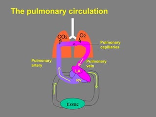
The pulmonary circulation has features designed for efficient gas exchange in the lungs:
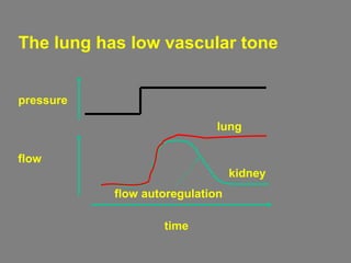
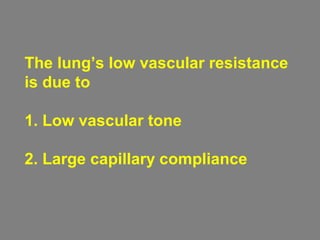
1. It accommodates the cardiac output through low vascular tone and high capillary compliance.
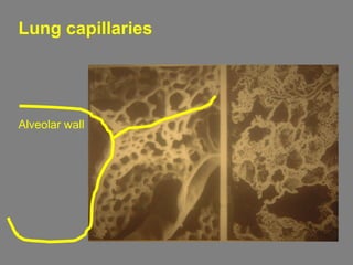
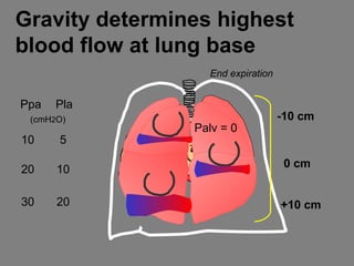
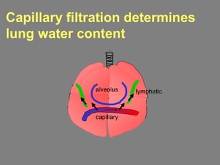
2. It keeps fluid filtration low near the alveoli through low capillary pressures and vascular-interstitial fluid sumps.
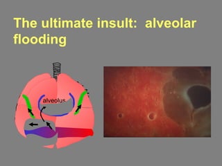
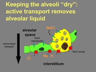
3. It keeps liquid out of the alveoli through active ion transport mechanisms and a high resistance epithelial barrier.


![Blood gas tension determines
blood gas content
Concentration of gas dissolved per
liter of blood (C) depends on gas
solubility (α) and Pgas in blood
CO2 = α x PO2, blood
CO2 = 0.03 x 100
= 3 ml O2/liter of blood
= 0.3 ml O2/100ml of blood
α, Solubility coefficient
(ml O2/[liter.mmHg])
Dissolved gas](https://image.slidesharecdn.com/pulcircslides-151102015549-lva1-app6892/85/Pul-circulation-9-320.jpg)
![The O2 carrying capacity of blood
= Hgb-bound O2 + dissolved O2
PO2, alv
O2 content in 1 liter of blood at PO2, blood of 100 mmHg
= (HgbO2 sat [%] x 1.34 x [Hgb]) + (.03 x PO2, blood)
= (0.98 x 1.34 x 150 [g/l]) + 3
= 200 ml O2/liter of blood
HgbO2 saturation = 1.34 ml at 100%PO2, blood
dissolved O2 in blood](https://image.slidesharecdn.com/pulcircslides-151102015549-lva1-app6892/85/Pul-circulation-11-320.jpg)


![The Starling equation
describes capillary filtration
FR = Lp x S [ (Pc – Pi) – σ (Πc – Πi) ]
FR filtration rate
S capillary surface area
Pc capillary pressure
Pi interstitial pressure
σ reflection coefficient
Πc plasma colloid osmotic pressure
Πi interstitial colloid osmotic pressure
alveolus
Pc
Pi
Πc
Πi](https://image.slidesharecdn.com/pulcircslides-151102015549-lva1-app6892/85/Pul-circulation-27-320.jpg)