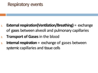
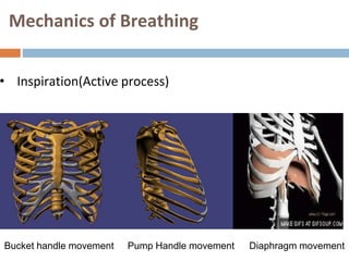
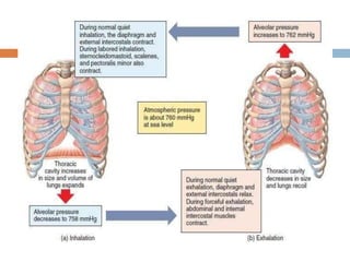
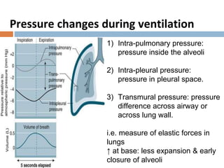
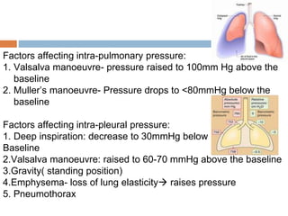
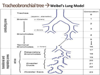
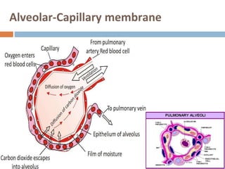
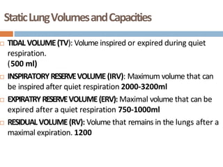
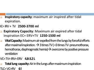
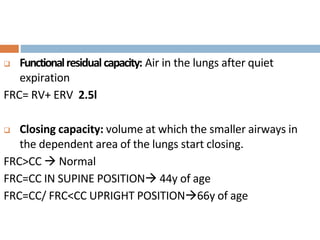
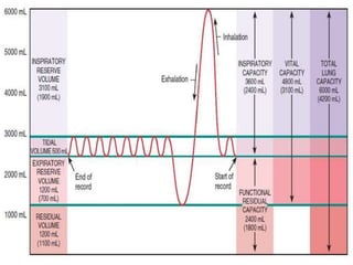
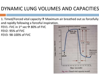
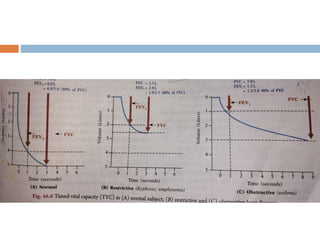
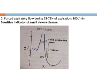
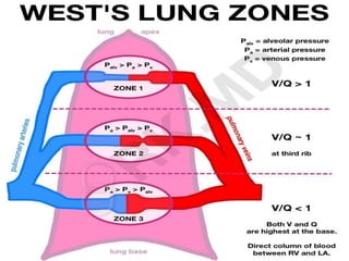
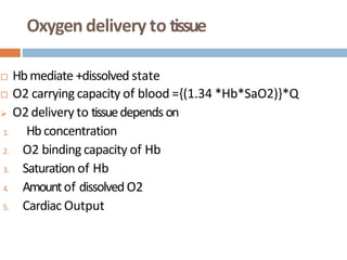
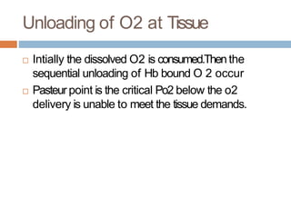
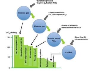
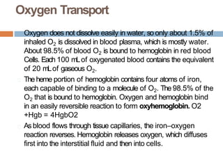
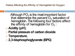
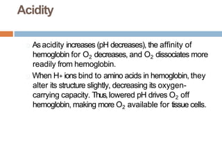
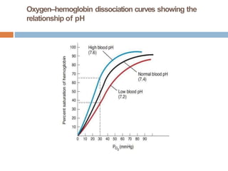
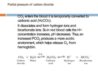
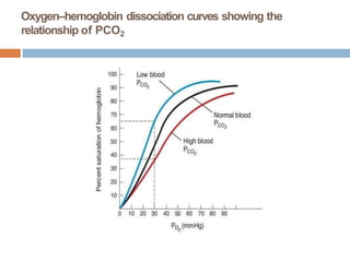
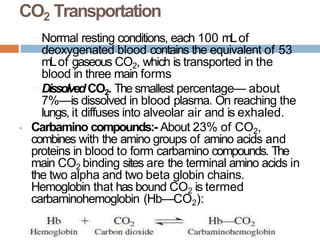
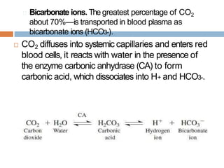
RESPIRATORY
PHYSIOLOGY
Chair Person: Dr. H V Airani
Moderator: Dr. Bhagyashree
Presenter: Dr. Aishwarya
Respiratory events
1. Externalrespiration(Ventilation/Breathing)= exchange
of gases between alveoli and pulmonary capillaries
2. Transport of Gases in the blood
3. Internal respiration= exchange of gases between
systemiccapillaries and tissue cells
Mechanics of Breathing
• Inspiration(Active process)
Bucket handle movement Pump Handle movement Diaphragm movement
• Expiration (Passive process)- Chest recoils
• Forced Expiration( Active process)
Contraction of abdominal muscles, Internal intercostal muscles
and accessory muscles of respiration.
Pressure changes during ventilation
1) Intra-pulmonary pressure:
pressure inside the alveoli
2) Intra-pleural pressure:
pressure in pleural space.
3) Transmural pressure: pressure
difference across airway or
across lung wall.
i.e. measure of elastic forces in
lungs
↑ at base: less expansion & early
closure of alveoli
Factors affecting intra-pulmonary pressure:
1. Valsalva manoeuvre- pressure raised to 100mm Hg above the
baseline
2. Muller’s manoeuvre- Pressure drops to <80mmHg below the
baseline
Factors affecting intra-pleural pressure:
1. Deep inspiration: decrease to 30mmHg below
Baseline
2.Valsalva manoeuvre: raised to 60-70 mmHg above the baseline
3.Gravity( standing position)
4.Emphysema- loss of lung elasticity raises pressure
5. Pneumothorax
TracheobronchialtreeWeibel’s Lung Model
Alveolar-Capillary membrane
StaticLungVolumesandCapacities
 TIDAL VOLUME(TV): Volume inspired or expired during quiet
respiration.
(500 ml)
 INSPIRATORYRESERVEVOLUME(IRV): Maximum volume that can
be inspired after quiet respiration 2000-3200ml
 EXPIRATRYRESERVEVOLUME(ERV): Maximal volume that can be
expired after a quiet respiration 750-1000ml
 RESIDUALVOLUME(RV): Volume that remains in the lungs after a
maximal expiration. 1200
 Inspiratory capacity: maximum air inspired after tidal
expiration.
IC=IRV+ TV 2500-3700 ml
 Expiratory Capacity: Maximum air expired after tidal
inspiration EC= ERV+TV 1250-1500 ml
 VitalCapacity:Maximumairexpelledfromthelungsbyforcefulefforts
aftermaximalinspiration.3timesTV(<3timesTV-pneumothorax,
hemothorax,diaphragmatichernia)overcomebypositivepressure
ventilation
VC=TV+IRV+ERV 4.8-3.2L
 Totallung capacity:Airinthelungsaftermaximuminspiration
TLC=VC+RV 6l
 Functionalresidualcapacity: Air in the lungs after quiet
expiration
FRC= RV+ ERV 2.5l
 Closing capacity: volume at which the smaller airways in
the dependent area of the lungs start closing.
FRC>CC  Normal
FRC=CC IN SUPINE POSITION 44y of age
FRC=CC/ FRC<CC UPRIGHT POSITION66y of age
DYNAMIC LUNG VOLUMES AND CAPACITIES
1. Timed/Forced vital capacity Maximum air breathed out as forcefully
and rapidly following a forceful inspiration.
FEV1- FVC in 1st sec 80% of FVC
FEV2- 95% of FVC
FEV3- 98-100% of FVC
2. Forced expiratory flow during 25-75% of expiration: 300l/min
Sensitive indicator of small airway disease
ELASTIC RESISTANCE & ALVEOLAR SURFACE TENSION
• Chest wall tend to expand outward( chest wall muscle tone)
Lungs tend to collapse ( ↑elastin & SURFACE TENSION)
• Surface tension: due to intermolecular attraction between surface
molecules COLLAPSE
 LAPLACE LAW: Distending pressure(P)= 2 x Surface tension(T)/
Radius(R)
Collapse occurs when Surface tension↑ & radius↓
 Surfactant↓ surface tension( concentration of surfactant ↑ when
alveolus becomes smaller)
COMPLIANCE
Change in the lung volume per unit change in airway pressure
C= ΔV/ΔP (Litres /sec)
Transpulmonary pressure- pressure needed to keep lung
inflated at a certain volume
Normal compliance:0.2-0.3l/cm of H2O
PRESSURE-VOLUME RELATIONSHIP IN THE LUNG
WORK OF BREATHING
 Elastic Resistance( 65%)- stretching the elastic tissue of lungs and chest
wall i.e. against elastic recoil
 Non-elastic resistance:
1. Viscous resistance(7%)- moving viscous material of lung tissue
2. Airway resistance (28%)- resistance to airflow
Airway Resistance:
Flow of air in a tube= pressure difference between the ends of tube P
F Resistance to flow R
Hence, R= P/F
Normal airway resistance- 1.5-2 cmH2O/l/sec
Factors affecting airway resistance:
1. Pressure differences
2. Flow of air
3. Type of flow
Poiseuille - Hagen formula
F= (PA – PB ) x (π/8) x (1/n) x r4 / l
4. Viscosity
5. Radius
6. Length
But smaller airway contributes 10-15% resistance only
As it is aligned more parallel and have more total surface area.
Ventilation-Perfusion Ratio
Ratio of alveolar ventilation to pulmonary blood flow
V/Q= 0.8
At apex- V>Q V/Q=3.4
( Dead space)
At Base- Q>VV/Q=0.63
(Physiological shunt)
ALVEOLAR VENTILATION (VA )
 Amount of air ventilating/minute
 VA = (TV-Dead space) – RR4.2L/Min
Alveolar ventilation affects Gaseous exchange by diffusion
between alveoli and blood
 Dead Space:
 Anatomical- air in conducting zone( nose and mouth to
terminal bronchioles) 150ml
 Physiological – Anatomical dead space+ volume of air in
alveoli that doesn’t get exchanged
Eg. 1. Alveoli ventilated but not receiving pulmonary blood
2. Over-ventilated alveoli
 Pathological dead space: Emphysema, Bronchiectasis,
Pulmonary embolism
SHUNT
 Wasted perfusion
 Absolute shunt- Anatomical : V/Q=O
Relative shunt- under ventilated lungs- V/Q<1
 Normal/physiological shunt- <5%
 Shunts above 15% Significant hypoxemia
Oxygen delivery to tissue
 Hbmediate +dissolved state
 O2 carrying capacity of blood ={(1.34 *Hb*SaO2)}*Q
 O2 delivery to tissuedepends on
1. Hb concentration
2. O2 binding capacity of Hb
3. Saturation of Hb
4. Amountof dissolved O2
5. Cardiac Output
Unloading of O2 at Tissue
 Intially the dissolved O2 is consumed.Thenthe
sequential unloading of Hb bound O 2 occur
 Pasteur point is the critical Po2 below the o2
delivery is unable to meet the tissue demands.
Oxygen Transport
Oxygen does not dissolve easily in water, so only about 1.5% of
inhaled O2 is dissolved in blood plasma, which is mostly water.
About 98.5% of blood O2 is bound to hemoglobin in red blood
Cells. Each 100 mLof oxygenated blood contains the equivalent
of 20 mLof gaseous O2.
🞑 Theheme portion of hemoglobin contains four atoms of iron,
each capable of binding to a molecule of O2. The 98.5% of the
O2 that is bound to hemoglobin. Oxygen and hemoglobin bind
in an easily reversible reaction to form oxyhemoglobin. O2
+Hgb = 4HgbO2
🞑 As blood flows through tissue capillaries, the iron–oxygen
reaction reverses. Hemoglobin releases oxygen, which diffuses
first into the interstitial fluid and then into cells.
Factors Affecting the Affinity of Hemoglobin for Oxygen
🞑Although PO2 is the most important factor
that determines the percent O2 saturation of
hemoglobin. The following four factors affect
the affinity of hemoglobin for O2 :
 Acidity (pH).
 Partial pressure of carbon dioxide
 Temperature.
 2,3-bisphosphoglycerate (BPG)
Acidity
🞑 Asacidity increases (pH decreases), the affinity of
hemoglobin for O2 decreases, and O2 dissociates more
readily from hemoglobin.
🞑 When H+ ions bind to amino acids in hemoglobin, they
alter its structure slightly, decreasing its oxygen-
carrying capacity. Thus,lowered pH drives O2 off
hemoglobin, making more O2 available for tissue cells.
Oxygen–hemoglobin dissociation curves showing the
relationship of pH
Partial pressure of carbon dioxide
🞑 CO2 enters the blood it is temporarily converted to
carbonic acid (H2CO3).
🞑 It dissociates and form hydrogen ions and
bicarbonate ions. Soin red blood cells the H+
concentration increases, pH decreases. Thus, an
increased PCO2 produces a more acidic
environment, which helps release O2 from
hemoglobin.
Oxygen–hemoglobin dissociation curves showing the
relationship of PCO2
Temperature
🞑Heat is a by-product of the metabolic
reactions of all cells, and the heat
released by contracting muscle fibers
tends to raise body temperature.
Metabolically active cells require more
O2 and liberate more acids and heat.
Oxygen–hemoglobin dissociation curves showing the effect of
temperature changes.
2,3-Bisphosphoglycerate (BPG)
(Diphosphoglycerate)
🞑 BPGis formed in red blood cells when they break down
glucose to produce ATPin a process called glycolysis.
When BPG combines with hemoglobin, it unloads or
decreases the bonding with oxygen.
CO2 Transportation
🞑 Normal resting conditions, each 100 mLof
deoxygenated blood contains the equivalent of 53
mLof gaseous CO2, which is transported in the
blood in three main forms
🞑 DissolvedCO2. The smallest percentage— about
7%—is dissolved in blood plasma. On reaching the
lungs, it diffuses into alveolar air and is exhaled.
• Carbamino compounds:- About 23% of CO2,
combines with the amino groups of amino acids and
proteins in blood to form carbamino compounds. The
main CO2 binding sites are the terminal amino acids in
the two alpha and two beta globin chains.
Hemoglobin that has bound CO2 is termed
carbaminohemoglobin (Hb—CO2):
🞑 Bicarbonate ions. The greatest percentage of CO2
about 70%—is transported in blood plasma as
bicarbonate ions (HCO3-).
 CO2 diffuses into systemic capillaries and enters red
blood cells, it reacts with water in the presence of
the enzyme carbonic anhydrase (CA) to form
carbonic acid, which dissociates into H+ and HCO3-.
CO2 Dissociation Curve
The arterial point (a) and the venous point (v)
Concentration of H+ ions or pH
 When Concentration of H+ ions increases, it
stimulates the peripheral chemoreceptors. H+ ions
diffuses with CO2 and form carbonic acid, to cross
the blood brain barrier then dissociates into H+ and
HCO3. There by H+ ions stimulates the central
chemoreceptors then the respiratory centers,
resulting a reduction in the level of CO2 in blood.
Thiswill inturn decrease concentration of H+ in
blood or increase the pH in to normal.
Concentration of oxygen In blood
When O2 concentration in blood decreases
Stimulates the peripheral chemoreceptors
Transmission of impulses to respiratory centers
Activation of respiratory centers
Increases the activities of respiration (rate and Depth)
Increase alveolar ventilation
Increases the uptake of O2

Thereby increases the level of O2 in blood
RESPIRATORY PHYSIOlogy presentation anaesthesia

RESPIRATORY PHYSIOlogy presentation anaesthesia

  • 1.
    RESPIRATORY PHYSIOLOGY Chair Person: Dr.H V Airani Moderator: Dr. Bhagyashree Presenter: Dr. Aishwarya
  • 2.
    Respiratory events 1. Externalrespiration(Ventilation/Breathing)=exchange of gases between alveoli and pulmonary capillaries 2. Transport of Gases in the blood 3. Internal respiration= exchange of gases between systemiccapillaries and tissue cells
  • 3.
    Mechanics of Breathing •Inspiration(Active process) Bucket handle movement Pump Handle movement Diaphragm movement
  • 4.
    • Expiration (Passiveprocess)- Chest recoils • Forced Expiration( Active process) Contraction of abdominal muscles, Internal intercostal muscles and accessory muscles of respiration.
  • 6.
    Pressure changes duringventilation 1) Intra-pulmonary pressure: pressure inside the alveoli 2) Intra-pleural pressure: pressure in pleural space. 3) Transmural pressure: pressure difference across airway or across lung wall. i.e. measure of elastic forces in lungs ↑ at base: less expansion & early closure of alveoli
  • 7.
    Factors affecting intra-pulmonarypressure: 1. Valsalva manoeuvre- pressure raised to 100mm Hg above the baseline 2. Muller’s manoeuvre- Pressure drops to <80mmHg below the baseline Factors affecting intra-pleural pressure: 1. Deep inspiration: decrease to 30mmHg below Baseline 2.Valsalva manoeuvre: raised to 60-70 mmHg above the baseline 3.Gravity( standing position) 4.Emphysema- loss of lung elasticity raises pressure 5. Pneumothorax
  • 8.
  • 9.
  • 10.
    StaticLungVolumesandCapacities  TIDAL VOLUME(TV):Volume inspired or expired during quiet respiration. (500 ml)  INSPIRATORYRESERVEVOLUME(IRV): Maximum volume that can be inspired after quiet respiration 2000-3200ml  EXPIRATRYRESERVEVOLUME(ERV): Maximal volume that can be expired after a quiet respiration 750-1000ml  RESIDUALVOLUME(RV): Volume that remains in the lungs after a maximal expiration. 1200
  • 11.
     Inspiratory capacity:maximum air inspired after tidal expiration. IC=IRV+ TV 2500-3700 ml  Expiratory Capacity: Maximum air expired after tidal inspiration EC= ERV+TV 1250-1500 ml  VitalCapacity:Maximumairexpelledfromthelungsbyforcefulefforts aftermaximalinspiration.3timesTV(<3timesTV-pneumothorax, hemothorax,diaphragmatichernia)overcomebypositivepressure ventilation VC=TV+IRV+ERV 4.8-3.2L  Totallung capacity:Airinthelungsaftermaximuminspiration TLC=VC+RV 6l
  • 12.
     Functionalresidualcapacity: Airin the lungs after quiet expiration FRC= RV+ ERV 2.5l  Closing capacity: volume at which the smaller airways in the dependent area of the lungs start closing. FRC>CC  Normal FRC=CC IN SUPINE POSITION 44y of age FRC=CC/ FRC<CC UPRIGHT POSITION66y of age
  • 14.
    DYNAMIC LUNG VOLUMESAND CAPACITIES 1. Timed/Forced vital capacity Maximum air breathed out as forcefully and rapidly following a forceful inspiration. FEV1- FVC in 1st sec 80% of FVC FEV2- 95% of FVC FEV3- 98-100% of FVC
  • 16.
    2. Forced expiratoryflow during 25-75% of expiration: 300l/min Sensitive indicator of small airway disease
  • 17.
    ELASTIC RESISTANCE &ALVEOLAR SURFACE TENSION • Chest wall tend to expand outward( chest wall muscle tone) Lungs tend to collapse ( ↑elastin & SURFACE TENSION) • Surface tension: due to intermolecular attraction between surface molecules COLLAPSE  LAPLACE LAW: Distending pressure(P)= 2 x Surface tension(T)/ Radius(R) Collapse occurs when Surface tension↑ & radius↓  Surfactant↓ surface tension( concentration of surfactant ↑ when alveolus becomes smaller)
  • 18.
    COMPLIANCE Change in thelung volume per unit change in airway pressure C= ΔV/ΔP (Litres /sec) Transpulmonary pressure- pressure needed to keep lung inflated at a certain volume Normal compliance:0.2-0.3l/cm of H2O
  • 19.
  • 21.
  • 22.
     Elastic Resistance(65%)- stretching the elastic tissue of lungs and chest wall i.e. against elastic recoil  Non-elastic resistance: 1. Viscous resistance(7%)- moving viscous material of lung tissue 2. Airway resistance (28%)- resistance to airflow Airway Resistance: Flow of air in a tube= pressure difference between the ends of tube P F Resistance to flow R Hence, R= P/F Normal airway resistance- 1.5-2 cmH2O/l/sec
  • 23.
    Factors affecting airwayresistance: 1. Pressure differences 2. Flow of air 3. Type of flow Poiseuille - Hagen formula F= (PA – PB ) x (π/8) x (1/n) x r4 / l 4. Viscosity 5. Radius 6. Length But smaller airway contributes 10-15% resistance only As it is aligned more parallel and have more total surface area.
  • 24.
    Ventilation-Perfusion Ratio Ratio ofalveolar ventilation to pulmonary blood flow V/Q= 0.8 At apex- V>Q V/Q=3.4 ( Dead space) At Base- Q>VV/Q=0.63 (Physiological shunt)
  • 26.
    ALVEOLAR VENTILATION (VA)  Amount of air ventilating/minute  VA = (TV-Dead space) – RR4.2L/Min Alveolar ventilation affects Gaseous exchange by diffusion between alveoli and blood  Dead Space:  Anatomical- air in conducting zone( nose and mouth to terminal bronchioles) 150ml  Physiological – Anatomical dead space+ volume of air in alveoli that doesn’t get exchanged
  • 27.
    Eg. 1. Alveoliventilated but not receiving pulmonary blood 2. Over-ventilated alveoli  Pathological dead space: Emphysema, Bronchiectasis, Pulmonary embolism
  • 28.
    SHUNT  Wasted perfusion Absolute shunt- Anatomical : V/Q=O Relative shunt- under ventilated lungs- V/Q<1  Normal/physiological shunt- <5%  Shunts above 15% Significant hypoxemia
  • 29.
    Oxygen delivery totissue  Hbmediate +dissolved state  O2 carrying capacity of blood ={(1.34 *Hb*SaO2)}*Q  O2 delivery to tissuedepends on 1. Hb concentration 2. O2 binding capacity of Hb 3. Saturation of Hb 4. Amountof dissolved O2 5. Cardiac Output
  • 30.
    Unloading of O2at Tissue  Intially the dissolved O2 is consumed.Thenthe sequential unloading of Hb bound O 2 occur  Pasteur point is the critical Po2 below the o2 delivery is unable to meet the tissue demands.
  • 32.
    Oxygen Transport Oxygen doesnot dissolve easily in water, so only about 1.5% of inhaled O2 is dissolved in blood plasma, which is mostly water. About 98.5% of blood O2 is bound to hemoglobin in red blood Cells. Each 100 mLof oxygenated blood contains the equivalent of 20 mLof gaseous O2. 🞑 Theheme portion of hemoglobin contains four atoms of iron, each capable of binding to a molecule of O2. The 98.5% of the O2 that is bound to hemoglobin. Oxygen and hemoglobin bind in an easily reversible reaction to form oxyhemoglobin. O2 +Hgb = 4HgbO2 🞑 As blood flows through tissue capillaries, the iron–oxygen reaction reverses. Hemoglobin releases oxygen, which diffuses first into the interstitial fluid and then into cells.
  • 33.
    Factors Affecting theAffinity of Hemoglobin for Oxygen 🞑Although PO2 is the most important factor that determines the percent O2 saturation of hemoglobin. The following four factors affect the affinity of hemoglobin for O2 :  Acidity (pH).  Partial pressure of carbon dioxide  Temperature.  2,3-bisphosphoglycerate (BPG)
  • 34.
    Acidity 🞑 Asacidity increases(pH decreases), the affinity of hemoglobin for O2 decreases, and O2 dissociates more readily from hemoglobin. 🞑 When H+ ions bind to amino acids in hemoglobin, they alter its structure slightly, decreasing its oxygen- carrying capacity. Thus,lowered pH drives O2 off hemoglobin, making more O2 available for tissue cells.
  • 35.
    Oxygen–hemoglobin dissociation curvesshowing the relationship of pH
  • 36.
    Partial pressure ofcarbon dioxide 🞑 CO2 enters the blood it is temporarily converted to carbonic acid (H2CO3). 🞑 It dissociates and form hydrogen ions and bicarbonate ions. Soin red blood cells the H+ concentration increases, pH decreases. Thus, an increased PCO2 produces a more acidic environment, which helps release O2 from hemoglobin.
  • 37.
    Oxygen–hemoglobin dissociation curvesshowing the relationship of PCO2
  • 38.
    Temperature 🞑Heat is aby-product of the metabolic reactions of all cells, and the heat released by contracting muscle fibers tends to raise body temperature. Metabolically active cells require more O2 and liberate more acids and heat.
  • 39.
    Oxygen–hemoglobin dissociation curvesshowing the effect of temperature changes.
  • 40.
    2,3-Bisphosphoglycerate (BPG) (Diphosphoglycerate) 🞑 BPGisformed in red blood cells when they break down glucose to produce ATPin a process called glycolysis. When BPG combines with hemoglobin, it unloads or decreases the bonding with oxygen.
  • 41.
    CO2 Transportation 🞑 Normalresting conditions, each 100 mLof deoxygenated blood contains the equivalent of 53 mLof gaseous CO2, which is transported in the blood in three main forms 🞑 DissolvedCO2. The smallest percentage— about 7%—is dissolved in blood plasma. On reaching the lungs, it diffuses into alveolar air and is exhaled. • Carbamino compounds:- About 23% of CO2, combines with the amino groups of amino acids and proteins in blood to form carbamino compounds. The main CO2 binding sites are the terminal amino acids in the two alpha and two beta globin chains. Hemoglobin that has bound CO2 is termed carbaminohemoglobin (Hb—CO2):
  • 42.
    🞑 Bicarbonate ions.The greatest percentage of CO2 about 70%—is transported in blood plasma as bicarbonate ions (HCO3-).  CO2 diffuses into systemic capillaries and enters red blood cells, it reacts with water in the presence of the enzyme carbonic anhydrase (CA) to form carbonic acid, which dissociates into H+ and HCO3-.
  • 43.
    CO2 Dissociation Curve Thearterial point (a) and the venous point (v)
  • 45.
    Concentration of H+ions or pH  When Concentration of H+ ions increases, it stimulates the peripheral chemoreceptors. H+ ions diffuses with CO2 and form carbonic acid, to cross the blood brain barrier then dissociates into H+ and HCO3. There by H+ ions stimulates the central chemoreceptors then the respiratory centers, resulting a reduction in the level of CO2 in blood. Thiswill inturn decrease concentration of H+ in blood or increase the pH in to normal.
  • 46.
    Concentration of oxygenIn blood When O2 concentration in blood decreases Stimulates the peripheral chemoreceptors Transmission of impulses to respiratory centers Activation of respiratory centers Increases the activities of respiration (rate and Depth) Increase alveolar ventilation Increases the uptake of O2  Thereby increases the level of O2 in blood