This document summarizes the anatomy and functions of salivary glands. It describes the major salivary glands - parotid, submandibular and sublingual glands - as well as minor salivary glands. The functions of saliva include protection, buffering, tooth integrity, antimicrobial activity, tissue repair, digestion and taste. Saliva production is nerve-mediated and its composition includes water, electrolytes, proteins, immunoglobulins and other components. The document also discusses salivary gland development, histology and clinical considerations.
Saliva is produced by the major and minor salivary glands and consists of both serous and mucous secretions. The three pairs of major salivary glands - the parotid, submandibular, and sublingual glands - provide over 90% of the total saliva produced. The parotid glands are the largest and purely serous, while the submandibular glands are predominantly mucous and mixed. The sublingual glands are the smallest but also predominantly mucous and mixed. Whole saliva contains secretions from the major and minor salivary glands as well as other components from the oral cavity.
This document summarizes the salivary glands. It defines salivary glands as exocrine glands that secrete saliva into the oral cavity. It classifies salivary glands as major (parotid, submandibular, sublingual) or minor based on size, and as serous, mixed, or mucous based on secretory cell type. The document describes the anatomy, histology, development and functions of the major salivary glands. It also discusses the structure of salivary glands including secretory end pieces, ductal system, and secretory cell types.
The document summarizes the normal anatomy and histology of the stomach as well as various pathologies that can affect the stomach. It describes the layers of the normal gastric wall and the different cell types present in each region. It then discusses various inflammatory conditions like acute and chronic gastritis, peptic ulcers, hyperplastic gastropathies including Menetrier's disease, and neoplastic conditions such as Zollinger-Ellison syndrome. For each condition, it provides details on pathogenesis, morphology, clinical features, and complications.
This document provides an overview of the anatomy and structure of the digestive tract. It begins with an introduction to the digestive tract and its two main divisions: the digestive tract itself, which is a tube running from the mouth to anus, and the accessory organs that help with digestion. It then describes each section of the digestive tract in detail, including the mouth, pharynx, esophagus, stomach, and small and large intestines. It discusses the layers of the tract wall and related structures like salivary glands and teeth. Finally, it focuses specifically on the anatomy of the stomach and small intestine.
This document discusses saliva and the salivary glands. It begins with an introduction to saliva's composition and functions. It then describes the major and minor salivary glands in terms of their location, morphology, blood supply, and development. The document discusses saliva secretion, including the formation of primary and final saliva. It also covers factors that influence salivary flow rate such as hormones, stimulation, and various physiological and pathological conditions. Overall, the document provides a comprehensive overview of saliva and the salivary glands.
Introduction
Suprahyoid muscle and its embryology
Relation of mylohyoid and digastric muscle
Submandibular gland and duct
Development and histology
Sublingual gland and duct ,it’s development and histology.
Submandibular ganglion and its relations
Clinical anatomy
Blood and nerve supply of submandibular and sublingual duct
Conclusion
References
Submandibular salivary gland dr chithraDr. Chithra P
This document provides information about the submandibular salivary gland. It discusses the gland's anatomy, development, blood supply, nerve supply and clinical evaluation. Key points include:
- The submandibular gland is the second largest major salivary gland located in the submandibular triangle below the mandible.
- It develops from an epithelial bud that branches during development forming a ductal system and acini.
- Anatomy includes a superficial and deep part divided by the mylohyoid muscle. Wharton's duct drains the gland opening on the floor of the mouth.
- Evaluation involves history, extraoral and intraoral examination including palpation and imaging
lecture 4 Diagnosis and management of salivary gland disordersLama K Banna
Maxillofacial Surgery
Dental Students Fifth Year First semester
Lecture Name Salivary gland
Diagnosis and management of salivary gland disorders
Al Azhar University Gaza Palestine
Dr. Lama El Banna
Salivary glands are divided into major and minor glands. The major glands are the parotid, submandibular, and sublingual glands. The parotid gland is the largest and is a serous gland located below and in front of each ear. The submandibular gland is mixed but predominantly serous, located under the jawbone. The sublingual gland is the smallest major gland and is predominantly mucous, located under the tongue. Minor salivary glands are found throughout the oral cavity and empty via short ducts directly into the mouth. Salivary glands produce saliva, a fluid that lubricates and protects tissues in the oral cavity.
ANATOMY AND PHYSIOLOGY OF HUMAN DIGESTIVE SYSTEMSAYANTANDUTTA49
The document discusses the digestive system and its components. It begins with an overview of digestion and the roles of the digestive system. It then describes the major organs that make up the primary digestive tract, including the mouth, salivary glands, esophagus, stomach, small intestine, and large intestine. It also mentions the accessory digestive organs that help in the digestive process, such as the teeth, tongue, liver, and pancreas.
This document provides information about saliva and the salivary glands. It discusses the development, classification, anatomy and functions of the major and minor salivary glands. The major salivary glands are the parotid, submandibular and sublingual glands. It describes the stages of development of the salivary glands beginning in the 6th week of fetal life and the histochemical nature of secretions. Diseases and age-related changes of the salivary glands are also mentioned.
The digestive system consists of the alimentary canal and accessory organs involved in ingestion, digestion, absorption and elimination of food. The alimentary canal includes the mouth, pharynx, esophagus, stomach, small intestine, large intestine, rectum and anal canal. Accessory organs that aid in digestion include the salivary glands, liver, gallbladder and pancreas.
This document discusses the anatomy and embryology of the major and minor salivary glands. It describes the three pairs of major salivary glands - the parotid, submandibular, and sublingual glands. It details their development during embryogenesis, classification based on size and secretory cell type, structure, blood supply, innervation and relations to surrounding tissues. The document also briefly discusses the numerous minor salivary glands found in the oral cavity.
The document provides information on the structure and function of the digestive system. It begins with an overview of the basic components of the alimentary canal including the mouth, salivary glands, pharynx, esophagus, stomach, small intestine, large intestine, rectum, anal canal, and accessory organs like the liver, pancreas, and bile duct. It then describes the layers of the alimentary canal including the peritoneum, muscle layers, submucosa, and mucosa. Specific structures like the tongue, teeth, stomach, small intestine and large intestine are examined in more detail. The functions of digestion and absorption in the mouth, stomach, small intestine, large intestine and accessory organs are
1. The document discusses benign and malignant disorders of salivary glands, including classifications, anatomy, developmental disorders, inflammatory conditions, obstructive disorders, cysts, autoimmune disorders, benign disorders, and classifications of salivary gland tumors.
2. It describes the major salivary glands, their blood supply, innervation and development. Inflammatory conditions discussed include acute and chronic sialadenitis, mumps, and irradiation-induced conditions. Obstructive disorders include sialolithiasis.
3. Benign disorders summarized are Frey's syndrome, sialorrhea, xerostomia, recurrent parotitis, sialodenosis, granulomatous s
The document discusses the embryology, anatomy, histology and physiology of salivary glands. It begins by describing the development of the major salivary glands from oral ectoderm between 6-8 weeks of gestation. It then covers the classification, locations and duct systems of the parotid, submandibular and sublingual glands. The histology section examines the serous and mucous acini, ductal system and myoepithelial cells. It concludes with the innervation and secretory processes of the salivary glands.
The document discusses the anatomy, histology, functions, and clinical considerations of the salivary glands. It describes the major and minor salivary glands, their secretory units, ductal systems, nerve supply, and blood supply. The properties and composition of saliva are discussed. Hyposalivation is defined and its clinical signs, symptoms, and management strategies are summarized.
This document discusses salivary glands and saliva. It begins by introducing the topic and listing the contents to be covered. It then classifies salivary glands based on their anatomical size, type of secretion, and location. The major glands are identified as the parotid, submandibular, and sublingual glands. Minor salivary glands are also discussed. The document further explores the constituents of saliva, formation and regulation of saliva, and prosthodontic implications. Key functions of saliva including protection, buffering, tissue repair, digestion, taste, and anticaries activity are summarized. Major organic constituents such as mucins, proteins, and enzymes are identified.
This presentation deals with the study of anatomy and physiology of nasal mucosa and the potential advantage of nasal administration of therapeutic agents...
The document discusses salivary glands and saliva. It defines salivary glands as exocrine glands that secrete saliva. It classifies salivary glands into major and minor glands and describes the anatomy and histology of the parotid, submandibular, and sublingual glands. It also outlines the composition of saliva, including water, electrolytes, enzymes, proteins, and other components. Saliva serves important functions like lubrication, digestion, and maintenance of oral health.
This document summarizes the key details about salivary glands. It describes the three major salivary glands - the parotid, submandibular, and sublingual glands. It explains their secretions help keep the mouth moist, provide protection, lubrication, and contain enzymes and immunoglobulin A. The glands have secretory elements like acini and alveoli that produce secretions which drain into a series of ducts that pour into the oral cavity. The document outlines the histological structure of the glands including their serous and mucous cells, and describes myoepithelial cells that help contract and squeeze out gland secretions.
Saliva and salivary glands by Dr.Nilesh VaidyaNilesh Vaidya
Saliva is a complex fluid produced by salivary glands that plays an important role in maintaining oral health. It is multifunctional, helping with swallowing, digestion, and protecting the oral cavity from dryness. Saliva is formed through secretion from the parotid, submandibular, sublingual glands and minor salivary glands in the oral cavity. It has digestive, protective, and cleansing functions that are important for oral and overall health.
The document provides information on the gastrointestinal tract. It discusses:
1. The gastrointestinal tract is responsible for digestion and absorption of food. It breaks down food into nutrients that can be used by the body.
2. The gastrointestinal tract consists of the gastrointestinal canal (mouth, esophagus, stomach, small and large intestines) and accessory organs like the liver, pancreas and gallbladder that help in the digestive process.
3. The document describes the layers of the gastrointestinal tract wall and the functions of the main organs in the digestive system.
This document discusses saliva and its implications in prosthodontics. It defines saliva as a clear fluid secreted by the parotid, submandibular, and sublingual salivary glands. Saliva has important functions such as lubrication, protection of teeth and digestion. Factors such as volume, pH, and composition of saliva impact denture retention through mechanisms like adhesion, cohesion, and surface tension. Disorders of saliva production like xerostomia can influence impression making and prosthodontic treatment.
GI System by Rupam Bhowmik.pptx help full for NORCET EXAMRupam Bhowmik
digestive system,human digestive system,system,gastrointestinal system,digestive system of human body,gastrointestinal system nursing,digestive system anatomy,digestive system anatomy and physiology,digestive system animation,gi,digestive system for kids,human systems,medical surgical nursing gastrointestinal system,gi tract,the digestive system,digestive system in english,digestive system model,enteric nervous system,digestive system intro
“Amidst Tempered Optimism” Main economic trends in May 2024 based on the results of the New Monthly Enterprises Survey, #NRES
On 12 June 2024 the Institute for Economic Research and Policy Consulting (IER) held an online event “Economic Trends from a Business Perspective (May 2024)”.
During the event, the results of the 25-th monthly survey of business executives “Ukrainian Business during the war”, which was conducted in May 2024, were presented.
The field stage of the 25-th wave lasted from May 20 to May 31, 2024. In May, 532 companies were surveyed.
The enterprise managers compared the work results in May 2024 with April, assessed the indicators at the time of the survey (May 2024), and gave forecasts for the next two, three, or six months, depending on the question. In certain issues (where indicated), the work results were compared with the pre-war period (before February 24, 2022).
✅ More survey results in the presentation.
✅ Video presentation: https://youtu.be/4ZvsSKd1MzE
KYC Compliance: A Cornerstone of Global Crypto Regulatory FrameworksAny kyc Account
This presentation explores the pivotal role of KYC compliance in shaping and enforcing global regulations within the dynamic landscape of cryptocurrencies. Dive into the intricate connection between KYC practices and the evolving legal frameworks governing the crypto industry.
Calculation of compliance cost: Veterinary and sanitary control of aquatic bi...Alexander Belyaev
Calculation of compliance cost in the fishing industry of Russia after extended SCM model (Veterinary and sanitary control of aquatic biological resources (ABR) - Preparation of documents, passing expertise)
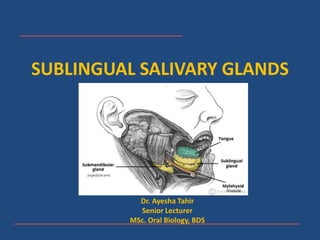
2. SUBLINGUAL SALIVARY GLANDS
• Smallest of the three
major salivary glands
• Mixed in secretion but
predominantly
mucous
• Composed of a major
sublingual gland along
with 8-30 mixed,
minor salivary glands
3. ANATOMY
• Located in the anterior part of the floor of the mouth
• It lies between the hyoglossus and mylohyoid muscles and lies
against the sublingual fossa of the mandible
4. DUCT AND SECRETIONS
• The secretions enter the oral cavity through a series of small ducts, the
ducts of Rivinus
• These ducts open into the sublingual fold in the floor of the mouth or;
• Through a larger duct- Bartholin’s Duct which opens at the sublingual
caruncle along with the submandibular duct
5.
6. BLOOD AND NERVE SUPPLY
• Blood Supply: Sublingual and Sub-mental
Arteries
• Parasympathetic Innervation: Chorda Tympani
branch of the Facial Nerve (CN VII)
• Pre-Ganglionic Fibres: via Lingual nerve to the
submandibular ganglion
• Post-Ganglionic Fibres: From SM Ganglion to the
suBmandibular and sublingual glands
7.
8. HISTOLOGY
• Predominantly mucous in nature
• Mucous tubules and serous demilunes resemble those in SM gland
• Serous end pieces are rare if present at all
• Ductal system is much less developed
• Intercalated ducts are short, difficult to recognize and may be
absent
• Striated ducts are usually absent
• Collecting ducts are rich in mitochondria, lack basal striations
• Sublingual saliva is rich in Sodium
9. MINOR SALIVARY GLANDS
• Estimated to be in between 600-1000 in number
• Small, discrete aggregates of secretory tissue
present in the submucosa, almost throughout the
oral cavity
• Mostly mucous except lingual serous glands
(Ebner’s glands) in tongue – secretion of digestive
enzymes and proteins
• Intercalated ducts are poorly developed
10. • Minor salivary gland secretion is typically rich in
mucins, antibacterial proteins and immunoglobulins
• Protection and moistening the oral cavity especially at
night
12. • Generalized loss of salivary glands parenchyma
• Lost salivary cells are often replaced by adipose
tissue
• Increase in fibrous and vascular elements
• Changes in ductal system:
– Increase in non-striated intralobular ducts
– Dilatation of extra lobular ducts
– Degenerative and metaplastic changes
• Decreased production of saliva
14. • Local and systematic
• Viruses: Infect and replicate within salivary glands e.g.
cytomegalovirus, Epstein-Bar virus
• Viral and bacterial infections may cause inflammation of the glands
• Blockages of ducts
• Ductal obstructions by formation of sialoliths
• Severing of a minor SG by trauma
• Benign and malignant tumours
• Diabetes
• Autoimmune disease e.g. Sjogren’s Syndrome
15. XEROSTOMIA (DRY MOUTH)
• Loss of salivary function or a reduction in the
volume of secreted saliva- dry mouth
• May occur as a side effect of medications (e.g.
antihypertensives, antidepressants) - central or
peripheral inhibition of salivary secretion
• Destruction of salivary gland- chemotherapy/
radiation therapy
• Autoimmune disease- Sjogren’s Disease
17. EFFECTS OF XEROSTOMIA
• Dry mouth- loss of protective effects
• Susceptibility to infections, difficulty and pain in
speech, eating, swallowing
• High susceptibility to caries
• TREATMENT MODALITIES:
– Frequent sipping of water/ artificial saliva
– Pharmacological therapy e.g. pilocarpine
– Genetic modification