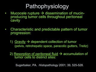
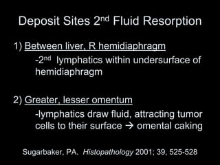
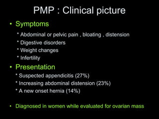
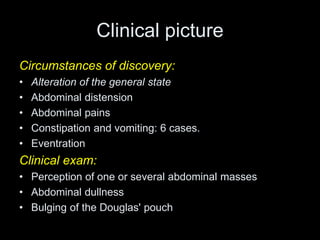
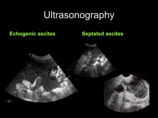
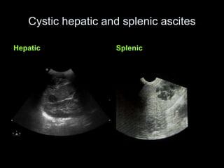
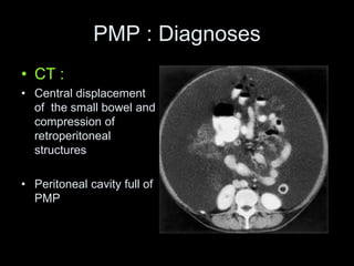
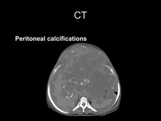
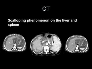
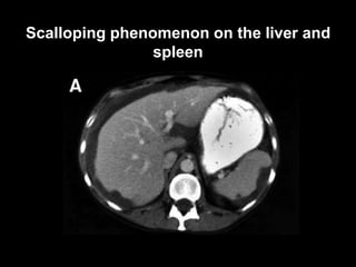
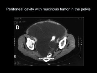
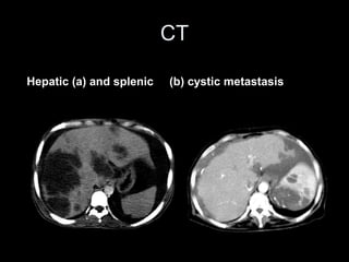
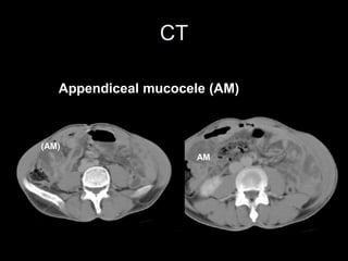
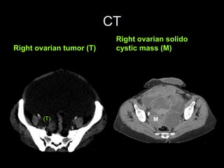
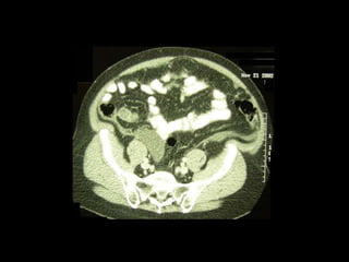
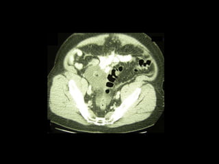
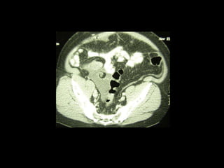
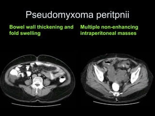
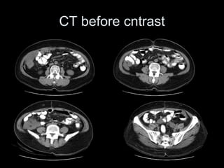
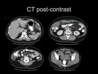
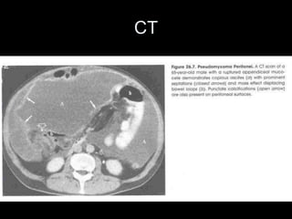
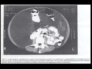
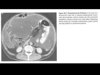
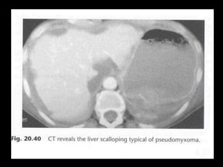
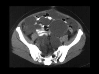
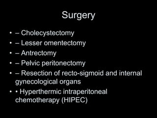
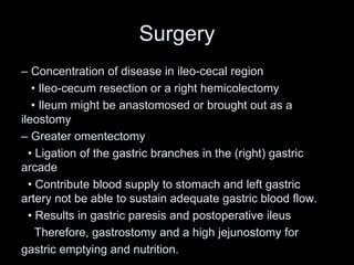
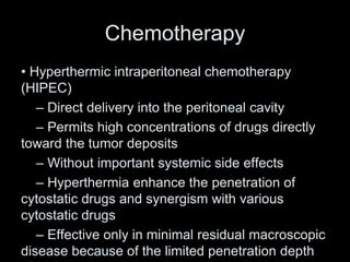
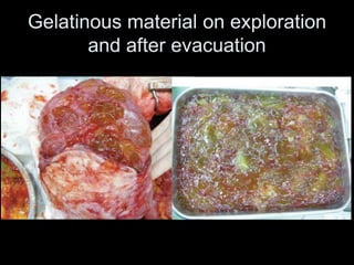
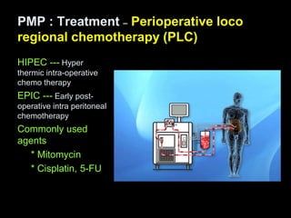
Hamed Rashad
Professor of surgery Banha faculty of medicine
Pseudomyxoma Peritonei and
Mucocele
Appendiceal Cancer
• 1% of appendectomies contain cancer
– 2/3 carcinoid (50% all GI carcinoids are appendiceal)
• Tan-yellow mass, surrounding desmoplastic reaction
• < 2 cm  no further treatment
• > 2 cm  R hemicolectomy
– Remainder: mucinous cystadenocarcinoma,
adenocarcinoma, adenocarcinoid
– Adenocarcinoma associated with metastatic disease
50% of time (likely 2nd appendiceal rupture, tumor
spread)
Appendiceal mucocele
• Dilated, mucin-filled appendix
• 0.3% of all appendectomies
• 4 histologic categories:
1) Retention cyst = simple mucocele (normal mucosa)
2) Mucosal hyperplasia
3) Mucinous cystadenoma (~ papillary adenoma of colon)
4) Mucinous cystadenocarcinoma
Contemporary Surgery, 2002; 58 (12), 609-613
Mucoceles: pathophysiology
• Obstruction of appendiceal lumen 
dilation with mucin
– Obstruction 2nd hyperplasia or fecalith
• Presentation depends on nature/degree of
obstruction:
– Slow, chronic  large mucocele formation
– Fecalith  acute inflammation, with mucinous
content
Mucoceles: natural history
• Asymptomatic (25%), vs. chronic RLQ
pain vs. acute appendicitis
• Mucocele can also lead to:
– Bowel obstruction 2nd intussusception  cecum
– Torsion  gangrenous appendix
– Rupture  mucinous ascites, pseudomyxoma
peritonei
– Risk factor for colon cancer (synchronous,
metachronous)
Differential Diagnosis
• Duplication cyst
• Mesenteric/omental cyst
• Ovarian cyst
• Periappendiceal abscess
Mucoceles: treatment
• Hyperplasia, mucinous cystadenoma
treated by simple appendectomy
• Malignant mucocele:
– Preop/intraop suspicion  R hemicolectomy,
or appendectomy and frozen section
– Post-op diagnosis  return for definitive
procedure
Pseudomyxoma peritonei
Introduction
• The Pseudomyxoma Peritonei is a rare
disease characterized by diffusion of a
gelatinous ascites whether associated or
not to mucinous implants in the peritoneal
cavity.
• It’s secondary to the rupture in the
peritoneal cavity of some mucinoid tumors
whose the origin is frequently appendiceal.
Pseudomyxoma Peritonei
• Extremely rare; 300 cases/yr in U.S.
• Diffuse, intraperitoneal collection of gelatinous
fluid with mucinous tumor implants on peritoneal
surfaces and omentum
• Strictly, etiology is 2nd grade I mucinous
cystadenocarcinoma of the appendix
• Ovarian, pancreatic cancer  similar picture
Epidemiology
• 2-3 times commoner in males than females
• Median ages at diagnoses 51-61 years old
• Present in 2 0ut of 10,000 laparotomy
PMP : Classification
• DPAM (Disseminated Peritoneal AdenoMucineses)
-- Abundant extracellular mucin
-- Little cytological atypia
• PMCA (Peritoneal Mucinous Carcinomatoses)
-- Carcinomatous cytologic features
• PMCA-I (Peritoneal Mucinous Carcimatoses-
Intemediate or discordant features)
-- Focal area of mucinous carcinoma
Ronnett, BM, et al., cancer,2001-92:85
Pathophysiology
• Mucocele rupture  dissemination of mucin-
producing tumor cells throughout peritoneal
cavity
• Characteristic and predictable pattern of tumor
progression:
1) Gravity  dependent collection of tumor
(pelvis, retrohepatic space, paracolic gutters, Treitz)
2) Resorption of peritoneal fluid  accumulation of
tumor cells to distinct sites:
Sugarbaker, PA. Histopathology 2001; 39, 525-528.
Deposit Sites 2nd Fluid Resorption
1) Between liver, R hemidiaphragm
-2nd lymphatics within undersurface of
hemidiaphragm
2) Greater, lesser omentum
-lymphatics draw fluid, attracting tumor
cells to their surface  omental caking
Sugarbaker, PA. Histopathology 2001; 39, 525-528
PMP : Clinical picture
• Symptoms
* Abdominal or pelvic pain , bloating , distension
* Digestive disorders
* Weight changes
* Infertility
• Presentation
* Suspected appendicitis (27%)
* Increasing abdominal distension (23%)
* A new onset hernia (14%)
• Diagnosed in women while evaluated for ovarian mass
Clinical picture
Circumstances of discovery:
• Alteration of the general state
• Abdominal distension
• Abdominal pains
• Constipation and vomiting: 6 cases.
• Eventration
Clinical exam:
• Perception of one or several abdominal masses
• Abdominal dullness
• Bulging of the Douglas' pouch
Ultrasonography
Echogenic ascites Septated ascites
Cystic hepatic and splenic ascites
Hepatic Splenic
PMP : Diagnoses
• CT :
• Central displacement
of the small bowel and
compression of
retroperitoneal
structures
• Peritoneal cavity full of
PMP
CT
Peritoneal calcifications
CT
Scalloping phenomenon on the liver and
spleen
Scalloping phenomenon on the liver and
spleen
Peritoneal cavity with mucinous tumor in the pelvis
CT
Hepatic (a) and splenic (b) cystic metastasis
CT
Appendiceal mucocele (AM)
(AM)
AM
CT
Right ovarian tumor (T)
Right ovarian solido
cystic mass (M)
(T) M
Pseudomyxoma peritpnii
Bowel wall thickening and
fold swelling
Multiple non-enhancing
intraperitoneal masses
CT before cntrast
CT post-contrast
Corresponding finding on laparotomy to previous CT
CT
Workup
• CT-guided aspiration of pelvic fluid
• Analysis: mucinous adenocarcinoma
• Taken to OR for exploratory laparoscopy
Treatment
• Traditional Surgical Treatment
– Consists of
• Cytoreductive surgery
• Removal of all mucinous ascites
• ± Additional modalities
– Long-term survival could not be achieved
by surgery alone
Surgery
– Aggressive cytoreductive surgery
• Parietal peritonectomy
– Greater omentectomy
– Splenectomy
– Stripping of the left diaphragm and the
right diaphragm
Surgery
• – Cholecystectomy
• – Lesser omentectomy
• – Antrectomy
• – Pelvic peritonectomy
• – Resection of recto-sigmoid and internal
gynecological organs
• • Hyperthermic intraperitoneal
chemotherapy (HIPEC)
Surgery
– Concentration of disease in ileo-cecal region
• Ileo-cecum resection or a right hemicolectomy
• Ileum might be anastomosed or brought out as a
ileostomy
– Greater omentectomy
• Ligation of the gastric branches in the (right) gastric
arcade
• Contribute blood supply to stomach and left gastric
artery not be able to sustain adequate gastric blood flow.
• Results in gastric paresis and postoperative ileus
Therefore, gastrostomy and a high jejunostomy for
gastric emptying and nutrition.
Chemotherapy
• Hyperthermic intraperitoneal chemotherapy
(HIPEC)
– Direct delivery into the peritoneal cavity
– Permits high concentrations of drugs directly
toward the tumor deposits
– Without important systemic side effects
– Hyperthermia enhance the penetration of
cytostatic drugs and synergism with various
cytostatic drugs
– Effective only in minimal residual macroscopic
disease because of the limited penetration depth
– Chemotherapeutic agents
• Doxorubicin (15mg/m2)
• Melphalan
• Mitomycin-C (12mg/m2)
• Cisplatin (50mg/m2)
• Gemcitabine
• Mitoxantrone
• Oxaliplatin
• Etoposide
• Irinotecan
• Paclitaxel
• Docetaxel
• 5-Fuorouracil (500 mg/m2)
• Carboplatin
– Carriers of chemotherapeutic agents
• Isotonic salt solutions and dextrose solutions
– Rapid absorption
– Inability to maintain a prolonged high
intraperitoneal fluid volume
–Most commonly used
• Hypotonic solutions
– Cisplatin accumulates in tumor cells &
enhances its cytotoxicity
– High incidence of unexplained postoperative
peritoneal bleeding with oxaloplatin
– Duration for perfusion
• 410C x 90 min
• 430C x 30–40 min
• 420C x 60 min
– Complications
• 30-45%
• Chemotherapy toxicity to kidneys, bone
marrow, liver,
lungs- 2-5%
• Organ damage secondary to hyperthermia
• Surgical complications – 25-30%
– Small bowel fistula
• Moratlity - 0-5%
Other treatment modality
– Laparoscopic approach
• • allows thorough exploration of the
abdomen
• • irrigation and aspiration of the thick
mucinous material, and the instillation of
mucolytic agents, 5% dextrose solution.
– Should be performed intra-operatively before
anastomoses,
to maximize uniform drug distribution and tumor exposure
Los G et al. Cancer Res 1992;52:1252-8
– In h/o prior surgery, adhesiolysis should be performed to
prevent nonuniform drug distribution
• All microscopic tumor residue is not eliminated
• Lead to recurrent disease
– Mitomycin as an intraperitoneal drug, either as single drug
or in combination
van Ruth S et al. Surg Oncol Clin N Am 2003;12:771-80.
HIPC
– Two techniques
• Open
– Small bowel floats in drug solution
• Closed
– No loss of heat or drug
– Entire peritoneum is exposed
– Allow administration of drug under
pressure
Sugarbaker Protocol
• Radical debulking of tumor load:
– appendix, peritoneum, omentum;
– additional viscera as indicated
– Curative therapy = all nodules > 2.5 mm
• Intraoperative heated mitomycin (44c)
• Post-operative 5-FU
• Reports of 80% 10 yr survival
Gelatinous material on exploration
and after evacuation
Laparoscopic view : showing gelatinous
material on peritoneal surface
Operation
• Rt hemicolectomy
• Harman’ procedure
• Debulking of tumour
– Mucinous tumor on parietal peritoneum
• Resection by high-power electrocautery with a
balltipped handpiece on high voltage pure cut
PMP : Treatment – Perioperative loco
regional chemotherapy (PLC)
HIPEC --- Hyper
thermic intra-operative
chemo therapy
EPIC --- Early post-
operative intra peritoneal
chemotherapy
Commonly used
agents
* Mitomycin
* Cisplatin, 5-FU
Further
• Other reports have 5-year survival of 50-
75%
• CEA, CA 19-9 of some use in surveillance
• Laparoscopic treatment reported by select
institutions (Cleveland Clinic)
Summary
• The Pseudomyxoma Peritonei is a dangerous
complication of the mucinous tumors dominated by the
appendiceal origin.
• the medical imaging is fundamental in the diagnosis and
the surveillance of this rare pathology. US permits a
better characterization of the ascites whereas the CT
visualizes the calcifications and the scalloping effect.
• A good knowledge of its particularities
physiopathological and radiological by the physician
radiologist is important to put in guard the surgeon.
Summary
• incidence is 2 per million per year with an unexplained
female dominance
• Redistribution phenomenon predicts location It is thought
to be peritoneal disseminated disease from a primary
appendiceal mucinous epithelial neoplasm unless (very
rarely) proven otherwise with a normal appendix and a
primary tumor elsewhere
• It is suspected in case of intraperitoneal mucus with
cellular content, found during laparotomy or at physical
examination (abdominal girth)
• combination of (aggressive) surgical debulking with
peritonectomy and hyperthermic intraperitoneal
chemotherapy seems to improve the outcome.
Summary
• • Patients with high-grade disease (peritoneal
mucinous carcinomatosis), disease extent with
more than 5 involved abdominal regions, and/or
small bowel involvement should receive
palliative treatment because they do not benefit
from aggressive treatment approaches
PMP : Summary
• Cytoreductive surgery with/without intra-
peritoneal chemotherapy may be the best
treatment strategy for PMP
• Surgery should be done to remain no or
minimal residual tumour
THANK YOU

Pseudomyxoma Peritonei the lect.ppt

  • 1.
    Hamed Rashad Professor ofsurgery Banha faculty of medicine Pseudomyxoma Peritonei and Mucocele
  • 2.
    Appendiceal Cancer • 1%of appendectomies contain cancer – 2/3 carcinoid (50% all GI carcinoids are appendiceal) • Tan-yellow mass, surrounding desmoplastic reaction • < 2 cm  no further treatment • > 2 cm  R hemicolectomy – Remainder: mucinous cystadenocarcinoma, adenocarcinoma, adenocarcinoid – Adenocarcinoma associated with metastatic disease 50% of time (likely 2nd appendiceal rupture, tumor spread)
  • 4.
    Appendiceal mucocele • Dilated,mucin-filled appendix • 0.3% of all appendectomies • 4 histologic categories: 1) Retention cyst = simple mucocele (normal mucosa) 2) Mucosal hyperplasia 3) Mucinous cystadenoma (~ papillary adenoma of colon) 4) Mucinous cystadenocarcinoma Contemporary Surgery, 2002; 58 (12), 609-613
  • 5.
    Mucoceles: pathophysiology • Obstructionof appendiceal lumen  dilation with mucin – Obstruction 2nd hyperplasia or fecalith • Presentation depends on nature/degree of obstruction: – Slow, chronic  large mucocele formation – Fecalith  acute inflammation, with mucinous content
  • 6.
    Mucoceles: natural history •Asymptomatic (25%), vs. chronic RLQ pain vs. acute appendicitis • Mucocele can also lead to: – Bowel obstruction 2nd intussusception  cecum – Torsion  gangrenous appendix – Rupture  mucinous ascites, pseudomyxoma peritonei – Risk factor for colon cancer (synchronous, metachronous)
  • 9.
    Differential Diagnosis • Duplicationcyst • Mesenteric/omental cyst • Ovarian cyst • Periappendiceal abscess
  • 11.
    Mucoceles: treatment • Hyperplasia,mucinous cystadenoma treated by simple appendectomy • Malignant mucocele: – Preop/intraop suspicion  R hemicolectomy, or appendectomy and frozen section – Post-op diagnosis  return for definitive procedure
  • 12.
  • 13.
    Introduction • The PseudomyxomaPeritonei is a rare disease characterized by diffusion of a gelatinous ascites whether associated or not to mucinous implants in the peritoneal cavity. • It’s secondary to the rupture in the peritoneal cavity of some mucinoid tumors whose the origin is frequently appendiceal.
  • 14.
    Pseudomyxoma Peritonei • Extremelyrare; 300 cases/yr in U.S. • Diffuse, intraperitoneal collection of gelatinous fluid with mucinous tumor implants on peritoneal surfaces and omentum • Strictly, etiology is 2nd grade I mucinous cystadenocarcinoma of the appendix • Ovarian, pancreatic cancer  similar picture
  • 15.
    Epidemiology • 2-3 timescommoner in males than females • Median ages at diagnoses 51-61 years old • Present in 2 0ut of 10,000 laparotomy
  • 16.
    PMP : Classification •DPAM (Disseminated Peritoneal AdenoMucineses) -- Abundant extracellular mucin -- Little cytological atypia • PMCA (Peritoneal Mucinous Carcinomatoses) -- Carcinomatous cytologic features • PMCA-I (Peritoneal Mucinous Carcimatoses- Intemediate or discordant features) -- Focal area of mucinous carcinoma Ronnett, BM, et al., cancer,2001-92:85
  • 17.
    Pathophysiology • Mucocele rupture dissemination of mucin- producing tumor cells throughout peritoneal cavity • Characteristic and predictable pattern of tumor progression: 1) Gravity  dependent collection of tumor (pelvis, retrohepatic space, paracolic gutters, Treitz) 2) Resorption of peritoneal fluid  accumulation of tumor cells to distinct sites: Sugarbaker, PA. Histopathology 2001; 39, 525-528.
  • 18.
    Deposit Sites 2ndFluid Resorption 1) Between liver, R hemidiaphragm -2nd lymphatics within undersurface of hemidiaphragm 2) Greater, lesser omentum -lymphatics draw fluid, attracting tumor cells to their surface  omental caking Sugarbaker, PA. Histopathology 2001; 39, 525-528
  • 19.
    PMP : Clinicalpicture • Symptoms * Abdominal or pelvic pain , bloating , distension * Digestive disorders * Weight changes * Infertility • Presentation * Suspected appendicitis (27%) * Increasing abdominal distension (23%) * A new onset hernia (14%) • Diagnosed in women while evaluated for ovarian mass
  • 20.
    Clinical picture Circumstances ofdiscovery: • Alteration of the general state • Abdominal distension • Abdominal pains • Constipation and vomiting: 6 cases. • Eventration Clinical exam: • Perception of one or several abdominal masses • Abdominal dullness • Bulging of the Douglas' pouch
  • 21.
  • 22.
    Cystic hepatic andsplenic ascites Hepatic Splenic
  • 23.
    PMP : Diagnoses •CT : • Central displacement of the small bowel and compression of retroperitoneal structures • Peritoneal cavity full of PMP
  • 24.
  • 25.
    CT Scalloping phenomenon onthe liver and spleen
  • 26.
    Scalloping phenomenon onthe liver and spleen
  • 27.
    Peritoneal cavity withmucinous tumor in the pelvis
  • 28.
    CT Hepatic (a) andsplenic (b) cystic metastasis
  • 29.
  • 30.
    CT Right ovarian tumor(T) Right ovarian solido cystic mass (M) (T) M
  • 34.
    Pseudomyxoma peritpnii Bowel wallthickening and fold swelling Multiple non-enhancing intraperitoneal masses
  • 35.
  • 36.
  • 37.
    Corresponding finding onlaparotomy to previous CT
  • 38.
  • 43.
    Workup • CT-guided aspirationof pelvic fluid • Analysis: mucinous adenocarcinoma • Taken to OR for exploratory laparoscopy
  • 44.
    Treatment • Traditional SurgicalTreatment – Consists of • Cytoreductive surgery • Removal of all mucinous ascites • ± Additional modalities – Long-term survival could not be achieved by surgery alone
  • 45.
    Surgery – Aggressive cytoreductivesurgery • Parietal peritonectomy – Greater omentectomy – Splenectomy – Stripping of the left diaphragm and the right diaphragm
  • 46.
    Surgery • – Cholecystectomy •– Lesser omentectomy • – Antrectomy • – Pelvic peritonectomy • – Resection of recto-sigmoid and internal gynecological organs • • Hyperthermic intraperitoneal chemotherapy (HIPEC)
  • 47.
    Surgery – Concentration ofdisease in ileo-cecal region • Ileo-cecum resection or a right hemicolectomy • Ileum might be anastomosed or brought out as a ileostomy – Greater omentectomy • Ligation of the gastric branches in the (right) gastric arcade • Contribute blood supply to stomach and left gastric artery not be able to sustain adequate gastric blood flow. • Results in gastric paresis and postoperative ileus Therefore, gastrostomy and a high jejunostomy for gastric emptying and nutrition.
  • 48.
    Chemotherapy • Hyperthermic intraperitonealchemotherapy (HIPEC) – Direct delivery into the peritoneal cavity – Permits high concentrations of drugs directly toward the tumor deposits – Without important systemic side effects – Hyperthermia enhance the penetration of cytostatic drugs and synergism with various cytostatic drugs – Effective only in minimal residual macroscopic disease because of the limited penetration depth
  • 49.
    – Chemotherapeutic agents •Doxorubicin (15mg/m2) • Melphalan • Mitomycin-C (12mg/m2) • Cisplatin (50mg/m2) • Gemcitabine • Mitoxantrone • Oxaliplatin • Etoposide • Irinotecan • Paclitaxel • Docetaxel • 5-Fuorouracil (500 mg/m2) • Carboplatin
  • 50.
    – Carriers ofchemotherapeutic agents • Isotonic salt solutions and dextrose solutions – Rapid absorption – Inability to maintain a prolonged high intraperitoneal fluid volume –Most commonly used • Hypotonic solutions – Cisplatin accumulates in tumor cells & enhances its cytotoxicity – High incidence of unexplained postoperative peritoneal bleeding with oxaloplatin
  • 51.
    – Duration forperfusion • 410C x 90 min • 430C x 30–40 min • 420C x 60 min – Complications • 30-45% • Chemotherapy toxicity to kidneys, bone marrow, liver, lungs- 2-5% • Organ damage secondary to hyperthermia • Surgical complications – 25-30% – Small bowel fistula • Moratlity - 0-5%
  • 53.
    Other treatment modality –Laparoscopic approach • • allows thorough exploration of the abdomen • • irrigation and aspiration of the thick mucinous material, and the instillation of mucolytic agents, 5% dextrose solution.
  • 54.
    – Should beperformed intra-operatively before anastomoses, to maximize uniform drug distribution and tumor exposure Los G et al. Cancer Res 1992;52:1252-8 – In h/o prior surgery, adhesiolysis should be performed to prevent nonuniform drug distribution • All microscopic tumor residue is not eliminated • Lead to recurrent disease – Mitomycin as an intraperitoneal drug, either as single drug or in combination van Ruth S et al. Surg Oncol Clin N Am 2003;12:771-80.
  • 55.
    HIPC – Two techniques •Open – Small bowel floats in drug solution • Closed – No loss of heat or drug – Entire peritoneum is exposed – Allow administration of drug under pressure
  • 56.
    Sugarbaker Protocol • Radicaldebulking of tumor load: – appendix, peritoneum, omentum; – additional viscera as indicated – Curative therapy = all nodules > 2.5 mm • Intraoperative heated mitomycin (44c) • Post-operative 5-FU • Reports of 80% 10 yr survival
  • 57.
    Gelatinous material onexploration and after evacuation
  • 58.
    Laparoscopic view :showing gelatinous material on peritoneal surface
  • 59.
    Operation • Rt hemicolectomy •Harman’ procedure • Debulking of tumour
  • 60.
    – Mucinous tumoron parietal peritoneum • Resection by high-power electrocautery with a balltipped handpiece on high voltage pure cut
  • 61.
    PMP : Treatment– Perioperative loco regional chemotherapy (PLC) HIPEC --- Hyper thermic intra-operative chemo therapy EPIC --- Early post- operative intra peritoneal chemotherapy Commonly used agents * Mitomycin * Cisplatin, 5-FU
  • 62.
    Further • Other reportshave 5-year survival of 50- 75% • CEA, CA 19-9 of some use in surveillance • Laparoscopic treatment reported by select institutions (Cleveland Clinic)
  • 63.
    Summary • The PseudomyxomaPeritonei is a dangerous complication of the mucinous tumors dominated by the appendiceal origin. • the medical imaging is fundamental in the diagnosis and the surveillance of this rare pathology. US permits a better characterization of the ascites whereas the CT visualizes the calcifications and the scalloping effect. • A good knowledge of its particularities physiopathological and radiological by the physician radiologist is important to put in guard the surgeon.
  • 64.
    Summary • incidence is2 per million per year with an unexplained female dominance • Redistribution phenomenon predicts location It is thought to be peritoneal disseminated disease from a primary appendiceal mucinous epithelial neoplasm unless (very rarely) proven otherwise with a normal appendix and a primary tumor elsewhere • It is suspected in case of intraperitoneal mucus with cellular content, found during laparotomy or at physical examination (abdominal girth) • combination of (aggressive) surgical debulking with peritonectomy and hyperthermic intraperitoneal chemotherapy seems to improve the outcome.
  • 65.
    Summary • • Patientswith high-grade disease (peritoneal mucinous carcinomatosis), disease extent with more than 5 involved abdominal regions, and/or small bowel involvement should receive palliative treatment because they do not benefit from aggressive treatment approaches
  • 66.
    PMP : Summary •Cytoreductive surgery with/without intra- peritoneal chemotherapy may be the best treatment strategy for PMP • Surgery should be done to remain no or minimal residual tumour
  • 67.

Editor's Notes