This document discusses the structure and features of prokaryotic and eukaryotic cells. It begins by describing the key differences between prokaryotic and eukaryotic cells, such as size, presence of organelles, and genetic material organization. The structure of prokaryotic cells is then explained, noting they lack membrane-bound organelles and have circular DNA in the cytosol. Eukaryotic cells are more complex, containing membrane-bound organelles including the nucleus which encloses the genetic material. Key organelles of animal and plant eukaryotic cells like the mitochondria, chloroplasts, and nuclei are then described in detail.
Molecular biology is the study of biology at a molecular level.
In broad sense, the study of gene structure and functions at the molecular level to understand the molecular basis of hereditary, genetic variation, and the expression patterns of genes.The field overlaps with other areas of biology and chemistry, particularly genetics and biochemistry.
This topic explains genomics and proteomics and types of genomics and proteoics and explains about positional cloning,microsatellites,SNP,VNTRS,HUMAN GENOME PRPJECT
Cell biology is the study of cell structure and function, and it revolves around the concept that the cell is the fundamental unit of life. Focusing on the cell permits a detailed understanding of the tissues and organisms that cells compose.
Molecular biology is a branch of science concerning biological activity at the molecular level.
The field of molecular biology overlaps with biology and chemistry and in particular, genetics and biochemistry.
A key area of molecular biology concerns understanding how various cellular systems interact in terms of the way DNA, RNA and protein synthesis function.
Molecular biology is the study of molecular underpinnings of the process of replication, transcription and translation of the genetic material.
There isn't one single person credited with discovering the mitochondria, as over the years a number of scientists have made important contributions to the study of the discovery of this important cellular structure:
1800s In 1857, Albert von Kölliker described what he called “granules” in the cells of muscles.
- Other scientists of the era also noticed these “granules” in other cell types.
1886 , when Richard Altman, a cytologist, identified the organelles using a dye technique, and dubbed them “bioblasts.” He postulated that the structures were the basic units of cellular activity.
1898, Carl Benda coined the term mitochondria. He derived the term from the Greek language for the words thread, mitos, and granule, chondros.
-Though mitochondria are an integral part of the cell, evidence shows that they evolved from primitive bacteria.
Analyzing the different organelles in eukaryotic & prokaryotic cells thro...Umair Raza
Analyzing the different organelles in Eukaryotic & Prokaryotic cells through their functions (List of organelles, structure, composition, diagram, charts, functions) by Engr. Umair Raza
Structure of prokariyotic and eukariyotic cells By KK Sahu SirKAUSHAL SAHU
Introduction
Origin
Prokaryotic cell structure
Eukaryotic cell structure
Comparision of prokaryotes and eukaryotes
Special points
Conclusion
The invention of electron microscope lead to the detailed study of cells.
Based on these studies, cells were classified as prokaryotes (Gr,pro-primitve; karyon-nucleus) and eukaryotes (Gr,eu-true; karyon-nucleus)
This presentation provides brief and relevant description of eukaryotic cell organisation. Well labeled figures and pictorial representations are made to give easy understanding to the readers. References are added at the end of the presentation so the readers can get detailed knowledge from the referred books.
- Video recording of this lecture in English language: https://youtu.be/lK81BzxMqdo
- Video recording of this lecture in Arabic language: https://youtu.be/Ve4P0COk9OI
- Link to download the book free: https://nephrotube.blogspot.com/p/nephrotube-nephrology-books.html
- Link to NephroTube website: www.NephroTube.com
- Link to NephroTube social media accounts: https://nephrotube.blogspot.com/p/join-nephrotube-on-social-media.html
Couples presenting to the infertility clinic- Do they really have infertility...Sujoy Dasgupta
Dr Sujoy Dasgupta presented the study on "Couples presenting to the infertility clinic- Do they really have infertility? – The unexplored stories of non-consummation" in the 13th Congress of the Asia Pacific Initiative on Reproduction (ASPIRE 2024) at Manila on 24 May, 2024.
Prix Galien International 2024 Forum ProgramLevi Shapiro
June 20, 2024, Prix Galien International and Jerusalem Ethics Forum in ROME. Detailed agenda including panels:
- ADVANCES IN CARDIOLOGY: A NEW PARADIGM IS COMING
- WOMEN’S HEALTH: FERTILITY PRESERVATION
- WHAT’S NEW IN THE TREATMENT OF INFECTIOUS,
ONCOLOGICAL AND INFLAMMATORY SKIN DISEASES?
- ARTIFICIAL INTELLIGENCE AND ETHICS
- GENE THERAPY
- BEYOND BORDERS: GLOBAL INITIATIVES FOR DEMOCRATIZING LIFE SCIENCE TECHNOLOGIES AND PROMOTING ACCESS TO HEALTHCARE
- ETHICAL CHALLENGES IN LIFE SCIENCES
- Prix Galien International Awards Ceremony
MANAGEMENT OF ATRIOVENTRICULAR CONDUCTION BLOCK.pdfJim Jacob Roy
Cardiac conduction defects can occur due to various causes.
Atrioventricular conduction blocks ( AV blocks ) are classified into 3 types.
This document describes the acute management of AV block.
Report Back from SGO 2024: What’s the Latest in Cervical Cancer?bkling
Are you curious about what’s new in cervical cancer research or unsure what the findings mean? Join Dr. Emily Ko, a gynecologic oncologist at Penn Medicine, to learn about the latest updates from the Society of Gynecologic Oncology (SGO) 2024 Annual Meeting on Women’s Cancer. Dr. Ko will discuss what the research presented at the conference means for you and answer your questions about the new developments.
Title: Sense of Smell
Presenter: Dr. Faiza, Assistant Professor of Physiology
Qualifications:
MBBS (Best Graduate, AIMC Lahore)
FCPS Physiology
ICMT, CHPE, DHPE (STMU)
MPH (GC University, Faisalabad)
MBA (Virtual University of Pakistan)
Learning Objectives:
Describe the primary categories of smells and the concept of odor blindness.
Explain the structure and location of the olfactory membrane and mucosa, including the types and roles of cells involved in olfaction.
Describe the pathway and mechanisms of olfactory signal transmission from the olfactory receptors to the brain.
Illustrate the biochemical cascade triggered by odorant binding to olfactory receptors, including the role of G-proteins and second messengers in generating an action potential.
Identify different types of olfactory disorders such as anosmia, hyposmia, hyperosmia, and dysosmia, including their potential causes.
Key Topics:
Olfactory Genes:
3% of the human genome accounts for olfactory genes.
400 genes for odorant receptors.
Olfactory Membrane:
Located in the superior part of the nasal cavity.
Medially: Folds downward along the superior septum.
Laterally: Folds over the superior turbinate and upper surface of the middle turbinate.
Total surface area: 5-10 square centimeters.
Olfactory Mucosa:
Olfactory Cells: Bipolar nerve cells derived from the CNS (100 million), with 4-25 olfactory cilia per cell.
Sustentacular Cells: Produce mucus and maintain ionic and molecular environment.
Basal Cells: Replace worn-out olfactory cells with an average lifespan of 1-2 months.
Bowman’s Gland: Secretes mucus.
Stimulation of Olfactory Cells:
Odorant dissolves in mucus and attaches to receptors on olfactory cilia.
Involves a cascade effect through G-proteins and second messengers, leading to depolarization and action potential generation in the olfactory nerve.
Quality of a Good Odorant:
Small (3-20 Carbon atoms), volatile, water-soluble, and lipid-soluble.
Facilitated by odorant-binding proteins in mucus.
Membrane Potential and Action Potential:
Resting membrane potential: -55mV.
Action potential frequency in the olfactory nerve increases with odorant strength.
Adaptation Towards the Sense of Smell:
Rapid adaptation within the first second, with further slow adaptation.
Psychological adaptation greater than receptor adaptation, involving feedback inhibition from the central nervous system.
Primary Sensations of Smell:
Camphoraceous, Musky, Floral, Pepperminty, Ethereal, Pungent, Putrid.
Odor Detection Threshold:
Examples: Hydrogen sulfide (0.0005 ppm), Methyl-mercaptan (0.002 ppm).
Some toxic substances are odorless at lethal concentrations.
Characteristics of Smell:
Odor blindness for single substances due to lack of appropriate receptor protein.
Behavioral and emotional influences of smell.
Transmission of Olfactory Signals:
From olfactory cells to glomeruli in the olfactory bulb, involving lateral inhibition.
Primitive, less old, and new olfactory systems with different path
Flu Vaccine Alert in Bangalore Karnatakaaddon Scans
As flu season approaches, health officials in Bangalore, Karnataka, are urging residents to get their flu vaccinations. The seasonal flu, while common, can lead to severe health complications, particularly for vulnerable populations such as young children, the elderly, and those with underlying health conditions.
Dr. Vidisha Kumari, a leading epidemiologist in Bangalore, emphasizes the importance of getting vaccinated. "The flu vaccine is our best defense against the influenza virus. It not only protects individuals but also helps prevent the spread of the virus in our communities," he says.
This year, the flu season is expected to coincide with a potential increase in other respiratory illnesses. The Karnataka Health Department has launched an awareness campaign highlighting the significance of flu vaccinations. They have set up multiple vaccination centers across Bangalore, making it convenient for residents to receive their shots.
To encourage widespread vaccination, the government is also collaborating with local schools, workplaces, and community centers to facilitate vaccination drives. Special attention is being given to ensuring that the vaccine is accessible to all, including marginalized communities who may have limited access to healthcare.
Residents are reminded that the flu vaccine is safe and effective. Common side effects are mild and may include soreness at the injection site, mild fever, or muscle aches. These side effects are generally short-lived and far less severe than the flu itself.
Healthcare providers are also stressing the importance of continuing COVID-19 precautions. Wearing masks, practicing good hand hygiene, and maintaining social distancing are still crucial, especially in crowded places.
Protect yourself and your loved ones by getting vaccinated. Together, we can help keep Bangalore healthy and safe this flu season. For more information on vaccination centers and schedules, residents can visit the Karnataka Health Department’s official website or follow their social media pages.
Stay informed, stay safe, and get your flu shot today!
Factory Supply Best Quality Pmk Oil CAS 28578–16–7 PMK Powder in Stockrebeccabio
Factory Supply Best Quality Pmk Oil CAS 28578–16–7 PMK Powder in Stock
Telegram: bmksupplier
signal: +85264872720
threema: TUD4A6YC
You can contact me on Telegram or Threema
Communicate promptly and reply
Free of customs clearance, Double Clearance 100% pass delivery to USA, Canada, Spain, Germany, Netherland, Poland, Italy, Sweden, UK, Czech Republic, Australia, Mexico, Russia, Ukraine, Kazakhstan.Door to door service
Hot Selling Organic intermediates
Factory Supply Best Quality Pmk Oil CAS 28578–16–7 PMK Powder in Stock
Prokaryotic and eukaryotic cells
1. NPTEL – Biotechnology – Fundamentals of Biotechnology
Joint initiative of IITs and IISc – Funded by MHRD Page 1 of 49
Lecture 3: Prokaryotic and Eukaryotic Cells
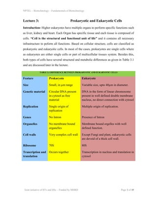
Introduction- Higher eukaryotes have multiple organs to perform specific functions such
as liver, kidney and heart. Each Organ has specific tissue and each tissue is composed of
cells. “Cell is the structural and functional unit of life” and it contains all necessary
infrastructure to peform all functions. Based on cellular structure, cells are classified as
prokaryotic and eukaryotic cells. In most of the cases, prokaryotes are single cells where
as eukaryotes are either single cells or part of multicellular tissues system. Besides this,
both types of cells have several structural and metabolic differences as given in Table 3.1
and are discussed later in the lecture.
TABLE 3.1 DIFFERENCE BETWEEN PROKARYOTIC AND EUKARYOTIC CELLS
Feature Prokaryote Eukaryote
Size Small, in µm range Variable size, upto 40µm in diameter.
Genetic material Circular DNA present
in cytosol as free
material
DNA in the form of linear chromosome
present in well defined double membrane
nucleus, no direct connection with cytosol
Replication Single origin of
replication
Multiple origin of replication.
Genes No Intron Presence of Intron
Organelles No membrane bound
organelles
Membrane bound orgelles with well
defined function.
Cell walls Very complex cell wall Except Fungi and plant, eukaryotic cells
are devoid of a thick cell wall.
Ribosome 70S 80S
Trancription and
translation
Occurs together Transcription in nucleus and translation in
cytosol
2. NPTEL – Biotechnology – Fundamentals of Biotechnology
Joint initiative of IITs and IISc – Funded by MHRD Page 2 of 49
Structure of Prokaryotic cells- A prokaryotic cell is much simpler and smaller than
eukarotic cells. It lacks membrane bound organelles including nucleus. A typical
prokaryotic cells is shown in Figure 3.1, A. The description of different structural feature
of prokaryotic cells is as follows-
1. Outer Flagella: A flagellum attached to the bacterial capsule is a central feature of
most of the prokaryotic cell especially of the motile bacteria. It provides motion or
locomotion to the bacteria and be responsible for chemotaxsis of bacteria. Movement of
bacteria towards a chemical gradient (such as glucose) is known as chemotaxsis.
Flagellum is a part of cell wall and its motion is regulated by motor proteins present
inside the cell. Flagellar motion is an energy consuming process and it is governed by an
ATPase present at the bottom of the shaft. It is made up of protein flagellin and reduction
or suppression of flagellar protein reduces bacterial infectivity (pathogenicity) and ability
to grow.
Figure 3.1: Structural details of a typical prokaryotic cell. (A) Whole cell and (B) composition of cell wall of gram negative and positive bacteria.
3. NPTEL – Biotechnology – Fundamentals of Biotechnology
Joint initiative of IITs and IISc – Funded by MHRD Page 3 of 49
2. Bacterial surface layers: Bacteria posses 3 anatomical barriers to protect the cells
from external damage. Bacterial capsule is the outer most layer and made up of high
molecular weight polysaccharides. It is impermeable to the water or other aqueous
solvent and it is responsible for antigenicity of bacterial cells. Cell wall in bacteria and its
response to gram staining is the basis of classification of bacterial species.
WHAT IS GRAM STAINING? Gram staining is developed by a Danish scientist Hans
Christian Gram. This technique differentiates bacterial strains based on their cell wall
composition, especially thickness of peptidoglycan layer. A detail staining procedure is
given in following paper (Use of the gram stain in microbiology. Beveridge, TJ (2001)
Biotech Histochem 76 (3): 111–8. Pubmed ID: 11475313). During the staining
procedure bacterial sample is stained with two dyes, crystal violet and safarin. During a
washing step with non-polar solvents such as alcohol or acetone (decolorization), gram –
ve bacteria leave the blue stain due to a thin peptidoglycan layer in cell wall whereas
gram +ve bacteria retains both stains and appear as Pink.
Cell wall composition in gram-ve and gram +ve bacteria is different. Bacterial cell wall
has different constituents and be responsible for their reactivity towards gram stain.
A. Peptidoglycan layer: peptidoglycan layer is thick in gram +ve bacteria and thin in
gram –ve bacteria. Peptidoglycan is a polymer of NAG (N-acetyl-glucosamine) and
NAM (N-acetyl-muramic acid) linked by a β-(1,4) linkage. Sugar polymer are attached to
peptide chain composed of amino acids, L-alanine, D-glutamic acid, L-lysine and D-
alanine. Peptide chain present in one layer cross linked to the next layer to form a mesh
work and be responsible for physical strength of the cell wall. Peptidoglycan synthesis is
targeted by antibiotics such as pencillin where as lysozyme (present in human saliva or
tears) degrades the peptidoglycan layer by cleaving glycosidic bond connecting NAG-
NAM to form polymer.
B. Lipoteichoic acids: Lipoteichoic acid (LTA) are only found in gram +ve bacteria cell
wall and it is an important antigenic determinant.
C. Lipopolysaccharides (LPS)- Lipopolysaccharides (LPS) are found only in gram –ve
bacterial cell wall and it is an important antigenic determinant.
4. NPTEL – Biotechnology – Fundamentals of Biotechnology
Joint initiative of IITs and IISc – Funded by MHRD Page 4 of 49
3. Cytosol and other organelles-Prokaryotic cells do not contain any membrane bound
organelle. The organelles are present in cytosol such as ribosome (70S), genetic material
where as electron transport chain complexes are embedded within the plasma membrane.
4. Chromosome and extra chromosomal DNA-Prokaryote cell contains genetic
material in the form of circular DNA, known as “bacterial chromosome”. It contains
genetic elements for replication, transcription and translation. Bacterial chromosome
follows a rolling circle mode of DNA replication. The genes present on chromosome
does not contains non coding region (introns) and it is co-translated to protein. Besides
main circle DNA, bacteria also contains extra chromosomal circular DNA known as
“plasmid”. Presence of plasmid containing resistance gene confers resistance towards
known antibiotics. Exchange of extra-chromosomal DNA between different bacterial
strains is one of the mechanisms responsible for spread of antibiotic resistance across the
bacterial population. Details of plasmid and its structural features will be discussed in a
later lecture.
5. NPTEL – Biotechnology – Fundamentals of Biotechnology
Joint initiative of IITs and IISc – Funded by MHRD Page 5 of 49
Struture of Eukaryotic cell- The eukaryotic cell is much more complex and it contains
many membrane bound organelles to perform specific functions. It contains a nucleus
isolated from cytosol and enclosed in a well defined double membrane. A typical
eukaryotic animal and plant cell is shown in Figure 3.2 and the difference between these
types of cells is given in Table 3.2.
A B
Figure 3.2 : Structure of Eukaryotic cell. (A) Animal Cell (B) Plant Cell
TABLE 3.2 DIFFERENCE BETWEEN ANIMAL AND PLANT CELLS
FEATURE PLANT CELL ANIMAL CELL
Cell wall Present Mostly absent
Size Large Comparatively small
Chlorophyll Present Absent
Vacuole Large Central Small and many in number
Mitochondria Few More
Lysosome Almost absent Present
Glyoxysomes Present Absent
Cytokinesis By Plate method By constriction
6. NPTEL – Biotechnology – Fundamentals of Biotechnology
Joint initiative of IITs and IISc – Funded by MHRD Page 6 of 49
The description of different structural feature of eukaryotic cell is as follows-
Different organelles of Eukryotic cells (Animal)
1. Cytosol-Cytosol is the liquid part filled inside the cell and it contains water, salt,
macromolecules (protein, lipid, RNA). It has an array of microtubule fiber running
through out the cytosol to give vesicular structure to its destination. Besides this, cytosol
exhibits “Sol” to “Gel” transition and such transition regulates multiple biochemical and
cellular processes.
2. Nucleus-Nucleus is the central processing unit of cell and homologous to the processor
in a typical computer (Figure 3.3, A). The liquid filled inside nucleus is called as
nucleoplasm. It is a viscous liquid containing nucleotides and enzymes to perform
replication, transcription, DNA damage repair etc. It contains genetic material (DNA) in
a complex fashion involving several proteins (histones) to pack into nuclear bodies or
chromosomes. The chromatin in eukarotic nucleus is divided into euchromatin or
heterochromatin. Euchromatin is a part of chromatin where DNA is loosely packed and
it is transcriptionally active to form mRNA where as Heterochromatin is more densily
packed and it is transcriptionally inactive. Nuclei in eukarytotic cells are present in a
double layer of membrane known as nuclear envelope (Figure 3.3, B). Outer membrane
of nuclear envelope is continuous with the rough endoplasmic reticulum and has
ribosome attached to it. The space between these two membranes is called as perinuclear
space. Nuclear envelope often has nuclear pore and as per calculation an average
nucleus has 3000-4000 pores per nuclear envelope.
7. NPTEL – Biotechnology – Fundamentals of Biotechnology
Joint initiative of IITs and IISc – Funded by MHRD Page 7 of 49
A B
Figure 3.3: Structural details of nucleus. (A) whole and (B) enlarged view of nuclear pore.
Nuclear pore is 100nm is diameter and consists of several proteins. It is a gateway for
transfer of material between nucleus and cytosol. RNA formed after transcription from
DNA within the nucleus and move out of the nucleus into the cytosol through nuclear
pore. Similarly protein from cytosol crosses nuclear pore to initiate replication,
transcription and other processes.
8. NPTEL – Biotechnology – Fundamentals of Biotechnology
Joint initiative of IITs and IISc – Funded by MHRD Page 8 of 49
Lecture 4: Prokaryotic and Eukaryotic Cells (Part II)
Summary of Previous Lecture: In the previous lecture we discussed the structure of
prokaryotic cells, differences between prokaryotic and eukaryotic cells and lastly we
started the discussion about the structure of eukaryotic cells. In continuation to previous
lecture, in the current lecture we will discuss remaining cellular organelles of eukaryotic
cells.
1. Mitochondria- It is popularly known as “power house of the cell” as the organelle is
actively involved in the generation of ATP to run the cellular activities. Mitochondria is a
double layered membrane-bound organelle with different structural properties (Figure
4.1, A). Outer membrane is smooth and cover the complete organelle with large number
of integral proteins, known as porins.
A B
Figure 4.1: Mitochondria. (A) Struture of mitochondria and (B) enlarged view of ATP Synthase.
9. NPTEL – Biotechnology – Fundamentals of Biotechnology
Joint initiative of IITs and IISc – Funded by MHRD Page 9 of 49
Porin allows free movement of molecules less than 5000da within and outside
mitochondria. Where as larger molecules or proteins moves into the mitochondria
through transporters involving signal peptides known as “mitochondrial targeting
sequence”. Inner membrane is folded into membrane projections to form cristae. Cristae
occupies major area of membrane surface and house machinery for anaerobic oxdidation
and electron transport chain to produce ATP. Due to presence of inner and outer
membrane, mitochondria can be divided into 2 compartments: first in between the inner
and outer membrane, known as intermembrane space and second inside the inner
membrane known as matrix. The proteins present in intermembrane space have a role in
executing “programmed cell death” or “apoptosis”. Matrix is the liquid part present in
the inner most compartment of the mitochondria and it contains ribosome, DNA, RNA,
enzymes to run Kreb’s cycle and other proteins. Mitochondrial DNA is circular and it has
full machinery to synthesize its own RNA (mRNA, rRNA and t-RNA) and proteins.
Marked differences exist between mitochondrial DNA and DNA present in nucleus and
these differences are not discussed here due to space constrain. Electron transport chain
components (complex I to complex V) are integral proteins, present in the inner
membrane of mitochondria. During metabolic reactions such as glycolysis, Kreb’s cycle
[metabolic reaction are discussed later] produces large amount of reducing equivalent in
the form of NADH2 and FADH2. Electron transport chain process reducing equivalent
and flow of the electron through different complexes (Complex I to Complex IV) causes
generation of proton gradient across the membrane. Proton expelled in the intermembrane
space returned back to the matrix through complex V (ATP synthase) to generates ATP.
ATP synthase (Figure 4.1, B) is a mushroom shaped multimeric protein complex, mainly
composed of two proteins Fo and F1. Fo is a membrane bound portion where as F1 is the
complex present into the lumen towards matrix. FoF1 complex of mitochondria harvest
the proton motive force to catalyze phosphorylation reaction involving ADP and
phosphate to generate ATP.
10. NPTEL – Biotechnology – Fundamentals of Biotechnology
Joint initiative of IITs and IISc – Funded by MHRD Page 10 of 49
Functions of mitochondria-
1. Production of ATP
2. Generation of Reactive Oxygen Species (ROS) in immune cells to kill infectious
agents.
3. Used to track tree of a family.
3. Role in programmed cell death or “apoptosis”
Apoptosis: Apoptosis is the programmed cell death involving a series of events involving
cellular metalloprotease known as caspases. In an adverse event of exposure of cell to the
cyto-toxic agent or environmental condition, it activates cell surface signaling to activate
cytosolic caspases. In addition, it disturbs mitochondrial membrane potential to cause the
release of CytC. Ultimately, these cellular events activates DNase activity within nucleus
and degrade genominc DNA to cause cell death.
2. Chloroplast-Chloroplasts are found in plant, algae and other lower invertebrates such
as euglena. Contrasting to mitochondria, chloroplast has outer membrane, an inner
membrane and then light pigment containing inner most thylakoid membrane (Figure 4.2,
A). Outer membrane is porous to the small molecules but protein or large molecules are
transported by TOC (translocon on the outer chloroplast membrane) complex. Movement
of material passed through outer membrane gets into the inner membrane through TIC
(translocon on the inner chloroplast membrane) complex. In between outer and inner
membrane is intermembrane space filled with aqueous liquid.
A B
Figure 4.2: (A) Struture of Chloroplast, (B) Arrangement of thylakoid membrane in chloroplast.
11. NPTEL – Biotechnology – Fundamentals of Biotechnology
Joint initiative of IITs and IISc – Funded by MHRD Page 11 of 49
The inner membrane of the chloroplast further folds to a flattend membrane system
known as thylakoids. The photosynthsis machinery such as light absorbing pigments,
electron carriers and ATP synthesizing machinery is present on inner membrane as
intergral protein complex. Thylakoid membranes are arranged like stack of coin to form
granum (Figure 4.2, B). The granum throughout the chloroplast are connected by tubule
to share the material. Over-all structure of chloroplast is similar to mitochondria but it has
few significant structural and biochemical differences. Thylakoid membrane contains
photosynthetic green colored pigment chlorophyll.
6CO2+6H2O+Solar Energy⟶C6H12O6+6O2………………………………………………(4.1)
Photosynthesis is an assimilation reaction involving CO2 and water to produce sugar in
the presence of solar energy (photons) that catalyzes fusion reaction as given Eq. 4.1. The
photo system present on thylakoid membrane consists of two photo system, photo
system-I (PS-I) and photo system complex II (PS-II). PS-II absorbs the photon from
solar energy to excite the electron to the higher energy state, and catalyze water break
down into the proton and oxygen. The electron pass through multiple electron carrier and
during this proton are exported out of the thylakoid membrane into the lumen. The proton
passes through ATP synthase and returns back into the stroma to generate ATP. The
electron from PS-II is eventually been received by PS-I and been excited after absorbing
photon from sun light to high energy state. The energy associated with these electrons are
used to generate NADPH in the stroma. Hence as a result of photosynthesis, solar energy
is trapped by photo synthesis apparatus to generate ATP and NADPH into the lumen.
Both of them are used to run Calvin cycle to assimilate environmental CO2 to form
sugar.
12. NPTEL – Biotechnology – Fundamentals of Biotechnology
Joint initiative of IITs and IISc – Funded by MHRD Page 12 of 49
Figure 4.3: Different Steps of Photosynthesis.
5. Organelles of Vesicular Trafficking System: The main function of these organelles
is to manage the distribution of material (food particles or proteins) throughout the cell. 3
different organelles such as endoplasmic reticulum, Golgi apparatus and lysosome, co-
ordinately work together to maintains vesicular transport of material across the cell
(Figure 4.3). Eukaryotic cell takes up the solid material from outside through a process
called “endocytosis” whereas uptake of liquid is through a process called as
“pinocytosis”. Similarly material is secreted out of the cells through “exocytosis”. In
addition, intravesicular system delivers protein synthesized in endoplasmic reticulum to
different organelles.
During endocytosis, material present outside the cells binds to the cells surface through
cell surface receptors and trap it in a membraneous structure called as endosome.
Endosomal vesicles are fused with the lysosomes to form late endosome. In late
endosome, with the help of lysosomal enzymes material is digested and then endosome is
fused with the Golgi bodies and deliver the content for further distribution. In the similar
manner, during secretion, vesicles originate from Golgi bodies and fuse with the plasma
membrane to release the content outside of the cell.
13. NPTEL – Biotechnology – Fundamentals of Biotechnology
Joint initiative of IITs and IISc – Funded by MHRD Page 13 of 49
Figure 4.3: Intra cellular vesicular trafficking system of cell. Figure 4.4: Endoplasmic reticulum.
Endoplasmic Reticulum- The vesicular network starts from nuclear membrane and
spread throughout the cytosol constitutes endoplasmic reticulum (Figure 4.4). There are
two different types of endoplasmic reticuli present in the cell, 1) Rough endoplasmic
reticulum (RER), and 2) smooth endoplasmic reticulum (SER). RER has ribosome
attached to it to give a rough appearance whereas smooth endoplasmic reticulum is
devoid of ribosomes. Protein synthesis on ribosome attached to RER are sorted into 3
different catagories, such as integral membrane proteins, proteins for secretion and
protein destined for different organelles. Proteins are synthesized with a n-signal peptide
and these signal peptides are recognized by signal recognition particle on their the target
organelles. For example, if a protein is synthesized with a signal peptide for
mitochondria, it will attach to signal recognition particle and receptor onto the outer
mitochondrial membrane to deliver the protein. The proteins without any signal peptide
tags are supposed to remain in the cytsol.
14. NPTEL – Biotechnology – Fundamentals of Biotechnology
Joint initiative of IITs and IISc – Funded by MHRD Page 14 of 49
Functions of endoplasmic reticulum:
1. Synthesis of steroid hormone in gonad cells.
2. Detoxification
3. Ca2+
sequestration
4. Synthesis of protein, phospholipid and carbohydrate.
5. Protein sorting to different organelles.
6. Protein modifications such as glycosylation etc.
Golgi Bodies- Golgi bodies were first visualized by a metallic stain invented by Camillo
golgi and it is made of flattend, disk like cisternae arranged in a stacked manner to give 3
distinct zones (Figure 4.5). Cis-face receives material or vesicles from endoplasmic
reticulum, medial Golgi is the actual place where protein are covalently modified with
the sugar. Trans Golgi is the face of Golgi towards plasma membrane and this site sorts
vesicle for their destined organelles or plasma membrane.
15. NPTEL – Biotechnology – Fundamentals of Biotechnology
Joint initiative of IITs and IISc – Funded by MHRD Page 15 of 49
Functions of golgi bodies
1. Protein sorting
2. Protein modifications (Glycosylation)
3. Proteolysis
A B
Figure 4.5: Scematic structure of (A) Golgi bodies and (B) Lysosome.
Lysosomes-Lysosomes are discovered by De Duve. They are membrane bound
organelles and an important component of intracellular vesicular system (Figure 4.5).
They are popularly known as suicidal bags due to their role in autophagy, a cellular
process probably operates in cells during starvation to meet their energy requirements.
[for more details of molecular mechanism of autophagy and underlying signaling
mechanism could be find here: Annu Rev Genet. 2009;43:67-93. Regulation
mechanisms and signaling pathways of autophagy]. Lysosome lumen is extremely
acidic and contains protease, cytolytic enzymes to degrade the ingested material.
Functions of lysosomes
1. Degradation of ingested food material for delivery through vesicular system.
2. Degradation of pathogenic bacteria
3. Degradation of old protein.
16. NPTEL – Biotechnology – Fundamentals of Biotechnology
Joint initiative of IITs and IISc – Funded by MHRD Page 16 of 49
Quiz
Q1: Which organelle is the destination for destruction of cellular proteins ?
Answer: Lysosome. The proteins that are either aged or misfolded are sent to lysosome
for degradation. Lysosome with the help of acidic environment and proteases degrade
protein into the smaller peptide. In addition, bacteria or other pathogenic organism also
follows the same path to degrade into smaller pieces for antigen presentation in immune
cells. Besides lysosome mediated protein degradation, proteins targeted for degradation
are also sent to the proteasome complex. Proteasome complex is a non-membranous
multimeric protein complex present in cytosol as free particles and they identify and
degrade aged and misfolded proteins. [Student can refer to following article for
further details of proteasome: Structural biology of the proteasome. Annu Rev
Biophys. 2013;42:29-49. doi: 10.1146].
Q2: Which organelle can be visualzed by basic dye Hematoxylin ?
Answer: Nucleus. Nucleus contains genomic DNA (deoxy-ribonucleic acid) and then
internal pH is acidic. As a result hematoxylin concentrates into the nucleus and visualize
genomic DNA. It can also stain circular DNA in mitochondria but sensitivity of the dye is
not optimal for visualization of mitochondrial DNA.
Q3: Treatment of mitochondria with molecule X destroys the proton gradient.
These molecular are called as ………………………………………………….
Ans: Uncoupler. The electron transport chain consists of 4 complexes involved in
relaying electron and complex V for harvesting proton gradient. Any molecule which can
destroy the membrane permeability via making pores will eventually destroy the protein
gradient and ultimately affects the ATP production.
Q4. Decribe the structural details of the molecular complex responsible for
harvesting proton gradient in the mitochondria?
Ans: ATP Synthase. Please go through the structural details of ATP synthase from web
and attempt to collect the information to describe the structure of ATP synthase.
17. NPTEL – Biotechnology – Fundamentals of Biotechnology
Joint initiative of IITs and IISc – Funded by MHRD Page 17 of 49
Lecture 5: Metabolism-I: Glycolysis
Introduction: In the previous lecture we discussed the structure of prokaryotic and
eukaryotic cells. Cellular integrity is maintained at the expense of energy produced by a
set of chemical reactions, collectively known as metabolism. It is a summation of two
different types of chemical processes:
Anabolism, the reactions which are responsible for formation of new compounds. It is
alternatively known as biosynthetic pathway.
Catabolism, the reactions which are responsible for utilization of organic nutrients to
produce energy in the form of ATP, NADH, FADH. ATP is the readily available form of
energy whereas NADH and FADH needs to go mitochondria for ATP generation.
Although carbohydrate, protein and fat undergo catabolism to produce energy but
carbohydrate is most preferred choice for this purpose and henceforth topic of choice to
discuss in the current course.
Carbohydrate Metabolism- Post digestion, food material is digested into the amino
acid, fatty acid and glucose. All these final digestion products are absorbed by intestine
and enter into the blood stream. Glucose enters into blood and distribute to the different
organs for storage purpose but liver is the prime site for storage. Glucose is converted
into the glycogen with the help of an enzyme glycogen synthase. Glucose is oxidized
into the glycolysis and Kreb’s cycle to produce ATP and other reducing equivalent to
produce energy.
Glycolysis- Glycolysis is central to carbohydrate metabolism and it is the universal
pathway found in prokaryotic or eukaryotic cells. It is a breakdown of 6 membered
glucose into two 3 membered carbon suger to feed Kreb’s cycle (in the presence of
oxygen) or to send for anaerobic oxidation (in the absence of oxygen). Hence, it plays a
crucial role for adopation of a living organism under differet types of stress conditions.
The glycolysis is a 10 step chemical reaction to enable glucose for its optimal oxidation.
All these reactions are given in Figure 5.1.
18. NPTEL – Biotechnology – Fundamentals of Biotechnology
Joint initiative of IITs and IISc – Funded by MHRD Page 18 of 49
STEP-1: Phosphorylation of glucose-Glucose produced after glycogen breakdown is
phosphorylated by glucokinase (in liver) or hexokinase in all other tissues especially in
muscles. In the phosphorylation reaction, phosphate (γ-phosphate) group of ATP is
transferred to glucose to form glucose-6-phosphate. The phosphorylation reaction of
glucose to produce glucose-6-phosphate marks the molecule for glycolysis. One molecule
of ATP is utilized in this step.
STEP 2: Conversion of glucose-6-phosphate to fructose-6-phosphate-Phosphorylated
sugar produced in step-1 is converted into the fructose-6-phosphate by the action of
phospho-hexose isomerase.
STEP 3: Phosphorylation of fructose-6-phosphate- In this step, sugar is further
phsophorylated at carbon 1 to produce fructose-1,6 bis phosphate by the action of
Phosphofructokinase. In the phosphorylation reaction, phosphate (γ-phosphate) group
of ATP is transferred to phosphorylated sugar to form fructose-1,6 bis phosphate. One
molecule of ATP is utilized in this step.
STEP 4: Clevage of fructose 1,6-bis phosphate-This step is catalyzed by enzyme
aldolase or fructose 1,6 bis aldolase to generate glyceraldehyde-3 phosphate (aldose)
and dihydroxy acetone phosphate (ketose).
STEP 1-4: First 4 reactions of enzymatic conversion of glucose (6 carbon sugar) to
glyceraldehydes-3 phosphate (aldose) and dihydroxy acetone phosphate (ketose) are
considered as preparative phase of glycolysis and during this phase, two major events
happen:
1. Commitment of Sugar for glycolysis- Phosphorylated products are negatively
charged and impermeable to the cell membrane through passive diffusion. Glycolysis
operates in cytosol and as a result first step of phosphorylation inhibits the passive
movement of the particular glucose moiety and drive it to participate in further steps of
glycolysis.
2. Activation of sugar- In the 1st
and 3rd
step of glycolysis, two phosphorylation
reactions add potential energy into the molecule and hence activate the sugar to
participate into the cleavage reaction to form two 3 carbon sugar moiety.
19. NPTEL – Biotechnology – Fundamentals of Biotechnology
Joint initiative of IITs and IISc – Funded by MHRD Page 19 of 49
STEP 5: Interconversion of the triose phosphates-Three carbon sugar formed in step 4
undergoes internal conversion and as glyceraldehyde-3 phosphate can readily be able
to enter into the next step, the ketose generated in step 4 is reversibly convereted into the
glyceraldehydes-3 phosphate by triose-3-phosphate isomerase.
Figure 5.1: Different Reactions of Glycolysis.
STEP 6: Glyceraldehyde-3-phosphate to 1,3 bis-phospho-glycerate-In this step, one
molecule of NADH is produced after oxidation of aldehyde group of glyceraldehyde-3-
phosphate with the help of enzyme glyceraldehyde-3-phosphate dehydrogenase.
STEP 7: In this step, phosphate group from 1,3 bis-phosphoglycerate is removed by
phosphoglycerate kinase with an acyl phosphate group transfer to ADP to generate ATP
molecule.
STEP 8: Conversion of 3-phosphoglycerate to 2-phosphoglycerate- In a two step
mechanism, phosphoglycerate mutase catalyzes a reversible shift of phosphoryl group to
form 2-phosphoglycerate.
20. NPTEL – Biotechnology – Fundamentals of Biotechnology
Joint initiative of IITs and IISc – Funded by MHRD Page 20 of 49
STEP 9: Dehydration of 2-phosphoglycerate to phosphoenol pyruvate- The enzyme
enolase catalyzes the dehydration reaction to produce phosphoenol pyruvate, a
compound with high phosphoryl group transfer potential.
STEP 10: In the last step of glycolysis, phosphate group from phosphoenol pyruvate is
transferred by pyruvate kinase with an acyl phosphate group transfer to ADP to generate
ATP molecule.
BOX 5.1 CALCULATION OF ATP PRODUCTION DURING
GLYCOLYSIS.
The balance sheet of ATP generation from one molecule of glucose is as follow-
STEPS OF GLYCOLYSIS Number of ATP
Generation (+) or
Investment (-)
1. Step 1-4
2. Generation of 2 molecules of glyceraldehyde-3
phosphate.
3. Step 6, generation of NADH, Each NADH in ETS
gives 3 ATP
4. Step 7, Generation of ATP
5. Step 10, Generation of ATP
- 2
2x3=6
2x1=2
2x1=2
NET BALANCE for oxidation of one glucose molecule. 6+2+2-2= 8 ATP molecules
21. NPTEL – Biotechnology – Fundamentals of Biotechnology
Joint initiative of IITs and IISc – Funded by MHRD Page 21 of 49
Regulation of Glycolysis-
1. Uptake of glucose from blood-The level of glucose present in a cell determines the
availability of sugar for oxidation via glycolysis. Glucose transport in cell is regulated by
several cell surface receptor which are under the control of insulin (Figure 5.2). Insulin
upregulates the level of glucose transporters Glut-3 or Glut-4 and increases the uptake of
glucose from blood stream. In addition, insulin also regulates breakdown of glycogen to
increase the amount of available glucose.
Figure 5.2: Regulation of uptake of glucose in the cell through action of insulin and cell surface receptors.
22. NPTEL – Biotechnology – Fundamentals of Biotechnology
Joint initiative of IITs and IISc – Funded by MHRD Page 22 of 49
2. Covalent Modification of Enzyme- Hexokinase, phosphofructokinase and pyruvate
kinase are key enzymes responsible for controlling glycolysis. Most of the typical protein
kinases are regulated by a reversible phosphorylation and dephosphorylation. In the
presence of low glucose in blood, pyruvate kinase is getting phosphorylated by cytosolic
enzymes and phosphoryated pyruvate kinase is less active. Similarly in the presence of
high blood glucose level, it remains as unphosphorylated and that relive the inhibition
caused by phosphorylation (Figure 5.3, A).
Figure 5.3: Regulation of glycolysis: (A) Covalent Modification (B) Alloteric regulation of enzymes of glycolysis.
3. Allosteric regulation- All the three crucial enzymes Hexokinase, phosphofructokinase
and pyruvate kinase of glycolysis are regulated allosterically. In an allosteric regulation,
an enzyme binds the allosteric molecules and this modulates the activity of the enzyme
either in positive or negative manner. In glycolysis, fructose 2,6 bis phosphate is
produced from fructose-6, phosphate by the enzyme phosphofructo kinase-2. fructose 2,6
bis phosphate is allosterically activating the enzymatic activity of phospho fructokinase
(PFK-1) and at the same time it is down regulating the activity of fructose 1,6 bis
phosphatase. In addition, ATP and citrate is inhibiting the activity of phospho
fructokinase where as ADP and AMP is allosterically enhancing the enzymatic activitiy.
23. NPTEL – Biotechnology – Fundamentals of Biotechnology
Joint initiative of IITs and IISc – Funded by MHRD Page 23 of 49
HOME ASSIGNMENT
1. Calculate the production of number of ATP molecules from oxidation of one
molecule of 1,6 bis-fructose ?
2. Calculate the production of number of ATP molecules from incomplete oxidation
(in the absence of oxygen) of one molecule of glucose ?
24. NPTEL – Biotechnology – Fundamentals of Biotechnology
Joint initiative of IITs and IISc – Funded by MHRD Page 24 of 49
Lecture 6: Metabolism-II (Kreb Cycle)
Kreb’s Cycle: Kreb’s Cycle is discovered by professor Hans Kreb and as it has all sugar
intermediates with three carbon. It is also known as tricarboxylic acid or citric acid cycle.
In higher eukaryotes, Kreb’s cycle operates inside the mitochondrial stroma with the
different enzymes. In the presence of oxygen, pyruvate formed during glycolysis enters
into the Kreb’s cycle for further oxidation to produce energy. But pyruvate can not enter
directly into the Kreb’s cycle, instead it needs further activation to form acetyl Co-A.
Production of Acetyl-CoA: It is a oxidative decarboxylation from pyruvate to release
CO2 and generation of acetyl CoA and reducing equivalent NADH. It is an irreversible
reaction catalyzed by pyruvate dehydrogenase complex. Similar to glycolysis,
irreversible decarboxylation commits the pyruvate for Kreb’s cycle. In addition, acetyl-
CoA is the reaction intermediate in fat metabolism and works as feeder point for Kreb’s
cycle (discussed more later).
Acetyl-CoA enters into the Kreb’s cycle and undergoes a chain of 8 different reactions to
produce energy. These steps are given in Figure 6.1.
STEP 1: Formation of citric acid- This reaction is catalyzed by citrate synthase where
acetyl CoA condense with oxaloacetate to form citric acid. During the reaction, citronyl-
CoA is produced due to joining of acetyl CoA and oxaloacetate. This high energy
intermediate undergoes hydrolysis to form citrate.
STEP 2: Formation of isocitrate- The reversible transformation of citrate to isocitrate
with cis-aconitate as an intermediate.This reaction is catalyzed by aconitase.
25. NPTEL – Biotechnology – Fundamentals of Biotechnology
Joint initiative of IITs and IISc – Funded by MHRD Page 25 of 49
STEP 3: Oxidation of Isocitrate to α-keto glutarate- This is the first step of Kreb’s
cycle where CO2 is produced with an additional oxidative decarboxylation of iso-citrate
to form α-keto glutarate catalyzed by isocitrate dehydrogenase. One molecule of
NADH is generated which will give 3 ATP molecule after oxidative phosphorylation.
Figure 6.1: Different Reactions of Kreb Cycle.
STEP 4: Oxidation of α-keto glutarate to succinyl CoA-This is the second oxidative
decarboxylation to produce succinyl CoA and CO2 in the presence of α-ketoglutarate
dehydrogenase complex. One molecule of NADH is generated which will give 3 ATP
molecule after oxidative phosphorylation. α-ketoglutarate dehydrogenase is a multimeric
enzyme complex comprised of 3 enzymes, E1, E2 and E3.
26. NPTEL – Biotechnology – Fundamentals of Biotechnology
Joint initiative of IITs and IISc – Funded by MHRD Page 26 of 49
STEP 5: Conversion of Succinyl CoA to Succinate- This is the first step where thio
ester linkage containing high energy compound is converted into a low energy product
with the help of succinyl CoA synthetase. The energy of thio ester bond is utilized by
the enzyme to produce GTP from condensation of GDP+Pi.
STEP 6: Oxidation of Succinate to fumarate-Succinate dehydrogenase, a flavo
protein catalyzes conversion of succinate to fumarate with the production of FADH. One
molecule of FADH is generated which will give 2 ATP molecule after oxidative
phosphorylation.
STEP 7: Conversion of fumarate to malate-The dehydration of fumarate causes release
of water molecule and generation of malate. This reaction is catalyzed by fumarase, a
streospecific enzyme which is capable of making distinction between trans and cis
isomer of the molecule.
STEP 8: Oxidation of malate to oxaloacetate- This is the last step of Kreb’s cycle
where malate is oxidized to oxaloacetate by malate dehydrogenase. One molecule of
NADH is generated which will give 3 ATP molecule after oxidative phosphorylation.
Oxaloacetate again recombines with new molecule of acetyl CoA to start another round
Kreb’s cycle.
27. NPTEL – Biotechnology – Fundamentals of Biotechnology
Joint initiative of IITs and IISc – Funded by MHRD Page 27 of 49
Regulation of Kreb’s Cycle- There are 4 rate limiting steps in kreb cycle and the points
where it can be regulated. These different steps are shown in Figure 6.2.
1. Conversion of pyruvate into the acetyl CoA is the first step which allow the entry of
sugar moiety into the kreb cycle. Pyruvate dehydrogenase complex is allosterically
inhibited by high ratio of ATP/ADP, NADH/NAD+
and acetyl CoA/CoA.
BOX 6.1 CALCULATION OF ATP PRODUCTION DURING KREB
CYCLE.
The balance sheet of ATP generation from one molecule of glucose is as follows-
Steps of Kreb Cycle Number of ATP produced
(+)
1. Production of Acetyl CoA
2. STEP 3, Generation of α-ketoglutarate
3. STEP 4, Generation of Succinyl CoA
4. STEP 5, Generation of GTP., GTP=ATP
5. STEP 6, Genration of fumarate, Generation of
FADH,
6. STEP 8, Generation of oxaloacetate,
3x1=3
3x1=3
3x1=3
1x1=1
2x1=2
3x1=3
NET BALANCE for oxidation of one pyruvate
molecule.
In glycolysis, two molecules of pyruvate is generated,
hence total
be generated.
3+3+3+1+2+3=15 ATP
molecules
2x15=30 molecules of ATP
will
2. First reaction of Kreb’s cycle, catalyzed by citrate synthase is inhibited by high level of
NADH, ATP and succinyl-CoA.
3. Isocitrate dehyrogenase is inhibited by high level of ATP, NADH where as Ca2+
and
ADP stimulate this step.
4. α-ketoglutarate dehydrogenase is inhibited by succinyl CoA and high level of NADH
where as Ca2+
stimulates this step.
28. NPTEL – Biotechnology – Fundamentals of Biotechnology
Joint initiative of IITs and IISc – Funded by MHRD Page 28 of 49
In addition, rate of glycolysis indirectly regulates the Kreb’s cycle through availability of
pyruvate in the feeding step. To maintain good co-ordination between two metabolic
pathways, citrate produced in first step of Kreb’s cycle allosterically
inhibitphosphofructokinase-1 in the glycolytic pathway.
Significance of Kreb Cycle:
1. As a master regulator of metabolism- Kreb cycle is centrally connected to metabolic
intermediates of carbohydrate, protein and lipid metabolism (Figure 6.3). It has several
branching points where it can communicate with either protein or lipid metabolism. Lipid
metabolism is connected to Kreb’s cycle through common intermediated as citrate and
acetyl Co-A.
Figure 6.2: Regulation of Kreb Cycle.
29. NPTEL – Biotechnology – Fundamentals of Biotechnology
Joint initiative of IITs and IISc – Funded by MHRD Page 29 of 49
Similarly, Protein metabolism shares intermediate at α-ketoglutarate, oxaloacetate. As a
result, Kreb’s cycle can allosterically or through product inhibition, regulates other
metabolic pathways. In addition, it can redistribute intermediates between metabolic
pathways and hence help in conversion of sugar to protein, lipid or vice-versa.
2. Role in Evolution- Kreb’s Cycle is directly associated with running of electron
transport chain and hence depends on availability of oxygen. Development of Kreb’s
cycle has evolved the organisms to adopt into the high oxygen environment.
Figure 6.3: Communication of Kreb’s cycle with other metabolic pathways.
30. NPTEL – Biotechnology – Fundamentals of Biotechnology
Joint initiative of IITs and IISc – Funded by MHRD Page 30 of 49
HOME ASSIGNMENT
1. Do a web search to list and describe crucial experiments performed to determine
that citric acid is a cylic pathway?
2. Make a list of glycolyis and Kreb’s cycle inhibitors used as drug ?
31. NPTEL – Biotechnology – Fundamentals of Biotechnology
Joint initiative of IITs and IISc – Funded by MHRD Page 31 of 49
Lecture 7: Anaerobic Oxidation and Fermentation
Anaerobic Oxidation-Glucose enters into the glycolysis produce pyruvate, which in turn
enters into the Kreb’s cycle for complete oxidation to produce maximum energy. The
primary requirement of the oxidative phosphorylation is presence of a well developed
electron transport chain to process reducing equivalents to produce ATP. In addition,
presence of oxygen is mandatory for this process. Hence, depending upon the
environmental conditions, pyruvate produced in glycolysis has multiple routes to follow
as given in Figure 7.1. As discussed before, in the presence of oxygen, pyruvate directly
enters into the kreb cycle to follow oxidative phosphorylation. In the absence of oxygen,
pyruvate accumulates in cytosol and is immediately processed into two routes: (1) direct
conversion to lactate with the help of cytosolic enzyme lactate dehyrogenase (LDH). (2)
conversion of pyruvate to alchol with acetaldehyde as a intermediate by the concerted
action of pyruvate decarboxylase and alchol dehyrogenase.
Figure 7.1: The distribution of Pyruvate during carbohydrate metabolism.
32. NPTEL – Biotechnology – Fundamentals of Biotechnology
Joint initiative of IITs and IISc – Funded by MHRD Page 32 of 49
These set of reactions operating in the absence of oxygen helps organism in many ways
and these possibilities are discussed later in the lecture. Now we will discuss the
mechanism of pyruvate conversion to lactic acid or alchol and the significance of these
pathways in adopting to the low oxygen environment.
Anaerobic reduction of Pyruvate to Lactate- Pyruvate is reduced to lactate with an
enzymatic action of lactate dehyrogenase. In this process, cell spend 1 molecule of
NADH and 1 molecule of NAD+ is generated. The NAD+ produced in this process will
be used to continue running glycolysis and other metabolic pathways.
Figure 7.2: Conversion of Pyruvate to Lactate.
The free energy change (-25.1 KJ/mol) of the pyruvate to lactate conversion favors
lactate formation inspite of no net gain of NADH, but it allows the glycolysis to keep
running in the absence of oxygen.
Pyruvate to Ethanol- It is a two step process, first conversion of pyruvate to
acetaldehyde and in the second step conversion of acetaldehyde to alchol. First step is a
decarboxylation reaction catalyzed by pyruvate decarboxylase where as second step is
reduction reaction catalyzed by alchol dehydrogenase.
The Over all equation of ethanol production from pyruvate is as follows
Glucose+2ADP+2Pi → 2 ethanol+2CO2+2ATP+2H2O
33. NPTEL – Biotechnology – Fundamentals of Biotechnology
Joint initiative of IITs and IISc – Funded by MHRD Page 33 of 49
Mechanism of pyruvate decarboxylation by pyruvate decarboxylase-Pyruvate
decarboxylase requires thiamine pyrophosphate (TPP) and Mg2+
as cofactors to catalyze
decarboxylation reaction (Figure 7.3). Thiamine pyrophosphate is a co-enzyme present in
pyruvate decarboxylase and responsible for stabilizing carbanion intermediate. The
sequence of event of reaction catalyzed by pyruvate decarboxylase is as follows-
1. Deprotination of TPP to form TPP carbanion.
2. Carbanion attacks on carbonyl group of pyruvate to form adduct.
3. Release of CO2.
4. Resonance stabilization of intermediates.
5. Protonation to generate hydroxyl methyl TPP.
6. Release of acetaldehyde and regeneration of TPP from hydroxyl-TPP for next round of
enzymatic catalysis.
Figure 7.3: Mechanistic details of conversion of pyruvate to acetaldehyde by pyruvate decarboxylase.
34. NPTEL – Biotechnology – Fundamentals of Biotechnology
Joint initiative of IITs and IISc – Funded by MHRD Page 34 of 49
Mechanism of Acetaldehyde to alchol-Alchol dehydrogenase is a dimeric metal
dependent dehydrogenase present in animal, plant and bacteria. The reaction mechanism
discussed below might have some modifications but over-all alchol dehyrogenase
follows it. The conversion of acetaldehyde to alchol by alchol dehydrogenase completes
in 4 steps:
1. Binding of substrate acetaldehyde to enzyme bound zinc,
2. binding of NADH
3. transfer of hydride ion from NADH to reduce acetaldehyde.
4. Reduced acetaldehyde intermediate acquires a proton from water to form alchol.
Figure 7.4: Mechanistic details of conversion of acetaldehyde to alchol by alchol dehydrogenase
35. NPTEL – Biotechnology – Fundamentals of Biotechnology
Joint initiative of IITs and IISc – Funded by MHRD Page 35 of 49
BOX 7.1 THE BALANCE SHEET OF DURING FERMENTATION OF
GLUCOSE TO ALCHOL
The balance sheet is as follow-
STEPS OF GLYCOLYSIS Number of ATP
Generation (+) or
Investment (-)
1. Step 1-4
2. Generation of 2 molecules of glyceraldehyde-3
phosphate.
3. Step 6, generation of NADH, Each NADH in ETS
gives 3 ATP
4. Step 7, Generation of ATP
5. Step 10, Generation of ATP
- 2
3x2=6 [If ETS will operate]
2x1=2
2x1=2
6. Oxidation of one glucose molecule. 6+2+2-2= 8
7. Pyruvate to acetaldehyde 0
8. Acetaldehyde to alchol, NADH -3x2=6
9. NET BALANCE 8-6=2 ATP molecule per
glucose
36. NPTEL – Biotechnology – Fundamentals of Biotechnology
Joint initiative of IITs and IISc – Funded by MHRD Page 36 of 49
Significance of Anaerobic Oxidation
In the absence of oxygen, cell becomes short of NAD+
as glycolysis convert all NAD+
into the NADH. Kreb’s cycle is not operating and to continue glycolysis to produce
energy, NAD+ is required. To meet the requirement of maintaining NAD+
pool,
metabolism has adopted a futile cycle approach where NADH produced in glycolysis will
eventually been utilized in anaerobic oxidation to convert the aldehyde to either lactic
acid or alchol. In higher vertebrate, under low oxygen pressure (such as during exercise
in muscle) anerobic oxidation produces large amount of lactic acid but once oxygen is
available lactic acid produced in muscle is sent to liver to regenerate glucose which will
be send back to muscle for oxidative phosphorylation. This cyclic event is known as Cori
cycle.
Quiz
Q1: The enzymes of glycolysis are found in ………………………………part of the
eukaryotic cell.
Q2: The released energy from kreb cycle is stored in the form of …………..
Q3: One molecule of glucose produces……………molecules of pyruvate.
Q4: Explain how presence of oxygen inhibits alchol production from yeast?
Q5: Which enzyme links glycolysis and kreb cycle?
37. NPTEL – Biotechnology – Fundamentals of Biotechnology
Joint initiative of IITs and IISc – Funded by MHRD Page 37 of 49
Lecture 8: Growth Media for different Expression Systems
Introduction: Growth and multiplication of host organisms used as expression system,
requires a suitable biochemical and biophysical conditions. The biochemical (nutritional)
conditions can be provided by the use of various nutrient media. Depending upon the
special needs, different types of media have been developed for expression system to
achieve growth, multiplication and over-expression of protein.
Growth Media for Bacterial Expression System
Bacterial expression systems are mainly utilized for protein over-expression because of
its rapid growth rate, low cost, ease of high-cell-density fermentation, and availability of
excellent genetic tools. Growth of bacterial expression system requires different types of
media based on the requirement which can be divided into either complex or defined
media. The complex media comprises of natural substances and rich in nutrients
therefore suitable for culturing fastidious organism (Table 8.1). On the other hand,
defined media are simple and made up of known components put together in the required
amounts (Table 8.2).
TABLE 8.1 COMMON MEDIA CONSTITUENTS FOR BACTERIAL GROWTH
Constituents Source
Amino-Nitrogen Peptone,protein hydrolysate, infusions and extracts
Growth Factors Blood,serum, yeast extract or vitamins, NAD
EnergySources Sugar, alcohols and carbohydrates
BufferSalts Phosphates, acetates and citrates
Mineral salts and Metals Phosphate, sulfate, magnesium, calcium,iron
Selective Agents Chemicals, antimicrobials and dyes
Indicator Dyes Phenol red, neutral red
Gelling agents Agar,gelatin,alginate,silicagel
38. NPTEL – Biotechnology – Fundamentals of Biotechnology
Joint initiative of IITs and IISc – Funded by MHRD Page 38 of 49
Preparation of Bacterial Expression media: The composition of the selected bacterial
expression media is given in Table 8.2. For preparation of bacterial media dissolve the
components in 1 liter of distilled water. Cover the top of the flask with cotton plug or
aluminium foil and autoclave the solution at 121°C for 20 minutes. The various
antibiotics or nutrient supplement should be added to the media when the temperature is
less than 50°C after autoclaving (Figure 8.1).
For making of solid media agar plates, 1.5% agar is added to the media and autoclaved.
After autoclaving allow the media to cool up-to 50°C.Transfer the warm media into a
petri dishes until it is 1/3 full, loosely close the lid and allow the media to solidify.
Figure 8.1:Equipments and media required for sterilization and growth of bacterial expression system.(A)autoclave
(B)autoclaved LB broth (C) E.coli grown in LB broth.
39. NPTEL – Biotechnology – Fundamentals of Biotechnology
Joint initiative of IITs and IISc – Funded by MHRD Page 39 of 49
TABLE 8.2: COMPOSITION OF SELECTED MEDIA FOR BACTERIAL GROWTH
Growth media Compositions Applications
M9 minimal media 0.6% disodium hydrogen phosphate
0.3% potassium dihydrogen phosphate,
0.05%, Sodium chloride
0.1% ammonium chloride
For cultivation and maintenance of
Escherichia coli (E. coli) strains.
M63 minimal media 0.2% ammonium sulfate
1.36% potassium dihydrogen
phosphate monobasic
0.00005% ferrous sulfate.7H2O
For cultivation and maintenance of E.
coli strains.
LB (Luria Bertani)
Miller broth
1% peptone
0.5% yeast extract
1% NaCl
For E.coli growth; plasmid DNA
isolation and protein production
LB (Luria Bertani)
Lennox Broth
1% peptone
0.5% yeast extract
0.5% NaCl
For E.coli growth; plasmid DNA
isolation and protein production
SOB medium 2% peptone
0.5% Yeast extract
10mM NaCl
2.5mM KCl, 20mM MgCl2
To make high efficiency competent
cells.
SOC medium SOB + 20mM glucose growth of competent cells.
2x YT broth (2x Yeast
extract and Tryptone)
1.6% peptone
1% yeast extract
0.5% NaCl
Phage DNA production
Terrific Broth) medium 1.2% peptone, 2.4% yeast extract
72 mM K2HPO4
17 mM KH2PO4
0.4% glycerol
For protein expression and plasmid
production.
Super Broth) medium 3.2% peptone, 2% yeast extract
0.5% NaCl
High yield plasmid DNA and protein
production
TYGPN media 2% Tryptone, 1% Yeast extract, 1ml
80% Glycerol, 1%Potassium Nitrate,
0.5% Sodium Phosphate dibasic
For rapid growth of E. coli.
40. NPTEL – Biotechnology – Fundamentals of Biotechnology
Joint initiative of IITs and IISc – Funded by MHRD Page 40 of 49
Growth Media for yeast Expression System
Yeast expression system offers advantages of speedy growth, easy genetic manipulation,
low cost media with the characteristics of higher eukaryotic systems such as post
translational modifications and secretory expression. Yeast expression system mainly
utilizes Saccharomyces cerevisiae (S. cerevisiae) and Pichia pastoris (P. pastoris) strains
for cloning and protein overexpression. For the growth, propagation and protein
overexpression of these strains specific formulations and ingredients are required (Table
8.3). The common type of yeast expression media are given in Table 8.3.
TABLE 8.3: SELECTED GROWTH MEDIA FOR YEAST EXPRESSION SYSTEM
Growth media Composition (For 1 litre) Applications
CSM Media CSM (without tryptophan)0.74gm,
Yeast Nitrogen Base6.66gm,
AmmoniumSulfate5gm,Glucose20gm,
Agar 20 gm
For making agar plates that
enable the growth of
Saccharomyces cerevisae
MaV203 competent cells.
YPD Broth 10gm Yeast extract
20gm Bacto peptone
20gm Dextrose (glucose)
Commonly used yeast media
for maintenance and
propagation of P. pastoris and
S. cerevisae .
YPGal 10gm Bacto Yeast Extract
20gm Bacto Peptone
100ml of 20% Galactose
15gm Bacto Agar
Standard medium for
S.cerevisae omitting glucose
repression
Standard Minimal
Medium (SD) /Yeast
Nitrogen Base (YNB)
6.7gm yeast nitrogen base with
ammonium sulfate and without amino
acids and 20gm dextrose plus any
amino acids or nucleotides required
for growth at ~50 ug/ml each).
Base medium for preparation
of minimal and synthetic
defined yeast media
Method of Preparation. Media preparation of yeast expression system is similar to the
microbiology media. As per the media composition, constituents are in 950 ml of water
and autoclave. Allow medium to cool to 50°C and then add 50ml of filtered sterile 40%
dextrose (glucose) so that the final concentration become 2% . Adjust the final volume to
1 litre, if necessary.
41. NPTEL – Biotechnology – Fundamentals of Biotechnology
Joint initiative of IITs and IISc – Funded by MHRD Page 41 of 49
Growth Media for Insect cell culture
In modern biotechnology insect cell culture is gaining a considerable attention for
production of recombinant proteins. Insect cell systems provide improved target protein
solubility and important post-translational modifications for increased activity.
Baculovirus Expression Vector System (BEVS) is the well known system to utilize the
insect cell lines for the production of recombinant proteins. The media used for insect cell
culture is a complex mixture of Amino acid, Monosaccharide, Vitamin, Inorganic ion,
trace elements, fetal bovine serum (FBS) and broad spectrum antibiotics. The popular
culture media required for the growth of various insect lines are given in Table 8.4.
TABLE 8.4: SELECTED GROWTH MEDIA FOR INSECT CELL CULTURE
Growth media Compositions#
Applications
Grace's Insect medium
supplemented
Unsupplemented media
actalbumin hydrolysate
yeastolate
Growth of Spodeptera
frugiperda cells, Sf9 and Sf21
cell lines
Hink’s TNM-FH Insect
Medium
supplemented Grace’s,
4.1 mM L-glutamine,
3.33g/Llactalbuminhydrolysate(LAH)
For the culture of cabbage
looper, Trichoplusia ni cells
IPL-41 Insect Medium
Modified
IPL-41 media
Calcium chloride
200mM L-glutamine
Sodium bicarbonate
Growth of Spodeptera
frugiperda cells, Sf9 and Sf21
cell lines
TC-100 Medium TC-100 Medium
200mM L-glutamine
Sodium bicarbonate
For the production of
baculovirus in lepidopteran
cell lines.
Mitsuhashi/Maramorosh
Insect Medium
Mitsuhashi/MaramoroshInsect Medium
Sodium bicarbonate
For Mosquito cell culture
especially Aedes aegypticus
Schneider’s Drosophila
Medium
Schneider’s Drosophila Medium
Calcium chloride
200mM L-glutamine
Sodium bicarbonate
For the in vitro culture of
Drosophila melanogaster
cells and tissues
#
NOTE: FBS and antibiotics solution are added after sterilization to make it
complete media
42. NPTEL – Biotechnology – Fundamentals of Biotechnology
Joint initiative of IITs and IISc – Funded by MHRD Page 42 of 49
General method of preparation: Dissolve dry powder of medium in cell culture grade
water (80% of the final volume). Mix powder until dissolved completely. Add required
amount of other component, mix completely and adjust the pH to 6.9-7.3 using 1N
NaOH or 1N HCl. Make up the final volume with cell culture grade water. Sterilize the
solution using a 0.22μm membrane filter. Finally, aseptically add antibiotics and serum to
the sterilized incomplete media.
Growth Media for mammalian cell culture:
The media used for animal cell culture is a complex mixture of Amino acid,
Monosaccharide, Vitamin, Inorganic ion , trace elements and broad spectrum
antibiotics. The other key ingredients of cell culture is a natural medium which may be
animal body fluids or medium of tissue extraction, including plasma, serum, lymph,
chicken embryos leaching solution, etc. Serum, usually bovine or calf is the most
commonly used natural medium. Serum provide a similar osmotic pressure and pH as of
body environment. Serum enhances the cell attachment and provides extra nutrients,
various hormones like growth factor that promotes healthy growth of the cell. In order to
monitor the status of media, phenol red is added as a pH indicator. This will turn yellow
if media becomes acidic otherwise media at pH 7.2-7.4 remains red.
TABLE 8.5 RECIPE OF MAMMALIAN CELL CULTURE COMPLETE MEDIA.
Components Composition
DMEM
Sodium bicarbonate
13.4 gm/ltr
3.7gm/ltr
Fetal bovine serum (FBS) 10%
100X Antibiotic (Pencillin –Streptomycin ) 1%
43. NPTEL – Biotechnology – Fundamentals of Biotechnology
Joint initiative of IITs and IISc – Funded by MHRD Page 43 of 49
Prepartion of cell culture medium
To explain the method of media preparation, we are taking the example of DMEM media
(Figure 8.2). Measure 80 - 90% of the final volume of cell culture grade water. Add 13.4
gm dry powder medium to the water and mix to dissolve it completely. For each liter of
DMEM, add 3.7g/L of sodium bicarbonate, mix completely and adjust the pH to 6.9 -7.1
using 1N NaOH or 1N HCl. Finally add cell culture grade water to the media to bring it
to the final volume. Sterilize the solution using a sterilized membrane filter with a pore
size of 0.22μm. Supplements, such as antibiotics and serum can be added to the sterilized
solution using aseptic technique.
Figure 8.2: Sterilization of cell culture medium (a) filteration unit set up for the filteration of DMEM media through 0.22μm
filter. (b) complete media containing 10% serum and 1% pencillin–streptomycin antibiotic.
44. NPTEL – Biotechnology – Fundamentals of Biotechnology
Joint initiative of IITs and IISc – Funded by MHRD Page 44 of 49
Lecture 9: Microbial Growth Kinetics
Introduction-Studying growth of a microorganism is the basis of biotechnological
exploitation of microflora for production of desired product. Optimization of growth of
microorganism in a particular media is desirable due to economical and availability of
particular growth constituent in a region. Despite this, some microorganisms have
specific requirement and they grow in a particular growth media. Common media for
growth of different microorganism, yeast and animal cells is discussed in future lecture.
In today’s lecture we will discuss bacterial cell division, methods of measuring growth,
different phase in bacterial growth and growth kinectics.
Modes of Bacterial Cell Division-
1. Binary division-binary division is the most common mode of cell division in bacteria
(Figure 9.1). In this mode of cell division, a single bacteria cell grows transversely with
the synthesis of chromosomal DNA. A transverse septum appears in the middle of the
cell body that divides the bacterial cell into the two with a distribution of chromosomal
DNA, ribosome and other cellular machinery.
Binary Division Budding
Figure 9.1: Different modes of cell division in bacteria.
45. NPTEL – Biotechnology – Fundamentals of Biotechnology
Joint initiative of IITs and IISc – Funded by MHRD Page 45 of 49
2. Budding-In this mode of cell division, chromosomal DNA divides to form two copies.
Sister chromosomal DNA moves to the one side of the cell and this portion of the cells
protrude from main body to form bud. Eventually bud grows in size and get separated
from main cell to develop a new cell.
3. Fragmentation-This mode of asexual division is more common in filamentous
bacteria. In this mode, filament of the growing cell gets fragmented into small bacillary
or coccoid cells, these cellular fragments eventually develop into new cell.
Measuring Bacterial growth- A number of methds have been developed to measure
bacterial growth in liquid media and in solid support media. A few are discussed below:
Microscopic count-bacterial cells can be counted easily on a “petroff-hausser counting
chamber” (Figure 9.2). The chamber has a ruling to make square (1/400 mm2
) of
equivalent volume. A glass slide is placed (~1/50mm height) to make a chamber filled
with bacterial cell suspension. Volume of each chamber is 1/20,000 mm3
. This chamber
can be used to observe bacteria with phase contrast microscope. For example, if each
chamber has 8 bacteria then there are 8x20,000,000 or 1.6x10⁸
bacteria/ml. A very high
or low concentration of bacterial sample can not be counted accurately.
Plate count method-In this method, a defined amount of bacterial culture suspension is
introduced onto solid support media to grow and give colonies. If number of colonies on
solid media is too high, then serial dilution of original stock can be plated on solid media
and number of colony can be counted with a colony counter. A manual colony counter
has lamp at the bottom, a grid to divide the bacterial culture plate and a magnifying glass
to visualize and count single colony. A plate with colony count of 30-300 can be used to
determine the number of bacteria present in original stock.
Number of bacteria per ml= Number of colonies counted on plate X dilution of sample
46. NPTEL – Biotechnology – Fundamentals of Biotechnology
Joint initiative of IITs and IISc – Funded by MHRD Page 46 of 49
Figure 9.2: Different methods of bacterial counting.
Turbidimetric methods-This method is based on light scattering principles of particulate
matter such as bacteria. A bacteria cell suspension is placed in test cuvette and
corresponding media in reference cuvette. The optical density or absorbance of the
bacterial suspension is used to measure the number of bacteria number. This method can
not distinguish between live or dead bacteria as both form contribute to the turbidity.
Nitrogen content and Dry weight- A bacterial cell mass can be measured by direct
measuremenof dry weight of culture or nitrogen content.
Growth cycle of bacteria- As discussed earlier, the most common method of bacteria
division is binary fission and by this method, one bacteria cell gives two daughter cells.
The time a bacteria takes to complete one division is called as generation time and it
depends on bacteria species and media properties.
47. NPTEL – Biotechnology – Fundamentals of Biotechnology
Joint initiative of IITs and IISc – Funded by MHRD Page 47 of 49
Hence, if we start from one bacteria, it divides after every generation time as follows-
Hence, After n generations, no of bacteria will be
N=1 x 2n
………………………………………………. Eq 9.1
But assume if number of bacteria at time 0 is No, then
N=No X 2n
……………………………………………..Eq 9.2
Log N=Log No+n log10 2……………………………...Eq 9.3
n= 3.3 (Log10 N-Log10 No)…………………………….Eq 9.4
Eq 9.2 can be used to determine number of bacteria, if initial number of bacteria and
number of generation is known where as Eq 9.4 can directly been used to calculate
number of generations.
48. NPTEL – Biotechnology – Fundamentals of Biotechnology
Joint initiative of IITs and IISc – Funded by MHRD Page 48 of 49
Bacterial growth in a liquid media is given in Figure 9.3 and it has 4 distinct phases:
1. Lag Phase-The single cell inoculation into the liquid media doesn’t start dividing as
per its generation time. During this phase bacteria gets adjusted to the new media and
grow in size instead of dividing into daughter cells. In this phase, bacteria synthesize the
most crucial enzymes or co-enzyme present in traces and required for optimal growth and
multiplication. In addition, cell is metabolically active and be busy in synthesizing large
amount of protoplasm. At the end of this phase, each bacterial cell divides and enter into
the next phase of active multiplication.
Figure 9.3: kinetics of Growth of bacteria.
49. NPTEL – Biotechnology – Fundamentals of Biotechnology
Joint initiative of IITs and IISc – Funded by MHRD Page 49 of 49
2. Log Phase-In this phase, bacterial cell population is involved in active division and
whole cell population is more or less homogenous in terms of chemical composition,
physiology and metabolic activity. A plot of number of cell (in log scale) against time
gives straight line. The growth of bacterial cell population is increasing at a constant rate
and continues until substrate concentration is not limiting.
3. Stationary Phase-Once substrate is limiting, the logarithmic phase of growth begins to
decline gradually with a constant number of cells to give a staright line. The population
remains constant because number of divisions are equal to the number of death events.
As substrate is limiting, death of old cell provides enough nutrient for remaining cells to
grow and multiply to maintain the constant number.
4. Death Phase-When substrate is not sufficient from dying cells, death rate of bacteria
superseed rate of growth and as a result number of bacteria declines sharply.
Quiz
Q1:In a culture of bacteria, a sample is taken at 10:00AM and contains 1000 cells per ml.
A second sample at 8:00PM has 10,000 cells per ml. what is the generation time ?
Q2: A scientist wants 20,000/ml E.Coli cells for his molecular biology experiment, he
has inoculated 1000 cells at around 8:00PM. The generation time of E.coli is 20 mins.
Please tell when he should harvest the culture?
Q3: What is the significance of lag phase in bacterial growth curve?
Q4: Which mode of cell division is most common in bacteria?
Q5: The unit of measurement in the turbidimetric method of measuring bacteria growth ?