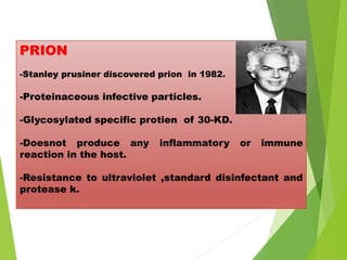
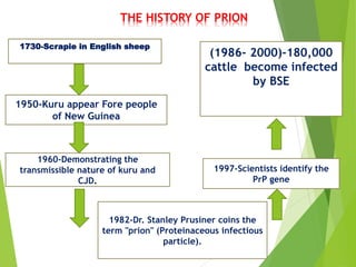
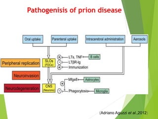
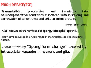
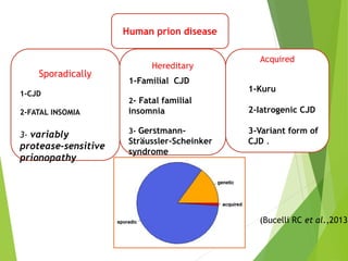
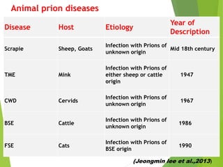
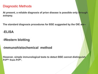
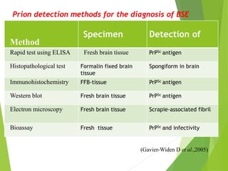
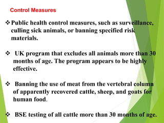
PRION DISEASE: AN OVERVIEW
Dr. Gazanfar Abass
M.V.Sc. Scholar(VPH)
IVRI
Overview
Introduction OF PRIONS
HISTORY OF PRION DISEASE
PATHOGENESIS OF PRION DISEASE
HUMAN PRION DISEASE
ANIMAL PRION DISEASE
DIAGNOSIS
Conclusions
PRION
-Stanley prusiner discovered prion in 1982.
-Proteinaceous infective particles.
-Glycosylated specific protien of 30-KD.
-Doesnot produce any inflammatory or immune
reaction in the host.
-Resistance to ultraviolet ,standard disinfectant and
protease k.
THE HISTORY OF PRION
1730-Scrapie in English sheep
1950-Kuru appear Fore people
of New Guinea
1960-Demonstrating the
transmissible nature of kuru and
CJD.
1982-Dr. Stanley Prusiner coins the
term "prion" (Proteinaceous infectious
particle).
1997-Scientists identify the
PrP gene
(1986- 2000)-180,000
cattle become infected
by BSE
Functions of normal Prion
protein
PrPC + Cu (Copper)
Antioxidant
Resistance to oxidative stress
Prevent neuronal dysfunction
(Brown et al., 2002)
MECHANISM OF PRION
DISEASE
α-helix
converted to β-
sheet structure
Prion acts as a
template.
Triggers a
chain reaction
Amyloid fibers
plaques
Transformation
PrPC PrPSc
DIFFERENCE BETWEEN
PrPC and PrPSc
PrPC PrPSc
Solubility Soluble Non soluble
Structure Alpha-helix Beta-helix
Multimerisation Monomeric Multimeric
Infectivity Non infectious Infectious
Susceptibility to
Proteinase K
Susceptible Resistant
Pathogenisis of prion disease
(Adriano Aguzzi et al.,2012)
PRION DISEASE(TSE)
Transmissible, progressive and invariably fatal
neurodegenerative conditions associated with misfolding and
aggregation of a host-encoded cellular prion protein.
(Imran et al., 2011)
-Also known as transmissable spongy encephalopathy.
-They have occurred in a wide range of mammalian species including
human.
-Characterized by “Spongiform change” caused by
intracellular vacuoles in neurons and glia.
Human prion disease
Acquired
Hereditary
Sporadically
1-CJD
2-FATAL INSOMIA
3- variably
protease-sensitive
prionopathy
1-Familial CJD
2- Fatal familial
insomnia
3- Gerstmann-
Sträussler-Scheinker
syndrome
1-Kuru
2-Iatrogenic CJD
3-Variant form of
CJD .
(Bucelli RC et al.,2013)
Disease Host Etiology
Year of
Description
Scrapie Sheep, Goats
Infection with Prions of
unknown origin
Mid 18th century
TME Mink
Infection with Prions of
either sheep or cattle
origin
1947
CWD Cervids
Infection with Prions of
unknown origin
1967
BSE Cattle
Infection with Prions of
unknown origin
1986
FSE Cats
Infection with Prions of
BSE origin
1990
Animal prion diseases
(Jeongmin lee et al.,2013)
Scrapie
In 1732, scrapie—disease was first reported in the UK, affecting the wool industry.
The name scrapie is derived from one of the clinical signs of
the condition- itching sensation in the animals.
CLINICAL SIGN
-Excessive lip smacking.
-Altered gaits.
-Convulsive collapse .
In 1900s, farmers in the UK started to feed cows with internal
organs or bones of sheep to benefit economically from the increase
in milk and meat production.
By the 1930s, other European countries and the United States
(USA) had adopted this practice. Based on the findings of
epidemiological studies on the origin of BSE, this later became the
main cause of prion disease transmission from sheep to cows across
the species barrier.
( Wilesmith JW et
al.,1991)
BSE(bovine spongyform encephalopathy)
-Late 1985: Fatal neurodegenerative disease in
cattle.
-Long incubation period (2.5 to 8years).
-Insidious onset
 Irritabilty, aggression
 Motor system impairment
 Difficulty in rising
 Decreased milk production
 Wasting
 Death (Hope j et al.,1988)
Cause of BSE
Ruminant tissue in food
chain
Scrapie
Meat and bone meal
(MBM)
Sporadic BSE in cattle
BSE Epidemic
 ~180,000 cumulative cases in UK.
 Peaked at 37,000 cases per annum in 1992.
 Progressive decline with introduction of feed
bans.
Transmissible Spongiform Encephalopathy as a Zoonotic Disease, Brown, P., et. al. ILSI, March
2003
(mammals)
three phases of BSE in
cattle
First phase:
- Low infectivity rate.
-NO threat to human.
Second phase
-High infectivity rate
-Risk to public health
-Symptoms not evident
to public health
The third phase:
Clinical symptoms
Then death follows shortly.
Cattle affected by BSE
experience progressive
degeneration of the
nervous system
Creutzfeldt-Jakob disease (CJD)
Identified by Creutzfeldt and Jakob (1920)
A progressive fatal neurodegenerative disease of
unknown cause.
Characterized by seizures, massive incoordination,
dementia.
Sporadic Cruetzfeldt-Jacob disease accounts for
85% of all CJD cases with annual worldwide
incidence of 1-2 cases/million population.
(Mead S et al.,2003)
 Variant Creutzfeldtt-Jakob Disease (vCJD), is caused by the
consumption of BSE infected meat products.
 First 10 cases of variant CJD were observed in 1996,ten
years after the outbreak of BSE in the UK.
 Variant CJD seems to affect mostly young patients below
30 year.
Variant Creutzfeldtt-Jakob Disease (vCJD)
COUNTRY CDC REPORT (OCT. 2009) WHO REPORT(FEB.2012)
UNITED KINGDOM 170 175
FRANCE 25 25
ITALY 2 2
SPAIN 5 5
IRELAND 4 4
UNITED STATES 3 3
NETHERLAND 3 3
PORTUGAL 2 2
CANADA 1 2
JAPAN 1 1
SAUDI ARABIA 1 1
TAIWAN - 1
TOTAL-217 TOTAL-224
Epidemiology of VCJD
KURU
Kuru is the first human prion disease that was shown to be
transmissible to chimpanzees by intracerebral introduction of brain
homogenates from kuru patients.
(Gajdusek DC et al.,1966)
-Fore linguistic group of Papua New Guinea. .
-Transmission- ritualistic cannibalism.
-Women and young children were more exposed to the risk material
than adult man.
- Kuru epidemic killed 1-2% of the population at its peak.
-With a ban on ritualistic cannibalism in the mid-1950s imposed by
Australian authorities, the incidence of the disease started to
decline steadily.
Deaths from kuru 1957–1982. Courtesy of late D. Carleton Gajdusek.
Kuru has 3 clinical stages
-Ambulant
-Sedentary
-Terminal
clinical signs.
-Headache and pain usually in the joints of legs.
-Cerebellar ataxia, tremors and and athetoid
movements are distinctive.
-Shivering amplifiable by cold was the symptom
on the basis of which the disease was named
"kuru.
Different prions affect different regions of the
brain
Cerebral cortex: the symptoms include
loss of memory and mental acuity, also
visual imparement (CJD).
Thalamus: Fatal Familial Insomnia
(FFI).
Cerebellum: lose the control of body
movements and difficulties to walk
(kuru, GSS).
Brain stem: In the mad cow disease
(BSE), the brain stem is affected.
Diagnostic Methods
At present, a reliable diagnosis of prion disease is possible only through
autopsy.
The standard diagnosis procedures for BSE suggested by the OIE are--
-ELISA
-Western blotting
-immunohistochemical method
However, simple immunological tests to detect BSE cannot distinguish
PrPSc from PrPC.
Method
Specimen Detection of
Rapid test using ELISA Fresh brain tissue PrPSc antigen
Histopathological test Formalin fixed brain
tissue
Spongiform in brain
Immunohistochemistry FFB-tissue PrPSc antigen
Western blot Fresh brain tissue PrPSc antigen
Electron microscopy Fresh brain tissue Scrapie-associated fibril
Bioassay Fresh tissue PrPSc and infectivity
Prion detection methods for the diagnosis of BSE
(Gavier-Widen D et al.,2005)
Control Measures
Public health control measures, such as surveillance,
culling sick animals, or banning specified risk
materials.
 UK program that excludes all animals more than 30
months of age. The program appears to be highly
effective.
 Banning the use of meat from the vertebral column
of apparently recovered cattle, sheep, and goats for
human food.
 BSE testing of all cattle more than 30 months of age.
CONCLUSION
BSE and vCJD are typical emerging epidemics.
SRM is the most important aspect in controlling.
Conditions or guidelines for trade” that can prevent the
disease from spreading from one country to another (SRM
standards set by the OIE ).
Risk reduction, strict precautionary principles should be
applied.
Ban on MBM and SBO.
Prion disease

Prion disease

  • 1.
    PRION DISEASE: ANOVERVIEW Dr. Gazanfar Abass M.V.Sc. Scholar(VPH) IVRI
  • 2.
    Overview Introduction OF PRIONS HISTORYOF PRION DISEASE PATHOGENESIS OF PRION DISEASE HUMAN PRION DISEASE ANIMAL PRION DISEASE DIAGNOSIS Conclusions
  • 3.
    PRION -Stanley prusiner discoveredprion in 1982. -Proteinaceous infective particles. -Glycosylated specific protien of 30-KD. -Doesnot produce any inflammatory or immune reaction in the host. -Resistance to ultraviolet ,standard disinfectant and protease k.
  • 4.
    THE HISTORY OFPRION 1730-Scrapie in English sheep 1950-Kuru appear Fore people of New Guinea 1960-Demonstrating the transmissible nature of kuru and CJD. 1982-Dr. Stanley Prusiner coins the term "prion" (Proteinaceous infectious particle). 1997-Scientists identify the PrP gene (1986- 2000)-180,000 cattle become infected by BSE
  • 5.
    Functions of normalPrion protein PrPC + Cu (Copper) Antioxidant Resistance to oxidative stress Prevent neuronal dysfunction (Brown et al., 2002)
  • 6.
    MECHANISM OF PRION DISEASE α-helix convertedto β- sheet structure Prion acts as a template. Triggers a chain reaction Amyloid fibers plaques
  • 7.
  • 8.
    DIFFERENCE BETWEEN PrPC andPrPSc PrPC PrPSc Solubility Soluble Non soluble Structure Alpha-helix Beta-helix Multimerisation Monomeric Multimeric Infectivity Non infectious Infectious Susceptibility to Proteinase K Susceptible Resistant
  • 9.
    Pathogenisis of priondisease (Adriano Aguzzi et al.,2012)
  • 10.
    PRION DISEASE(TSE) Transmissible, progressiveand invariably fatal neurodegenerative conditions associated with misfolding and aggregation of a host-encoded cellular prion protein. (Imran et al., 2011) -Also known as transmissable spongy encephalopathy. -They have occurred in a wide range of mammalian species including human. -Characterized by “Spongiform change” caused by intracellular vacuoles in neurons and glia.
  • 11.
    Human prion disease Acquired Hereditary Sporadically 1-CJD 2-FATALINSOMIA 3- variably protease-sensitive prionopathy 1-Familial CJD 2- Fatal familial insomnia 3- Gerstmann- Sträussler-Scheinker syndrome 1-Kuru 2-Iatrogenic CJD 3-Variant form of CJD . (Bucelli RC et al.,2013)
  • 12.
    Disease Host Etiology Yearof Description Scrapie Sheep, Goats Infection with Prions of unknown origin Mid 18th century TME Mink Infection with Prions of either sheep or cattle origin 1947 CWD Cervids Infection with Prions of unknown origin 1967 BSE Cattle Infection with Prions of unknown origin 1986 FSE Cats Infection with Prions of BSE origin 1990 Animal prion diseases (Jeongmin lee et al.,2013)
  • 13.
    Scrapie In 1732, scrapie—diseasewas first reported in the UK, affecting the wool industry. The name scrapie is derived from one of the clinical signs of the condition- itching sensation in the animals. CLINICAL SIGN -Excessive lip smacking. -Altered gaits. -Convulsive collapse .
  • 14.
    In 1900s, farmersin the UK started to feed cows with internal organs or bones of sheep to benefit economically from the increase in milk and meat production. By the 1930s, other European countries and the United States (USA) had adopted this practice. Based on the findings of epidemiological studies on the origin of BSE, this later became the main cause of prion disease transmission from sheep to cows across the species barrier. ( Wilesmith JW et al.,1991)
  • 15.
    BSE(bovine spongyform encephalopathy) -Late1985: Fatal neurodegenerative disease in cattle. -Long incubation period (2.5 to 8years). -Insidious onset  Irritabilty, aggression  Motor system impairment  Difficulty in rising  Decreased milk production  Wasting  Death (Hope j et al.,1988)
  • 16.
    Cause of BSE Ruminanttissue in food chain Scrapie Meat and bone meal (MBM) Sporadic BSE in cattle
  • 17.
    BSE Epidemic  ~180,000cumulative cases in UK.  Peaked at 37,000 cases per annum in 1992.  Progressive decline with introduction of feed bans.
  • 18.
    Transmissible Spongiform Encephalopathyas a Zoonotic Disease, Brown, P., et. al. ILSI, March 2003 (mammals)
  • 20.
    three phases ofBSE in cattle First phase: - Low infectivity rate. -NO threat to human. Second phase -High infectivity rate -Risk to public health -Symptoms not evident to public health The third phase: Clinical symptoms Then death follows shortly.
  • 21.
    Cattle affected byBSE experience progressive degeneration of the nervous system
  • 22.
    Creutzfeldt-Jakob disease (CJD) Identifiedby Creutzfeldt and Jakob (1920) A progressive fatal neurodegenerative disease of unknown cause. Characterized by seizures, massive incoordination, dementia. Sporadic Cruetzfeldt-Jacob disease accounts for 85% of all CJD cases with annual worldwide incidence of 1-2 cases/million population. (Mead S et al.,2003)
  • 23.
     Variant Creutzfeldtt-JakobDisease (vCJD), is caused by the consumption of BSE infected meat products.  First 10 cases of variant CJD were observed in 1996,ten years after the outbreak of BSE in the UK.  Variant CJD seems to affect mostly young patients below 30 year. Variant Creutzfeldtt-Jakob Disease (vCJD)
  • 24.
    COUNTRY CDC REPORT(OCT. 2009) WHO REPORT(FEB.2012) UNITED KINGDOM 170 175 FRANCE 25 25 ITALY 2 2 SPAIN 5 5 IRELAND 4 4 UNITED STATES 3 3 NETHERLAND 3 3 PORTUGAL 2 2 CANADA 1 2 JAPAN 1 1 SAUDI ARABIA 1 1 TAIWAN - 1 TOTAL-217 TOTAL-224 Epidemiology of VCJD
  • 25.
    KURU Kuru is thefirst human prion disease that was shown to be transmissible to chimpanzees by intracerebral introduction of brain homogenates from kuru patients. (Gajdusek DC et al.,1966) -Fore linguistic group of Papua New Guinea. . -Transmission- ritualistic cannibalism. -Women and young children were more exposed to the risk material than adult man. - Kuru epidemic killed 1-2% of the population at its peak. -With a ban on ritualistic cannibalism in the mid-1950s imposed by Australian authorities, the incidence of the disease started to decline steadily.
  • 26.
    Deaths from kuru1957–1982. Courtesy of late D. Carleton Gajdusek.
  • 27.
    Kuru has 3clinical stages -Ambulant -Sedentary -Terminal clinical signs. -Headache and pain usually in the joints of legs. -Cerebellar ataxia, tremors and and athetoid movements are distinctive. -Shivering amplifiable by cold was the symptom on the basis of which the disease was named "kuru.
  • 28.
    Different prions affectdifferent regions of the brain Cerebral cortex: the symptoms include loss of memory and mental acuity, also visual imparement (CJD). Thalamus: Fatal Familial Insomnia (FFI). Cerebellum: lose the control of body movements and difficulties to walk (kuru, GSS). Brain stem: In the mad cow disease (BSE), the brain stem is affected.
  • 29.
    Diagnostic Methods At present,a reliable diagnosis of prion disease is possible only through autopsy. The standard diagnosis procedures for BSE suggested by the OIE are-- -ELISA -Western blotting -immunohistochemical method However, simple immunological tests to detect BSE cannot distinguish PrPSc from PrPC.
  • 30.
    Method Specimen Detection of Rapidtest using ELISA Fresh brain tissue PrPSc antigen Histopathological test Formalin fixed brain tissue Spongiform in brain Immunohistochemistry FFB-tissue PrPSc antigen Western blot Fresh brain tissue PrPSc antigen Electron microscopy Fresh brain tissue Scrapie-associated fibril Bioassay Fresh tissue PrPSc and infectivity Prion detection methods for the diagnosis of BSE (Gavier-Widen D et al.,2005)
  • 31.
    Control Measures Public healthcontrol measures, such as surveillance, culling sick animals, or banning specified risk materials.  UK program that excludes all animals more than 30 months of age. The program appears to be highly effective.  Banning the use of meat from the vertebral column of apparently recovered cattle, sheep, and goats for human food.  BSE testing of all cattle more than 30 months of age.
  • 32.
    CONCLUSION BSE and vCJDare typical emerging epidemics. SRM is the most important aspect in controlling. Conditions or guidelines for trade” that can prevent the disease from spreading from one country to another (SRM standards set by the OIE ). Risk reduction, strict precautionary principles should be applied. Ban on MBM and SBO.

Editor's Notes

  • #33 Animal prion diseases other than scrapie and CWD have been controlled by the withdrawal of source of contamination from animals' diet. However,