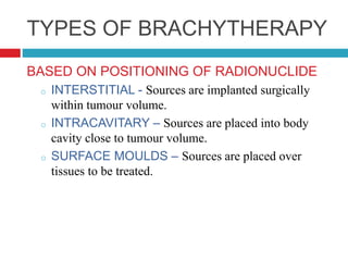
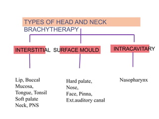
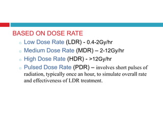
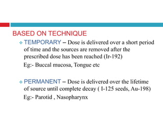
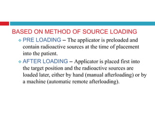
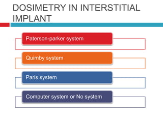
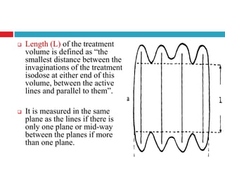
This document discusses brachytherapy techniques for head and neck cancers. It describes different types of brachytherapy based on positioning of the radionuclide (interstitial, intracavitary, surface moulds), dose rate (LDR, MDR, HDR, PDR), and technique (temporary, permanent). It also discusses dosimetry systems like Patterson-Parker, Quimby, Paris and computerized planning. Key aspects of treatment planning, delivery, and post-treatment care are summarized. Advantages include localized high dose with rapid falloff and organ preservation, while limitations include inaccessibility and quality dependence on implant. American Brachytherapy Society guidelines emphasize accurate assessment and dental
the role of brachytherapy in oral cavity carcinoma.
physics of brachytherapy
radiobiology of brachytherapy
clinical application in tongue, buccal mucosa cancer
the role of brachytherapy in oral cavity carcinoma.
physics of brachytherapy
radiobiology of brachytherapy
clinical application in tongue, buccal mucosa cancer
Side effects of radiation in head and neck cancerAnagha pachat
this presentation describes how radiation effects normal structures in head and neck region and about the late and acute toxicities which may occur if the radiation exceeds tolerance dose as per QUANTEC
Conventional radiotherapy treatments are delivered with radiation beams that are of uniform intensity across the field (within the flatness specification limits). Wedges or compensators are used to modify the intensity profile to offset contour in irregularities and produce more uniform composite dose distributions such as in techniques using wedges. This process of changing beam intensity profile to meet the goals of a composite plan is called intensity modulation
IMRT refers to a radiation therapy technique in which nonuniform fluence is delivered to the patient from any given position of the treatment beam to optimize the composite dose distribution. The optimal fluence profiles for a given set of beam directions are determined through inverse planning. The fluence files thus generated are electronically transmitted to the linear accelerator, which is computer controlled, to deliver intensity modulated beams (IMBs) as calculated.
Title: Sense of Taste
Presenter: Dr. Faiza, Assistant Professor of Physiology
Qualifications:
MBBS (Best Graduate, AIMC Lahore)
FCPS Physiology
ICMT, CHPE, DHPE (STMU)
MPH (GC University, Faisalabad)
MBA (Virtual University of Pakistan)
Learning Objectives:
Describe the structure and function of taste buds.
Describe the relationship between the taste threshold and taste index of common substances.
Explain the chemical basis and signal transduction of taste perception for each type of primary taste sensation.
Recognize different abnormalities of taste perception and their causes.
Key Topics:
Significance of Taste Sensation:
Differentiation between pleasant and harmful food
Influence on behavior
Selection of food based on metabolic needs
Receptors of Taste:
Taste buds on the tongue
Influence of sense of smell, texture of food, and pain stimulation (e.g., by pepper)
Primary and Secondary Taste Sensations:
Primary taste sensations: Sweet, Sour, Salty, Bitter, Umami
Chemical basis and signal transduction mechanisms for each taste
Taste Threshold and Index:
Taste threshold values for Sweet (sucrose), Salty (NaCl), Sour (HCl), and Bitter (Quinine)
Taste index relationship: Inversely proportional to taste threshold
Taste Blindness:
Inability to taste certain substances, particularly thiourea compounds
Example: Phenylthiocarbamide
Structure and Function of Taste Buds:
Composition: Epithelial cells, Sustentacular/Supporting cells, Taste cells, Basal cells
Features: Taste pores, Taste hairs/microvilli, and Taste nerve fibers
Location of Taste Buds:
Found in papillae of the tongue (Fungiform, Circumvallate, Foliate)
Also present on the palate, tonsillar pillars, epiglottis, and proximal esophagus
Mechanism of Taste Stimulation:
Interaction of taste substances with receptors on microvilli
Signal transduction pathways for Umami, Sweet, Bitter, Sour, and Salty tastes
Taste Sensitivity and Adaptation:
Decrease in sensitivity with age
Rapid adaptation of taste sensation
Role of Saliva in Taste:
Dissolution of tastants to reach receptors
Washing away the stimulus
Taste Preferences and Aversions:
Mechanisms behind taste preference and aversion
Influence of receptors and neural pathways
Impact of Sensory Nerve Damage:
Degeneration of taste buds if the sensory nerve fiber is cut
Abnormalities of Taste Detection:
Conditions: Ageusia, Hypogeusia, Dysgeusia (parageusia)
Causes: Nerve damage, neurological disorders, infections, poor oral hygiene, adverse drug effects, deficiencies, aging, tobacco use, altered neurotransmitter levels
Neurotransmitters and Taste Threshold:
Effects of serotonin (5-HT) and norepinephrine (NE) on taste sensitivity
Supertasters:
25% of the population with heightened sensitivity to taste, especially bitterness
Increased number of fungiform papillae
Factory Supply Best Quality Pmk Oil CAS 28578–16–7 PMK Powder in Stockrebeccabio
Factory Supply Best Quality Pmk Oil CAS 28578–16–7 PMK Powder in Stock
Telegram: bmksupplier
signal: +85264872720
threema: TUD4A6YC
You can contact me on Telegram or Threema
Communicate promptly and reply
Free of customs clearance, Double Clearance 100% pass delivery to USA, Canada, Spain, Germany, Netherland, Poland, Italy, Sweden, UK, Czech Republic, Australia, Mexico, Russia, Ukraine, Kazakhstan.Door to door service
Hot Selling Organic intermediates
These lecture slides, by Dr Sidra Arshad, offer a quick overview of physiological basis of a normal electrocardiogram.
Learning objectives:
1. Define an electrocardiogram (ECG) and electrocardiography
2. Describe how dipoles generated by the heart produce the waveforms of the ECG
3. Describe the components of a normal electrocardiogram of a typical bipolar leads (limb II)
4. Differentiate between intervals and segments
5. Enlist some common indications for obtaining an ECG
Study Resources:
1. Chapter 11, Guyton and Hall Textbook of Medical Physiology, 14th edition
2. Chapter 9, Human Physiology - From Cells to Systems, Lauralee Sherwood, 9th edition
3. Chapter 29, Ganong’s Review of Medical Physiology, 26th edition
4. Electrocardiogram, StatPearls - https://www.ncbi.nlm.nih.gov/books/NBK549803/
5. ECG in Medical Practice by ABM Abdullah, 4th edition
6. ECG Basics, http://www.nataliescasebook.com/tag/e-c-g-basics
Report Back from SGO 2024: What’s the Latest in Cervical Cancer?bkling
Are you curious about what’s new in cervical cancer research or unsure what the findings mean? Join Dr. Emily Ko, a gynecologic oncologist at Penn Medicine, to learn about the latest updates from the Society of Gynecologic Oncology (SGO) 2024 Annual Meeting on Women’s Cancer. Dr. Ko will discuss what the research presented at the conference means for you and answer your questions about the new developments.
Flu Vaccine Alert in Bangalore Karnatakaaddon Scans
As flu season approaches, health officials in Bangalore, Karnataka, are urging residents to get their flu vaccinations. The seasonal flu, while common, can lead to severe health complications, particularly for vulnerable populations such as young children, the elderly, and those with underlying health conditions.
Dr. Vidisha Kumari, a leading epidemiologist in Bangalore, emphasizes the importance of getting vaccinated. "The flu vaccine is our best defense against the influenza virus. It not only protects individuals but also helps prevent the spread of the virus in our communities," he says.
This year, the flu season is expected to coincide with a potential increase in other respiratory illnesses. The Karnataka Health Department has launched an awareness campaign highlighting the significance of flu vaccinations. They have set up multiple vaccination centers across Bangalore, making it convenient for residents to receive their shots.
To encourage widespread vaccination, the government is also collaborating with local schools, workplaces, and community centers to facilitate vaccination drives. Special attention is being given to ensuring that the vaccine is accessible to all, including marginalized communities who may have limited access to healthcare.
Residents are reminded that the flu vaccine is safe and effective. Common side effects are mild and may include soreness at the injection site, mild fever, or muscle aches. These side effects are generally short-lived and far less severe than the flu itself.
Healthcare providers are also stressing the importance of continuing COVID-19 precautions. Wearing masks, practicing good hand hygiene, and maintaining social distancing are still crucial, especially in crowded places.
Protect yourself and your loved ones by getting vaccinated. Together, we can help keep Bangalore healthy and safe this flu season. For more information on vaccination centers and schedules, residents can visit the Karnataka Health Department’s official website or follow their social media pages.
Stay informed, stay safe, and get your flu shot today!
Lung Cancer: Artificial Intelligence, Synergetics, Complex System Analysis, S...Oleg Kshivets
RESULTS: Overall life span (LS) was 2252.1±1742.5 days and cumulative 5-year survival (5YS) reached 73.2%, 10 years – 64.8%, 20 years – 42.5%. 513 LCP lived more than 5 years (LS=3124.6±1525.6 days), 148 LCP – more than 10 years (LS=5054.4±1504.1 days).199 LCP died because of LC (LS=562.7±374.5 days). 5YS of LCP after bi/lobectomies was significantly superior in comparison with LCP after pneumonectomies (78.1% vs.63.7%, P=0.00001 by log-rank test). AT significantly improved 5YS (66.3% vs. 34.8%) (P=0.00000 by log-rank test) only for LCP with N1-2. Cox modeling displayed that 5YS of LCP significantly depended on: phase transition (PT) early-invasive LC in terms of synergetics, PT N0—N12, cell ratio factors (ratio between cancer cells- CC and blood cells subpopulations), G1-3, histology, glucose, AT, blood cell circuit, prothrombin index, heparin tolerance, recalcification time (P=0.000-0.038). Neural networks, genetic algorithm selection and bootstrap simulation revealed relationships between 5YS and PT early-invasive LC (rank=1), PT N0—N12 (rank=2), thrombocytes/CC (3), erythrocytes/CC (4), eosinophils/CC (5), healthy cells/CC (6), lymphocytes/CC (7), segmented neutrophils/CC (8), stick neutrophils/CC (9), monocytes/CC (10); leucocytes/CC (11). Correct prediction of 5YS was 100% by neural networks computing (area under ROC curve=1.0; error=0.0).
CONCLUSIONS: 5YS of LCP after radical procedures significantly depended on: 1) PT early-invasive cancer; 2) PT N0--N12; 3) cell ratio factors; 4) blood cell circuit; 5) biochemical factors; 6) hemostasis system; 7) AT; 8) LC characteristics; 9) LC cell dynamics; 10) surgery type: lobectomy/pneumonectomy; 11) anthropometric data. Optimal diagnosis and treatment strategies for LC are: 1) screening and early detection of LC; 2) availability of experienced thoracic surgeons because of complexity of radical procedures; 3) aggressive en block surgery and adequate lymph node dissection for completeness; 4) precise prediction; 5) adjuvant chemoimmunoradiotherapy for LCP with unfavorable prognosis.
ARTIFICIAL INTELLIGENCE IN HEALTHCARE.pdfAnujkumaranit
Artificial intelligence (AI) refers to the simulation of human intelligence processes by machines, especially computer systems. It encompasses tasks such as learning, reasoning, problem-solving, perception, and language understanding. AI technologies are revolutionizing various fields, from healthcare to finance, by enabling machines to perform tasks that typically require human intelligence.
- Video recording of this lecture in English language: https://youtu.be/lK81BzxMqdo
- Video recording of this lecture in Arabic language: https://youtu.be/Ve4P0COk9OI
- Link to download the book free: https://nephrotube.blogspot.com/p/nephrotube-nephrology-books.html
- Link to NephroTube website: www.NephroTube.com
- Link to NephroTube social media accounts: https://nephrotube.blogspot.com/p/join-nephrotube-on-social-media.html
TEST BANK for Operations Management, 14th Edition by William J. Stevenson, Ve...kevinkariuki227
TEST BANK for Operations Management, 14th Edition by William J. Stevenson, Verified Chapters 1 - 19, Complete Newest Version.pdf
Pulmonary Thromboembolism - etilogy, types, medical- Surgical and nursing man...VarunMahajani
Disruption of blood supply to lung alveoli due to blockage of one or more pulmonary blood vessels is called as Pulmonary thromboembolism. In this presentation we will discuss its causes, types and its management in depth.
MANAGEMENT OF ATRIOVENTRICULAR CONDUCTION BLOCK.pdfJim Jacob Roy
Cardiac conduction defects can occur due to various causes.
Atrioventricular conduction blocks ( AV blocks ) are classified into 3 types.
This document describes the acute management of AV block.
Surgical Site Infections, pathophysiology, and prevention.pptx
Head and neck; brachytherapy.pptx final
1. BRACHYTHERAPY IN HEAD AND
NECK
DR AKHILESH
DEPARTMENT OF RADIATION
ONCOLOGY
REGIONAL CANCER CENTRE ,
TRIVANDRUM
2. BRACHYTHERAPY –
Method of radiaton therapy in which an encapsulated
source or group of such sources is utilized to deliver
gamma or beta radiation at a distance of up to a few
centimetres, either by surface, intracavitary or
interstitial application.
4. TYPES OF BRACHYTHERAPY
BASED ON POSITIONING OF RADIONUCLIDE
o INTERSTITIAL - Sources are implanted surgically
within tumour volume.
o INTRACAVITARY – Sources are placed into body
cavity close to tumour volume.
o SURFACE MOULDS – Sources are placed over
tissues to be treated.
5. TYPES OF HEAD AND NECK
BRACHYTHERAPY
INTERSTITIAL SURFACE MOULD INTRACAVITARY
Lip, Buccal
Mucosa,
Tongue, Tonsil
Soft palate
Neck, PNS
Hard palate,
Nose,
Face, Pinna,
Ext.auditory canal
Nasopharynx
6. BASED ON DOSE RATE
o Low Dose Rate (LDR) - 0.4-2Gy/hr
o Medium Dose Rate (MDR) – 2-12Gy/hr
o High Dose Rate (HDR) - >12Gy/hr
o Pulsed Dose Rate (PDR) – involves short pulses of
radiation, typically once an hour, to simulate overall rate
and effectiveness of LDR treatment.
7. BASED ON TECHNIQUE
TEMPORARY – Dose is delivered over a short period
of time and the sources are removed after the
prescribed dose has been reached (Ir-192)
Eg:- Buccal mucosa, Tongue etc
PERMANENT – Dose is delivered over the lifetime
of source until complete decay ( I-125 seeds, Au-198)
Eg:- Parotid , Nasopharynx
8. BASED ON METHOD OF SOURCE LOADING
PRE LOADING – The applicator is preloaded and
contain radioactive sources at the time of placement
into the patient.
AFTER LOADING – Applicator is placed first into
the target position and the radioactive sources are
loaded later, either by hand (manual afterloading) or by
a machine (automatic remote afterloading).
10. INTENT OF TREATMENT
RADICAL : Brachytherapy alone as treatment.
BOOST : EBRT Brachytherapy to boost dose to the
primary.
SALVAGE THERAPY : Recurrent cases who have
been irradiated before or who are unfit for surgery.
11. SELECTION CRITERIA
Easily accessible lesions
Early stage diseases (Ideal implant ≤ 5 cm)
Well localized tumor to organ of origin
No nodal or distant metastases
No local infections or inflammation
Proliferative/ ulcerative lesions preferred.
Favorable histology
12. Brachytherapy alone contraindicated –
Non accessible tumour location
Tumour limits are ill-defined
Tumor is abutting or has invaded bone
13. No randomized trials
Recommendations by experts
American Brachytherapy Society (ABS)
recommendations
GEC-ESTRO recommendations
For head and neck
brachytherapy
14. PRE TREATMENT WORKUP
Detailed CLINICAL EXAMINATION of the head and
neck region
Examination under general anesthesia preferably in
combination with panendoscopy – Posterior lesion
Computerized tomography (CT) and magnetic resonance
imaging (MRI) - Optional
Dental preparation.
15. Prerequisites for brachytherapy
treatment
TARGET VOLUME :-
Brachytherapy catheters should be placed about 1 to
1.5 cm apart as equidistant and parallel as possible, to
encompass the CTV with a margin determined on the
basis of the clinical parameters.
Pre-planning :-
Measure the tumour carefully.
Plan the exact number of radiation sources to be used
with their length and separation.
16. Treatment Delivery
High Dose Rate Brachytherapy
Two fractions given every day
6 hours apart
Dose: 300-400cGy per fraction
17. RADICAL :- Equivalent of 60-66Gy of low dose
rate brachytherapy, 400cGy per fraction bid X 12
BOOST :- Equivalent of 20-30Gy of low dose rate
brachytherapy, 400cGy per fraction bid X 8-10
TOTAL DOSE
18. The most common technique is afterloading
with Ir192
Eg:- Lip, Buccal Mucosa,
Tongue, Tonsil
Soft palate
Neck, PNS
INTERSTITIAL
BRACHYTHERAPY
21. PATERSON PARKER SYSTEM
Deliver uniform dose(+/- 10%) to a plane or volume.
Row of parallel needles with ends crossed by needles at
right angles.
Needles should be in parallel rows at spacing not greater
than 1 cm
Crossing needles should ideally cross the active needle
ends, but should not be more than 1 cm from the active
ends.
22. Depending on the size of the lesion a single plane, double
plane, or volume implant can be used to cover the tumor
with a 1-cm margin.
For tumors :-
• <1 cm in thickness - single plane implant
• 1-2.5 cm thickness - double plane implant
• > 2.5 cm thickness - volume implant
23. Areas covered by
implants with
1. Two crossed
ends
2. One crossed end
3.No crossed ends
24. Paterson Parker tables
The P-P tables are designed to
give milligram hours / 1000
roentgens for various implant sizes
both for planar and volume
implants depending on the surface
area of implant.
CORRECTIONS
(Total correction factor = 0.90)
Gamma-factor originally 8.4
Rcm2/mg-h,now 8.25 Rcm2/mg-h.
R to cGy factor = 0.957
Oblique filtration
Correction for tissue attenuation and
scattering
25. Rules for planar implant
Area of plane
(cm2)
% Activity in
periphery
% Activity in
the center
<25 2/3 1/3
25-100 ½ ½
>100 1/3 2/3
Deduct 10% of the area for each uncrossed end
26. For volume implants
Sources on each surface must
be spaced as evenly and not
more than 1.0 to 1.5 cm
apart.
The belt should contain not
less than 8 needles and the
core not less than 4 needles.
For each Uncrossed ends :
7.5% reduction in volume.
27. P-P tables give cumulated source strength per unit dose
(in mgh per 1000cGy) for given implant or volume.
To obtain the total dose strength, the table value is
multiplied by the desired dose rate.
Dose derived from the table value – STATED DOSE
Stated dose is 10% larger than minimum dose and 10%
lesser than maximum dose ( Uniformity Criterion) in the
treatment region.
28. QUIMBY SYSTEM
Uniform distribution of sources of equal linear activity of
Ra-226.
surface applicators.
No appropriate guidelines for multiple planar implants.
Volume implants: the stated dose is the minimum dose
within the volume.
Two plane implants hotter near center, cooler at edges
Central minimum dose typically 25- 30% hotter than
prescription dose
Corrections are the same as the Patterson-Parker system
29. PARIS SYSTEM
Predictive Implant System
Define a target volume
Define its three dimensions. ie., length (L), width (W)
and thickness (T) . TV= L x W x T
Number of source planes depends upon the thickness.
If the thickness exceeds 12 mm, there must be 2 source
planes.
the source pattern, the number of sources and the source
spacing are easily determined
30. GENERAL RULES :-
Sources must be straight, parallel and equidistant from each
other ( no crossing).
Regular geometric pattern with equal separation( 5mm and
20mm
The linear activity of the sources must be uniform along the
length of each line and identical for all the lines used.
31. Central plane : plane
perpendicular to the sources ,
which is at right angles to the
long axis of the sources, and is
situated mid-way along their
length.
32. Basal Dose Rate provides a measure of the dose rate in
the centre of the treated volume .
Arithmetic mean of all the basal dose rates
Single plane: midway
Triangular plane: centroid of each triangle
Square : centre of each square
Calculated from the position of the sources in the central
plane and is at the points of minimum dose rate between
a pair or group of sources
34. Reference Dose Rate - dose which encompasses the
tumour volume.
85% of the basal dose rate
Dose rate used for calculating the total time of the implant.
Treatment volume - volume enclosed by the 85%
reference isodose ( approx. 0.65 times the length of
sources)
Sources should be 20-30% longer at each end than the
target volume.
35. Hyperdose Sleeve - defined as the volume of the tissue
receiving twice the reference dose rate (170% of the basal
dose).
36.
37. Length (L) of the treatment
volume is defined as “the
smallest distance between the
invaginations of the treatment
isodose at either end of this
volume, between the active
lines and parallel to them”.
It is measured in the same
plane as the lines if there is
only one plane or mid-way
between the planes if more
than one plane.
38. Thickness (t) of the
treatment volume is
defined as “the smallest
distance between two
parallel planes which are
tangents to those
invaginations which give
the target volume its least
thickness”.
39. Width (w) is defined as
“the maximum width of the
reference isodose in the
central plane” .
It is equal to the distance
between the most lateral
sources plus 37% of the
separation between the
sources added on to each
side.
40. Safety margin is defined as
“the minimum distance
measured between the
reference isodose and a line
joining the points where
any two sources intersect
the central plane”.
41. COMPUTER SYSTEM
Single-stepping source High Dose Rate Remote After-
loading machine (Single miniature Ir-192 source)
3D imaging for defining target volumes and for guiding
applicator insertion. (CT/MRI)
Three dimensional isodose distributions.
Optimization.( Dwell positions & Dwell times)
Dose–Volume Histogram (DVH) indices for quantifying
dose delivery and implant quality
43. Similar to Paris and Quimby, Implant that is hotter in the
centre than the periphery.
This dose inhomogeniety accepted – more dose needed to
the centre to sterilize the tumour.
No crossing sources – active length of line sources
should be suitably longer (=40% longer) than the length
of target volume.
45. HDR brachytherapy using a mould, without need of
invasive needles.
Surface mould is a custom made device, which attaches
to the patient and supports applicators or radioactive
sources at a fixed distance from the skin surface ( 5-
20mm).
Dosimetry system – Paterson parker system or Paris
system
46. Distance between source to skin surface – depend on
depth of treatment.
Dose to the treated area varies by +10% due to the non-
uniform isodoses produced by the spaced, discrete
sources.
47. Nasopharynx
Salvage therapy –
well circumscribed and
superficial local recurrences
limited to nasopharynx without
involvement of underlying
bone.
INTRACAVITARY
BRACHYTHERAPY
48. Nucletron Rotterdam applicator
Specialized form of surface mould
Made of soft silicone
Consists of 2 approximately parallel applicators and a
Paris system of basal points can be set up to give an
initial dose calculation.
49. Catheter removal in interstitial
brachytherapy
Removed in the operating room
An intravenous access is recommended
Presence of two persons is mandatory
In case of bleeding - bimanual compression for ten
minutes.
50. Post-treatment patient care and follow-
up
Should be regularly followed up.
Every monthly for atleast 1-2 years
Every 3 monthly thereafter
Most common complication – soft tissue necrosis
51. Advantage of Brachytherapy
Delivers localized dose to the tumor
Rapid dose fall off outside the target volume allows
excellent normal tissue sparing
Less integral dose as compared to 3DCRT & IMRT
High biological efficacy
Decreased risk of tumor population
Elimination of set up errors as the source maintains a
fixed relationship to target volume
52. Advantage of Brachytherapy
High tolerance: Tolerable acute intense reaction
High control rate
Better cosmetics: May avoid disfigurement and
mutilating surgery
Minimal radiation morbidity
Day care procedure
Organ preservation
Reirradiation for localized recurrence
53. Limitation of brachytherapy
Difficult for inaccessible regions
Limited for small tumors (T1-T2)
Nodal disease cannot be covered simultaneously
Invasive procedures, require GA
Greater conformation –small errors in placement of
sources lead to extreme changes from the intended dose
distribution
Quality of implant is operator dependent
54. General concepts based on ABS
recommendation
Use of brachytherapy as a component of the treatment of
head-and-neck tumors.
No definite evidence on use of concomitant
chemotherapy; risk of increased mucosal toxicity
compromising treatment – appears to be useful for the
treatment of recurrences.
55. Sequencing of EBRT and brachytherapy - obtain
shrinkage with EBRT before applying brachytherapy in
advanced tumors.
Brachytherapy boost - placement of radio-opaque
markers before starting EBRT can help delineate the target
volume, before any shrinkage occurs.
56. Dental Preparation
Teeth with deep caries or poor periodontal support -
removed and complete healing obtained before starting
RT.
A prosthesis (made of acrylic resin) including lead
shielding (2mm thick) should be made for brachytherapy
of the lips, tongue, and floor of mouth, to reduce dose to
the mandible and prevent osteoradionecrosis.
57. Important Facts to be Noted in H&N
Brachytherapy Based on ABS
recommendations
CLINICAL :-
Accurate assessment of : tumor dimensions, neck node,
lesion type via clinical examination.
Feasibility for Brachytherapy: Mouth opening, dental
status, proximity of bones to tumor.
Requirement of dental shields/spacers.
Requirement of tracheostomy.
Fitness for anaesthesia
58. PHYSICAL :-
Dose distribution :- non-homogenous (due to complex
geometry)
Minor displacement - significant hot/cold spots;
increased morbidity/recurrence.
Peripheral fall off- cause under-dosage of a site
especially at borders
Interstitial edema - produce alteration in dose
distribution calculated to an extent of 10-15%.
59. The isodose distribution should be computer optimized
to conform to the CTV.
The dwell times can be adjusted to minimize dose
inhomogeneity.
Optimization should not be used as a substitute for
good catheter placement.
60. Treatment Monitoring
To detect potential displacement of radioactive sources
or catheters.
Adequate antalgic and anti inflammatory coverage
Mouthwashes and nutritional support through a
nasogastric tube
Antibiotics may be useful
patient must also be taught - watch out for
inflammatory reactions - occur after the removal of the
implants, start about 7 days later, increase until the
third week, are stable over one week, and then decrease
and finally disappear at the end of the sixth week.
61. BIOLOGICAL :-
o Total duration of EBRT + Brachytherapy should be
kept as short as possible (<8 weeks) to minimize tumor
cell repopulation
o Interval between EBRT and Brachytherapy should be
as short as possible (<1–2 weeks) depending on degree
of recovery from mucositis
o Interval between twice daily HDR fractions should be
as long as possible (minimum of 6 hours)
o Previous irradiation history for dose calculation
62. ABS Recommendations -recurrent head &
neck cancer
Strongly emphasizes on using brachytherapy for
recurrent tumors
The extent of disease should be carefully studied with
CT, MRI, or PET scan as necessary.
Complication risks are increased in patients with
previous surgery, skin or mucosal ulceration, deep soft
tissue necrosis, bone exposure, or severe fibrosis
Meticulous implant technique and adequate doses are
necessary
63. Generally larger margins are required for recurrent
tumors, especially if additional EBRT is not applied.
Because of the paucity of data – No specific
recommendations for the indications for HDR
brachytherapy in recurrent head and neck tumors.
64.
65. PARAMETER
SYSTEM
Manchester Quimby Paris
Dose 6000-8000 R 5000-6000 R 6000-7000 R
Dose Rate 40-60 R/hr 60-70 R/hr 25-90 cGy/hr
Prescription Point 10% above absolute
minimum dose
On bisector
(planar) or
periphery (volume)
85% of minimum
central dose
Linear Activity Variable :
0.66 & 0.33
mgRaEq/cm
Constant :
1.0 mgRaEq/cm
Constant
Activity Distribution
Single Plane Varies with area Uniform Uniform
Volume Implant Varies with shape Uniform Multiple Uniform
Planes
Source Spacing Constant at 1 cm Constant at 1-2 cm Constant at 0.5-2
cm
Crossing needles Yes No No
66. LDR HDR PDR
Predictable clinical
effects
Short treatment time Emulates LDR
Superior radiobiologic
role
Dose optimization Optimized dose
distribution
Less morbidity, control
is best
No radiation hazards Potential radiation
safety hazards
Well practiced since
long
Small applicator Superior radiobiologic
role
Minimum intersession
variability in dose
distribution.
Radiation hazards