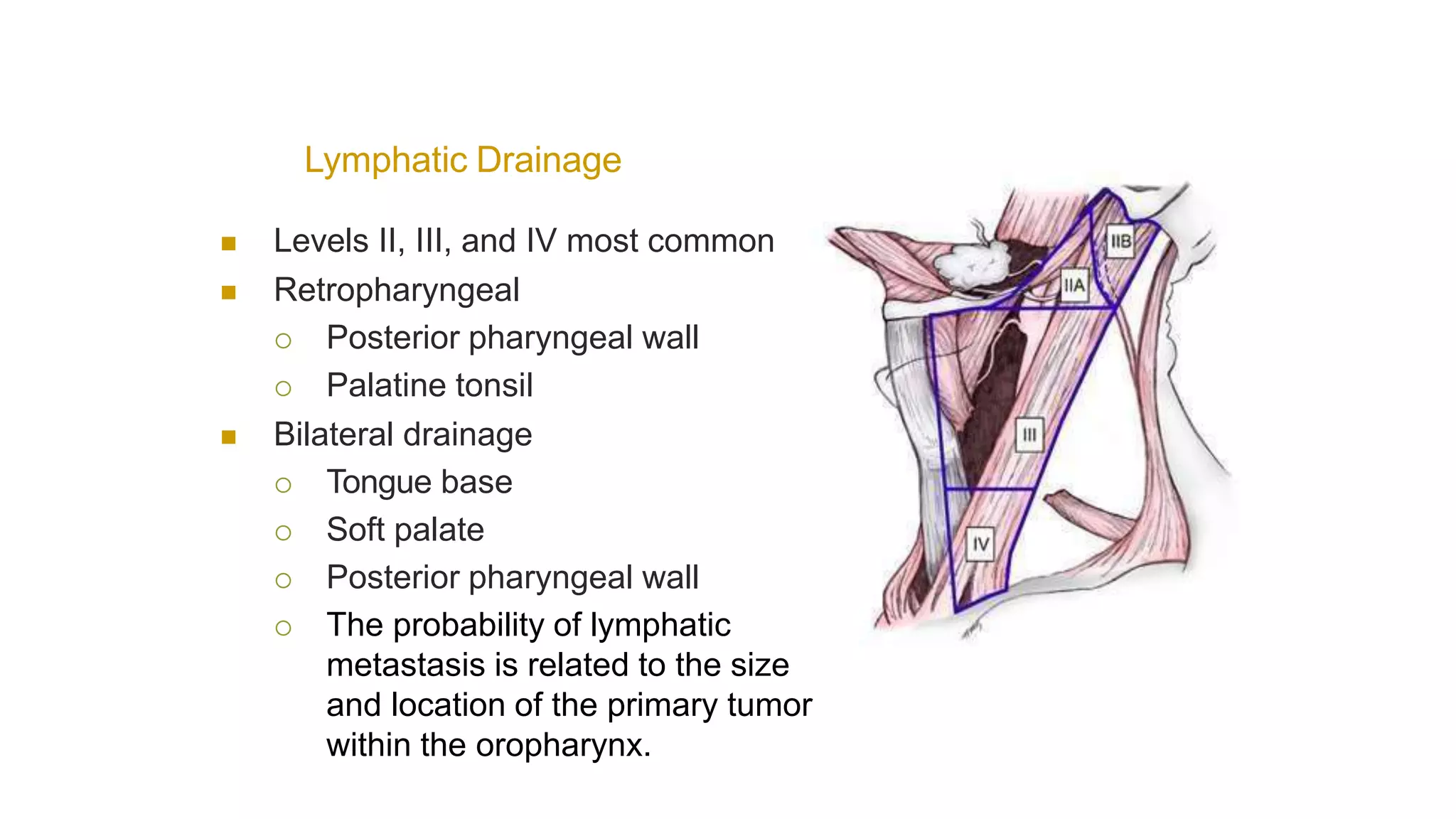
MANAGEMENT OF OROPHARYNGEAL
CANCERS
Dr Kiran Kumar BR
• The oropharynx is the
portion of the
continuity of the
pharynx extending
from the plane of the
superior surface of
the soft palate to the
superior surface of
the hyoid bone
(or vallecula).
Oropharynx
 Base of the tongue,
Vallecula,
 The inferior (anterior)
surface of the soft
palate
 The uvula,
 The anterior and
posterior tonsillar
pillars,
 The glossotonsillar
sulci,
 The pharyngeal tonsils,
and
 The lateral and
posterior pharyngeal
walls.
Lymphatic Drainage
 Levels II, III, and IV most common
 Retropharyngeal
 Posterior pharyngeal wall
 Palatine tonsil
 Bilateral drainage
 Tongue base
 Soft palate
 Posterior pharyngeal wall
 The probability of lymphatic
metastasis is related to the size
and location of the primary tumor
within the oropharynx.
DISTRIBUTION OF CLINICAL METASTATIC NECK NODES
FROM HEAD AND NECK SQUAMOUS CELL CARCINOMAS
Epidemiology
 Account for approximately 10-12% of head and
neck malignancies worldwide.
 Incidence is high in developing countries 4.8 in
100,000.
 There is increase in HPV associated oropharyngeal
cancers.
 Peak incidence in 6th or 7th decades oflife.
 More common in men(4:1).
Risk Factors
Patient related-
Age and gender: predisposition in males especially
those who are smokers and older than 50 years.
Lifestyle: cigarette smoking, alcohol, tobacco; tobacco
and alcohol are synergistic risk factors.
HPV- associated orpharyngeal cancer
• HPV is a circular double stranded DNAvirus
• Found to be associated with cervical cancer in 1983.
• Appr. 150 types of HPV have been identified
• HPV 16 is most common type asso. with more than 90% of all
oropharyngeal cancers.
HPV structure
• HPV genome encodes 3
oncoproteins(E5,E6,E7),regula
tory genes(E1,E2),capsid
proteins(L1,L2).
• Oncogenesis is primarily
mediated via the E6 and E7
proteins. HPV E6 complexes
promote ubiquitin-mediated
destruction of p53.
• Loss of cellular p53 function
results in dysregulation of the
G1/S and G2/M checkpoints.
• E7 is believed to be the major
transforming oncogene during
early carcinogenesis, with E6
functioning later.
Clinical characteristics-HPV associated
• more likely to occur among men than women (3:1)
• most of whom (80%) will not have a smoking history.
• diagnosed in individuals who are 5 to 10 years younger than HPV-
unassociated oropharyngeal cancers
• p16 IHC testing is an accurate surrogate for HPV infection
Response of HPV associated cancers
• Patients with HPV-associated oropharyngeal cancers have
significantly better outcomes compared to HPV-unassociated
oropharyngeal tumors.
• In RTOG 0129, HPV status was independently associated with
improved outcomes. Three-year overall survival was 82% in HPV-
positive patients compared with 54% in HPV-negative patients
Pathology
• Squamous cell carcinoma (SCC) accounts for >90% of
oropharyngeal cancer
• Minor salivary gland carcinomas
• Lymphoma
• Plasmacytoma
• Sarcoma
• Melanoma
Route of spread
Local
Oropharynx cancers may invade
vallecula/larynx,
 parapharyngeal area into the pterygoid muscles/plates,
nasopharynx, and oral cavity (oral tongue and retromolar
trigone)
Perineural invasion especially along branches of CN V and VII
Lymph node involvement is seen in 55% of oropharynx
cancers.
• most common location for lymph node metastases from
oropharyngeal cancers is the ipsilateral level II.
• The typical order of metastatic progression is systematic,
from the upper jugular chain nodes superiorly (level I/II; first
echelon), to mid-cervical (level III), and to lower cervical
nodes (level IV), inferiorly. Retropharyngeal nodes represent
a nodal drainage for oropharynx cancers.
• Contralateral nodes are at risk for tumors near midline,
advanced T- and N-stage diseases
Distant Metastasis in patients with advanced-stage disease
• The most common site of hematogenous metastasis is Lung.
• Seen in about 20% cases.
• However, a primary lung cancer should always be excluded.
Clinical Features
 Asymptomatic neck node
 Pain
 Dysphagia
 Otalgia
 Foreign body sensation
 Hemoptysis
 Weight loss
Otalgia
 T, tumor
 N, node
 M, metastasis
OropharyngealCancer
Staging
 Tx: primary site cannot be evaluated
 T0: no evidence of carcinoma
 Tis: carcinoma in-situ
 T1: tumor < 2cm in greatestdimension
 T2: tumor 2-4cm in greatestdimension
 T3: tumor > 4cm in greatestdimension
 T4
 T4a: invades larynx, deep/extrinsic tongue
muscles, medial pterygoid, hard palate, or
mandible
 T4b: invades lateral pterygoid, pterygoid plates,
lateral nasopharynx, skull base, or carotid
 T, tumor
 N, node
 M, metastasis
Invasion of pre-epiglottic fat
(i.e. laryngeal involvement)
Invasion of medial
pterygoid muscle
 T4a: invades larynx, deep/extrinsic tongue muscles, medial
pterygoid, hard palate, or mandible
 T4b: invades lateral pterygoid, pterygoid plates, lateral
nasopharynx, skull base, or carotid
 T4
 T4a: invades larynx, deep/extrinsic tongue muscles, medial
pterygoid, hard palate, or mandible
 T4b: invades lateral pterygoid, pterygoid plates, lateral
nasopharynx, skull base, or carotid
Encasement of
carotid artery
Involvement of
foramen ovale
 T, tumor
 N, node
 M, metastasis
Staging
 Nx: Regionallymph nodes cannotbe assessed
 N0: No regional lymph nodemetastasis
 N1: single ipsilateral node <3cm
 N2
 N2a: single ipsilateral node involved, ˃3but
• ˂6cm
 N2b: multiple ipsilateral nodes, < 6cm
 N2c: bilateral or contralateral nodal involvement, none ˃6cm
 N3: nodal involvement >6cm
 T, tumor
 N, node
 M, metastasis
Staging
 Mx: distant metastasis cannot be
evaluated
 M0: no distant metastasis
 M1: distant metastasis present
Diagnostic workup
• Complete physical examination-examination of oral cavity,
oropharynx, neck.
•Mirror examination
• Endoscopic Examination
 Fiberoptic laryngoscopy is done to assess local extent of
dosease.
 Evaluation of the upper aerodigestive tract is crucial to
evaluate the primary site of disease and the presence of
synchronous primaries.
Laboratory
tests
Includes:
1) Complete blood count
2) Basic blood chemistry
3) Hepatic and metabolic panels
4) Testing for HPV in the biopsy specimen (p16 IHC testing)
 Biopsy
 Tissue from either primary tumor or neck lymphadenopathy
is crucial for pathologic diagnosis.
Imaging-
 CECT Face & Neck
 Chest X-ray
 Ultrasound whole abdomen
 CECT Thorax (if indicated)
 FDG-PET/CT scan
CT Scan
• Scan slice thickness <5 mm is desirable to optimize the
detection of smaller pathologically involved lymph nodes.
• Pathologically involved lymph nodes are characterized on
CT imaging as those that are enlarged, enhance with
contrast, and have a necrotic centre.
• Primary tumors appear as contrast-enhancing masses,
distorting normal anatomic relationships.
• Whereas ulceration and invasion into surrounding organs
are readily assessed, submucosal spread is often difficult to
characterize with CT.
• Thoracic CT should be performed routinely to assess for
pulmonary spread of oropharyngeal cancer patients with N2
or greater nodal disease, as well as those with advanced
primary tumors due to risk of pulmonary metastases.
Magnetic ResonanceImaging
• Magnetic resonanceimaging (MRI) canbe auseful imaging tool for
oropharyngeal tumors.
• Squamous cell carcinoma appears aslow signal in T1MRI and
corresponding high signal in T2sequences.
• Theability of MRI to differentiate tumor from soft tissues is
particularly useful when determination of the extent of base of
tongue or oral tongue invasion isneeded.
• Additionally, MRI is useful in patients with compromised renal
function who are not able to receive iodine-based CTcontrastagents.
PositronEmissionTomography
• Positron emission tomography (PET) and/or PET/CT
imaging incorporating tumor physiology in conjunction with
anatomic information are now routinely recommended for
the initial staging of oropharyngeal cancer patients.
• PET-based imaging can assess not only the locoregional
burden of disease but also detect and quantify distant
metastases.
• It has high sensitivity approaching 100%, and about 60%
specificity.
• As per NCCN guidelines FDG-PET/CT should generally
considered in Stage III-IV disease.
Treatment
modalities
• Surgery
• Radiotherapy
• Chemotherapy
• Targeted Therapy
• Stage I and stage II tumors are considered as early stage,
whereas stage III and IV (nonmetastatic) are considered
locoregionally advanced disease.
• Early-stage tumors are usually well controlled with a single
local modality, either radiotherapy or surgery.
• For locoregionally advanced disease, two appropriate
treatment strategies are used:
(a) either surgery followed by radiation therapy with or
without chemotherapy based on pathologic risk factors or (b)
radiotherapy usually given with chemotherapy.
Surgery
• Base of Tongue
• Surgery plays a limited role in the management of base of
tongue tumors given the inherent morbidity of a near-total or
total glossectomy, which is required for large and/or midline
tumors.
• For select, well-lateralized base of tongue tumors with
minimal cervical lymphadenopathy, a partial glossectomy
can be performed.
• Given the high propensity for occult microscopic nodal
involvement, bilateral cervical lymph node dissection is often
performed.
• Base of tongue tumors in close proximity to the laryngeal
apparatus, such as those arising in the vallecula, often
require a supraglottic or total laryngectomy to achieve
adequate margins of resection.
• Traditional surgical approaches for base of tongue tumors
include :
• The midline mandibulotomy (splitting the lip, mandible, and
oral tongue midline),
• The lateral mandibulotomy (dividing the mandible near the
angle and approaching the base of tongue from the side),
• The floor drop procedure (elevating the inner periosteum
from the mandible from angle to angle, which releases the
entire floor of mouth and oral tongue into the neck, exposing
the base of tongue).
TonsilCancers
• For small (<1 cm) early-stage tonsil cancers confined to the anterior
pillar, a wide local excision can achieve adequate tumor-free margins,
whereas tumors involving the palatine tonsil often require a radical
tonsillectomy.
• For both of these situations, the tonsil is approached transorally, with
primary closure.
• Larger tumors with extension onto the tongue, onto the mandible or
into surrounding tissue often require a composite resection, usually
including resection of the tonsil, tonsillar fossa, pillars, a portion of the
soft palate, tongue, and mandible.
• For tumors not adjacent or adherent to the mandible, a midline
mandibulotomy approach is used.
• For tumors adherent to the mandible, a partial mandibulectomy is
used.
• Complications from surgery depend on the extent of resection, with
impairment in swallowing possible by removal of part of the tongue or
soft palate.
SoftPalateCancers
• Surgical resection is rarely recommended as initial therapy
for soft palate tumors.
• Resection of the soft palate is often associated with
significant reflux into the nasopharynx during swallowing,
even with the use of custom prostheses.
• Additionally, because of the midline location, primary
disease spreads bilaterally to the neck with frequency high
enough to require elective treatment.
• However, when surgery is performed, the tumors are
approached transorally and a full-thickness wide local
resection is performed for tumors limited to the soft palate.
• A more extensive composite resection is required if disease
extends to surrounding structures.
• Flaps or prostheses are used to preserve velopharyngeal
competence. Nasal speech is also often a consequence.
TransoralLaserSurgery
• Small series report favorable outcomes for selected patients
with stage I through stage IV oropharyngeal tumors treated
with transoral laser microsurgery with or without neck
dissection, followed by adjuvant radiotherapy or
chemoradiotherapy.
• Positive margin rates are variable (3% to 24%) and appear
to vary based on primary site, being more common in base
of tongue tumors.
• Complications include postoperative hemorrhage (5% to
10%).
Temporary tracheostomy placement is relatively common
(17% to 30%) and needed for exposure, airway control, or
aspiration following extensive resection.
• High rates of locoregional control following this procedure
have been reported, primarily for stage I/II patients (87% to
100%), although for stage III/IV patients, local recurrence is
more common (20% to 30%).
TransoralRoboticSurgery
• The most common robotic surgical system, the da Vinci Surgical
System, is comprised of three surgical instruments and a binocular
endoscope controlled by robotic arms and inserted under direct or
endoscopic guidance by the surgeon from a patient-side apparatus.
• The operative environment is visualized virtually, in a three-
dimensional (3D) environment created via a computer that links the
environment provided by the binocular endoscope to the position of
the instruments.
• The surgeon’s movements are translated into the micromovements
of the instruments. The advantages of this system include motion
scaling, which can increase precision as well as reduce hand tremor
and fatigue.
• When the system is used for transoral surgeries, an assistant is
often positioned by the patient’s head.
• There are no prospective randomized studies supporting the
use of transoral robotic surgery (TORS) for oropharyngeal
tumor resection over conventional surgery.
• Until mature prospective multi-institutional series and
randomized data are available, the true utility of transoral
laser microsurgery and TORS remains unknown.
• Although early results are favorable and associated with
shorter hospital stays, long-term data are needed.
• Additionally, standard oncologic principles limiting the
number of modalities used to minimize treatment related
side effects should be carefully considered prior to
widespread adoption of the surgical techniques.
Neck Management
• The type of neck dissection (comprehensive or selective) is
defined according to preoperative clinical staging, is
determined at the discretion of the surgeon, and is based on
the initial preoperative staging as follows:
N0= Selective neck dissection, atleast Levels II-IV
N1-N2a-c= comprehensive neck dissection
N3= Comprehensive neck dissection
Radiotherapy
• Definitive chemoradiotherapy is the treatment of choice.
• For early-stage oropharyngeal cancers, the use of radiation
therapy as a single modality is associated with good
outcomes and functional preservation.
• Patients with locoregionally advanced oropharyngeal cancer,
concurrent chemoradiotherapy is the standard treatment.
Resection is generally not recommended given the
associated surgical morbidity.
Chemotherapy
• Used in concurrent setting along with radiotherapy
• Recurrent and metastatic tumors
• As neoadjuvant chemotherapy, but there is no definitive role.
Radiotherapy
techniques
• Two dimensional planning
• 3D planning
• IMRT
Conventionalborders
CarcinomaBaseoftonguelower
NeckField
3DPlanning
• Patients should undergo simulation, preferably CT based, to
allow for optimal radiotherapy planning.
• A peripheral IV should be placed prior to simulation for the
delivery of low osmolar iodinated contrast to optimize the
distinction between vascular structures and lymph nodes.
• Patients are positioned supine, extended head position is
preferable.
• Care should be taken to ensure that the mask is tight and
should not allow movement of the nose, chin or, forehead.
Chemotherapy
RoleofTargetedTherapy
• EGFR inhibitors have been tried in H&N cancers
• Weekly Cetuximab loading doseof 400 mg/m2 followed by 250
mg/m2 for locoregionally advanced head and neck cancer patients
• Thecombination therapy wasfound to improve locoregional
control, disease-free survival, and overall survival
CETUXIMAB IN HPV+ OPC
Follow up
• Every 1 to 3 months for the first year post RT
• Every 2 to 4 months in the second year post RT, and every 4
to 6 months in the next 3 to 5 years.
• The intensity of the examinations within the first 2 years
coincides with the likelihood of recurrence in the interval.
Given that radiation to the neck commonly causes
hypothyroidism, thyroid-stimulating hormone levels should
be evaluated every 6 months.
• Follow-up imaging should be performed within the first 3
months of treatment
ReirradiationforLocoregionallyConfinedRecurrentorSecond Primary
Disease
• Surgical resection is recommended, although this is possible
only in a small proportion of patients.
• Following surgery in those with high-risk pathologic features
or in those who are not surgical candidates,a second course
of full-dose radiotherapy with chemotherapy has been
shown to result in long-term survival in approximately 20% of
patients.
THANKYOU

ORO PHARYNX.pptx

  • 1.
  • 2.
    • The oropharynxis the portion of the continuity of the pharynx extending from the plane of the superior surface of the soft palate to the superior surface of the hyoid bone (or vallecula).
  • 3.
    Oropharynx  Base ofthe tongue, Vallecula,  The inferior (anterior) surface of the soft palate  The uvula,  The anterior and posterior tonsillar pillars,  The glossotonsillar sulci,  The pharyngeal tonsils, and  The lateral and posterior pharyngeal walls.
  • 4.
    Lymphatic Drainage  LevelsII, III, and IV most common  Retropharyngeal  Posterior pharyngeal wall  Palatine tonsil  Bilateral drainage  Tongue base  Soft palate  Posterior pharyngeal wall  The probability of lymphatic metastasis is related to the size and location of the primary tumor within the oropharynx.
  • 5.
    DISTRIBUTION OF CLINICALMETASTATIC NECK NODES FROM HEAD AND NECK SQUAMOUS CELL CARCINOMAS
  • 6.
    Epidemiology  Account forapproximately 10-12% of head and neck malignancies worldwide.  Incidence is high in developing countries 4.8 in 100,000.  There is increase in HPV associated oropharyngeal cancers.  Peak incidence in 6th or 7th decades oflife.  More common in men(4:1).
  • 7.
    Risk Factors Patient related- Ageand gender: predisposition in males especially those who are smokers and older than 50 years. Lifestyle: cigarette smoking, alcohol, tobacco; tobacco and alcohol are synergistic risk factors.
  • 8.
    HPV- associated orpharyngealcancer • HPV is a circular double stranded DNAvirus • Found to be associated with cervical cancer in 1983. • Appr. 150 types of HPV have been identified • HPV 16 is most common type asso. with more than 90% of all oropharyngeal cancers.
  • 9.
    HPV structure • HPVgenome encodes 3 oncoproteins(E5,E6,E7),regula tory genes(E1,E2),capsid proteins(L1,L2). • Oncogenesis is primarily mediated via the E6 and E7 proteins. HPV E6 complexes promote ubiquitin-mediated destruction of p53. • Loss of cellular p53 function results in dysregulation of the G1/S and G2/M checkpoints. • E7 is believed to be the major transforming oncogene during early carcinogenesis, with E6 functioning later.
  • 11.
    Clinical characteristics-HPV associated •more likely to occur among men than women (3:1) • most of whom (80%) will not have a smoking history. • diagnosed in individuals who are 5 to 10 years younger than HPV- unassociated oropharyngeal cancers • p16 IHC testing is an accurate surrogate for HPV infection
  • 12.
    Response of HPVassociated cancers • Patients with HPV-associated oropharyngeal cancers have significantly better outcomes compared to HPV-unassociated oropharyngeal tumors. • In RTOG 0129, HPV status was independently associated with improved outcomes. Three-year overall survival was 82% in HPV- positive patients compared with 54% in HPV-negative patients
  • 14.
    Pathology • Squamous cellcarcinoma (SCC) accounts for >90% of oropharyngeal cancer • Minor salivary gland carcinomas • Lymphoma • Plasmacytoma • Sarcoma • Melanoma
  • 15.
    Route of spread Local Oropharynxcancers may invade vallecula/larynx,  parapharyngeal area into the pterygoid muscles/plates, nasopharynx, and oral cavity (oral tongue and retromolar trigone) Perineural invasion especially along branches of CN V and VII
  • 16.
    Lymph node involvementis seen in 55% of oropharynx cancers. • most common location for lymph node metastases from oropharyngeal cancers is the ipsilateral level II. • The typical order of metastatic progression is systematic, from the upper jugular chain nodes superiorly (level I/II; first echelon), to mid-cervical (level III), and to lower cervical nodes (level IV), inferiorly. Retropharyngeal nodes represent a nodal drainage for oropharynx cancers. • Contralateral nodes are at risk for tumors near midline, advanced T- and N-stage diseases
  • 17.
    Distant Metastasis inpatients with advanced-stage disease • The most common site of hematogenous metastasis is Lung. • Seen in about 20% cases. • However, a primary lung cancer should always be excluded.
  • 18.
    Clinical Features  Asymptomaticneck node  Pain  Dysphagia  Otalgia  Foreign body sensation  Hemoptysis  Weight loss
  • 19.
  • 20.
     T, tumor N, node  M, metastasis OropharyngealCancer Staging  Tx: primary site cannot be evaluated  T0: no evidence of carcinoma  Tis: carcinoma in-situ  T1: tumor < 2cm in greatestdimension  T2: tumor 2-4cm in greatestdimension  T3: tumor > 4cm in greatestdimension  T4  T4a: invades larynx, deep/extrinsic tongue muscles, medial pterygoid, hard palate, or mandible  T4b: invades lateral pterygoid, pterygoid plates, lateral nasopharynx, skull base, or carotid
  • 21.
     T, tumor N, node  M, metastasis Invasion of pre-epiglottic fat (i.e. laryngeal involvement) Invasion of medial pterygoid muscle  T4a: invades larynx, deep/extrinsic tongue muscles, medial pterygoid, hard palate, or mandible  T4b: invades lateral pterygoid, pterygoid plates, lateral nasopharynx, skull base, or carotid
  • 22.
     T4  T4a:invades larynx, deep/extrinsic tongue muscles, medial pterygoid, hard palate, or mandible  T4b: invades lateral pterygoid, pterygoid plates, lateral nasopharynx, skull base, or carotid Encasement of carotid artery Involvement of foramen ovale
  • 23.
     T, tumor N, node  M, metastasis Staging  Nx: Regionallymph nodes cannotbe assessed  N0: No regional lymph nodemetastasis  N1: single ipsilateral node <3cm  N2  N2a: single ipsilateral node involved, ˃3but • ˂6cm  N2b: multiple ipsilateral nodes, < 6cm  N2c: bilateral or contralateral nodal involvement, none ˃6cm  N3: nodal involvement >6cm
  • 24.
     T, tumor N, node  M, metastasis Staging  Mx: distant metastasis cannot be evaluated  M0: no distant metastasis  M1: distant metastasis present
  • 25.
    Diagnostic workup • Completephysical examination-examination of oral cavity, oropharynx, neck. •Mirror examination • Endoscopic Examination  Fiberoptic laryngoscopy is done to assess local extent of dosease.  Evaluation of the upper aerodigestive tract is crucial to evaluate the primary site of disease and the presence of synchronous primaries.
  • 26.
    Laboratory tests Includes: 1) Complete bloodcount 2) Basic blood chemistry 3) Hepatic and metabolic panels 4) Testing for HPV in the biopsy specimen (p16 IHC testing)
  • 27.
     Biopsy  Tissuefrom either primary tumor or neck lymphadenopathy is crucial for pathologic diagnosis. Imaging-  CECT Face & Neck  Chest X-ray  Ultrasound whole abdomen  CECT Thorax (if indicated)  FDG-PET/CT scan
  • 28.
    CT Scan • Scanslice thickness <5 mm is desirable to optimize the detection of smaller pathologically involved lymph nodes. • Pathologically involved lymph nodes are characterized on CT imaging as those that are enlarged, enhance with contrast, and have a necrotic centre. • Primary tumors appear as contrast-enhancing masses, distorting normal anatomic relationships. • Whereas ulceration and invasion into surrounding organs are readily assessed, submucosal spread is often difficult to characterize with CT. • Thoracic CT should be performed routinely to assess for pulmonary spread of oropharyngeal cancer patients with N2 or greater nodal disease, as well as those with advanced primary tumors due to risk of pulmonary metastases.
  • 29.
    Magnetic ResonanceImaging • Magneticresonanceimaging (MRI) canbe auseful imaging tool for oropharyngeal tumors. • Squamous cell carcinoma appears aslow signal in T1MRI and corresponding high signal in T2sequences. • Theability of MRI to differentiate tumor from soft tissues is particularly useful when determination of the extent of base of tongue or oral tongue invasion isneeded. • Additionally, MRI is useful in patients with compromised renal function who are not able to receive iodine-based CTcontrastagents.
  • 30.
    PositronEmissionTomography • Positron emissiontomography (PET) and/or PET/CT imaging incorporating tumor physiology in conjunction with anatomic information are now routinely recommended for the initial staging of oropharyngeal cancer patients. • PET-based imaging can assess not only the locoregional burden of disease but also detect and quantify distant metastases. • It has high sensitivity approaching 100%, and about 60% specificity. • As per NCCN guidelines FDG-PET/CT should generally considered in Stage III-IV disease.
  • 31.
  • 32.
    • Stage Iand stage II tumors are considered as early stage, whereas stage III and IV (nonmetastatic) are considered locoregionally advanced disease. • Early-stage tumors are usually well controlled with a single local modality, either radiotherapy or surgery. • For locoregionally advanced disease, two appropriate treatment strategies are used: (a) either surgery followed by radiation therapy with or without chemotherapy based on pathologic risk factors or (b) radiotherapy usually given with chemotherapy.
  • 33.
    Surgery • Base ofTongue • Surgery plays a limited role in the management of base of tongue tumors given the inherent morbidity of a near-total or total glossectomy, which is required for large and/or midline tumors. • For select, well-lateralized base of tongue tumors with minimal cervical lymphadenopathy, a partial glossectomy can be performed. • Given the high propensity for occult microscopic nodal involvement, bilateral cervical lymph node dissection is often performed. • Base of tongue tumors in close proximity to the laryngeal apparatus, such as those arising in the vallecula, often require a supraglottic or total laryngectomy to achieve adequate margins of resection.
  • 34.
    • Traditional surgicalapproaches for base of tongue tumors include : • The midline mandibulotomy (splitting the lip, mandible, and oral tongue midline), • The lateral mandibulotomy (dividing the mandible near the angle and approaching the base of tongue from the side), • The floor drop procedure (elevating the inner periosteum from the mandible from angle to angle, which releases the entire floor of mouth and oral tongue into the neck, exposing the base of tongue).
  • 35.
    TonsilCancers • For small(<1 cm) early-stage tonsil cancers confined to the anterior pillar, a wide local excision can achieve adequate tumor-free margins, whereas tumors involving the palatine tonsil often require a radical tonsillectomy. • For both of these situations, the tonsil is approached transorally, with primary closure. • Larger tumors with extension onto the tongue, onto the mandible or into surrounding tissue often require a composite resection, usually including resection of the tonsil, tonsillar fossa, pillars, a portion of the soft palate, tongue, and mandible. • For tumors not adjacent or adherent to the mandible, a midline mandibulotomy approach is used. • For tumors adherent to the mandible, a partial mandibulectomy is used. • Complications from surgery depend on the extent of resection, with impairment in swallowing possible by removal of part of the tongue or soft palate.
  • 36.
    SoftPalateCancers • Surgical resectionis rarely recommended as initial therapy for soft palate tumors. • Resection of the soft palate is often associated with significant reflux into the nasopharynx during swallowing, even with the use of custom prostheses. • Additionally, because of the midline location, primary disease spreads bilaterally to the neck with frequency high enough to require elective treatment. • However, when surgery is performed, the tumors are approached transorally and a full-thickness wide local resection is performed for tumors limited to the soft palate. • A more extensive composite resection is required if disease extends to surrounding structures. • Flaps or prostheses are used to preserve velopharyngeal competence. Nasal speech is also often a consequence.
  • 37.
    TransoralLaserSurgery • Small seriesreport favorable outcomes for selected patients with stage I through stage IV oropharyngeal tumors treated with transoral laser microsurgery with or without neck dissection, followed by adjuvant radiotherapy or chemoradiotherapy. • Positive margin rates are variable (3% to 24%) and appear to vary based on primary site, being more common in base of tongue tumors. • Complications include postoperative hemorrhage (5% to 10%). Temporary tracheostomy placement is relatively common (17% to 30%) and needed for exposure, airway control, or aspiration following extensive resection. • High rates of locoregional control following this procedure have been reported, primarily for stage I/II patients (87% to 100%), although for stage III/IV patients, local recurrence is more common (20% to 30%).
  • 38.
    TransoralRoboticSurgery • The mostcommon robotic surgical system, the da Vinci Surgical System, is comprised of three surgical instruments and a binocular endoscope controlled by robotic arms and inserted under direct or endoscopic guidance by the surgeon from a patient-side apparatus. • The operative environment is visualized virtually, in a three- dimensional (3D) environment created via a computer that links the environment provided by the binocular endoscope to the position of the instruments. • The surgeon’s movements are translated into the micromovements of the instruments. The advantages of this system include motion scaling, which can increase precision as well as reduce hand tremor and fatigue. • When the system is used for transoral surgeries, an assistant is often positioned by the patient’s head.
  • 39.
    • There areno prospective randomized studies supporting the use of transoral robotic surgery (TORS) for oropharyngeal tumor resection over conventional surgery. • Until mature prospective multi-institutional series and randomized data are available, the true utility of transoral laser microsurgery and TORS remains unknown. • Although early results are favorable and associated with shorter hospital stays, long-term data are needed. • Additionally, standard oncologic principles limiting the number of modalities used to minimize treatment related side effects should be carefully considered prior to widespread adoption of the surgical techniques.
  • 40.
    Neck Management • Thetype of neck dissection (comprehensive or selective) is defined according to preoperative clinical staging, is determined at the discretion of the surgeon, and is based on the initial preoperative staging as follows: N0= Selective neck dissection, atleast Levels II-IV N1-N2a-c= comprehensive neck dissection N3= Comprehensive neck dissection
  • 41.
    Radiotherapy • Definitive chemoradiotherapyis the treatment of choice. • For early-stage oropharyngeal cancers, the use of radiation therapy as a single modality is associated with good outcomes and functional preservation. • Patients with locoregionally advanced oropharyngeal cancer, concurrent chemoradiotherapy is the standard treatment. Resection is generally not recommended given the associated surgical morbidity.
  • 42.
    Chemotherapy • Used inconcurrent setting along with radiotherapy • Recurrent and metastatic tumors • As neoadjuvant chemotherapy, but there is no definitive role.
  • 43.
    Radiotherapy techniques • Two dimensionalplanning • 3D planning • IMRT
  • 44.
  • 46.
  • 47.
    3DPlanning • Patients shouldundergo simulation, preferably CT based, to allow for optimal radiotherapy planning. • A peripheral IV should be placed prior to simulation for the delivery of low osmolar iodinated contrast to optimize the distinction between vascular structures and lymph nodes. • Patients are positioned supine, extended head position is preferable. • Care should be taken to ensure that the mask is tight and should not allow movement of the nose, chin or, forehead.
  • 50.
  • 52.
    RoleofTargetedTherapy • EGFR inhibitorshave been tried in H&N cancers • Weekly Cetuximab loading doseof 400 mg/m2 followed by 250 mg/m2 for locoregionally advanced head and neck cancer patients • Thecombination therapy wasfound to improve locoregional control, disease-free survival, and overall survival
  • 53.
  • 54.
    Follow up • Every1 to 3 months for the first year post RT • Every 2 to 4 months in the second year post RT, and every 4 to 6 months in the next 3 to 5 years. • The intensity of the examinations within the first 2 years coincides with the likelihood of recurrence in the interval. Given that radiation to the neck commonly causes hypothyroidism, thyroid-stimulating hormone levels should be evaluated every 6 months. • Follow-up imaging should be performed within the first 3 months of treatment
  • 55.
    ReirradiationforLocoregionallyConfinedRecurrentorSecond Primary Disease • Surgicalresection is recommended, although this is possible only in a small proportion of patients. • Following surgery in those with high-risk pathologic features or in those who are not surgical candidates,a second course of full-dose radiotherapy with chemotherapy has been shown to result in long-term survival in approximately 20% of patients.
  • 56.

Editor's Notes

  • #54 NOV 15 2018 IN LANCET