The current research aimed at fabricating plant extract mediated biosynthesized silver nanoparticles (AgNPs) utilizing thorn extract of Bombax ceiba (TEBC). The synthesized AgNPs was characterized by UV spectroscopy where the surface plasmonic resonance peak (SPR) was located at 222 nm. The scanning electron microscopy (SEM) studies demonstrated that the morphology of fabricated nanomaterials was primarily cylindrical of average size of 20-30 nm with some spindles of size >50 nm. The anti-microbial evaluation against Staphylococcus aureus revealed that AgNPs exhibited notable activity with ZOI of 27.2 mm at MIC of 25 μg/mL. The outcome of this research evidently signified that the biofabricated AgNPs using TEBC may be a new greener approach or technology to formulate anti-bacterial nanodrugs in future.
Nowadays synthesis of nanomaterials by using bio-root is limelight of modern nanotechnology. In the present investigation, we have isolated four strains viz: KFRI 330 (A), KFRI 332 (B), KFRI 351 (C) and KFRI 352 (D) of Beauveria bassiana from the forest soils in Kerala. Spore count was tested for all the strains of B. bassiana stored in the laboratory. Silver nanoparticles were synthesized from the four strains of B. bassiana and the formation of nanoparticles was observed within 48 hours. The synthesized silver nanoparticle has been characterized by UV-Vis spectroscopy, FT-IR and TEM analysis. The appearance of UV-Vis Peak (SPR 440 nm) revealed the reduction of silver metal ions to silver nanoparticles by using the fungal strains. The possible bio-molecules involved in nanoparticles synthesis was identified by HPLC analysis. The functional groups involved in the silver nanoparticles synthesis were identified. The amide group is responsible for the synthesis of silver nanoparticles. From the TEM analysis, the size of the AGNPs has been measured as 4-70 nm (mean 10.7±0.04 nm). It was evident from the HPLC result that primary amines act on capping as a well as a stabilizing agent.
In the present work, green synthesis of copper oxide nanoparticles has wide interest due to its inherent features such as eco-friendly and low costs. Here we propose a cost effective and eco-friendly green synthesis of copper oxide nanoparticles using aloe barbadensis and copper acetate. Aloe barbadensis has antioxidant property that helps in the formation of nanoparticles. Aloe barbadensis extract was obtained by heating it in distilled water and mixed to copper acetate to form copper oxide nanoparticles. The synthesised nanoparticles were analysed using UV-Visible spectroscopy, X-ray diffraction studies (XRD) and Fourier-transform infrared spectroscopy (FTIR).
Green Synthesis of ZnO Nanoparticles using Jasminum Fluminense Leaf Extract a...ijtsrd
The main objective of the present study is to deal with the green synthesis of Zinc Oxide [ZnO] nanoparticles utilizing aqueous leaf extract of Jasminum Fluminense.` Zinc acetate [Zn(O2CCH3)2(H2O)2] and sodium hydroxide [NaOH] were used as starting materials and Jasminum Fluminense [Nithyamalli] leaf extract is used as precursor in the synthesis of ZnO nanoparticles. The resultant nanopowder was characterized by using Ultraviolet [UV] “ Visible Spectroscopy, Fourier Transform Infrared Spectroscopy [FTIR] and Transmission Electron Microscopic [TEM] studies. Formation of ZnO nanoparticles has been confirmed by UV-visible spectroscopy and the TEM analysis spectacles that the synthesized ZnO nanoparticles are of face centered cubic (fcc) structure and the size is found to be around 20 nm. FTIR spectral analysis indicated the leaf extract acts as the reducing and capping agents on the surface of ZnO nanoparticles. This simplistic and green approach may provide a useful tool to hefty extent in the synthesis of ZnO nanoparticles. These synthesized nanostructures illustrate novel applications in many fields such as cosmetics, optoelectronics, sensors, transducers and biomedical science because it is environmentally friendly and does not involve any harmful substances. S. Amudha"Green Synthesis of ZnO Nanoparticles using Jasminum Fluminense Leaf Extract and its Spectral and Optical Characterization Studies" Published in International Journal of Trend in Scientific Research and Development (ijtsrd), ISSN: 2456-6470, Volume-2 | Issue-4 , June 2018, URL: http://www.ijtsrd.com/papers/ijtsrd12908.pdf http://www.ijtsrd.com/physics/nanotechnology/12908/green-synthesis-of-zno-nanoparticles-using-jasminum-fluminense-leaf-extract-and-its-spectral-and-optical-characterization-studies/s-amudha
Biosynthesis and characterization of silver nanoparticles using ficus benghal...eSAT Publishing House
IJRET : International Journal of Research in Engineering and Technology is an international peer reviewed, online journal published by eSAT Publishing House for the enhancement of research in various disciplines of Engineering and Technology. The aim and scope of the journal is to provide an academic medium and an important reference for the advancement and dissemination of research results that support high-level learning, teaching and research in the fields of Engineering and Technology. We bring together Scientists, Academician, Field Engineers, Scholars and Students of related fields of Engineering and Technology
Green synthesis of zinc oxide nano particles using flower extract cassia dens...IJERD Editor
Green synthesis of metal nanoparticles is an interesting issue of the nanoscience and
nanobiotechnology. There is a growing attention to biosynthesis the metal nanoparticles using organisms.
Among these organisms, plants seem to be the best and they are suitable for large scale biosynthesis of
nanoparticles. Nanoparticles produced by plants are more stable, and the rate of synthesis is faster than that in
the case of other organisms. The present investigation was carried out to green synthesis of zinc oxide
nanoparticles by using the medicinal plant cassia densistipulata taub. The flower was collected from the campus
of Anantapuramu, Andhra Pradesh and their petals were separated. The petals were taken and cleaned with
dimeneralized water and soaked for an hour on dry cloth to remove moisture from the petals.
Synthesis of Zinc Nanoparticles was done by mixing 5gms of Zinc Nitrate with 50ml of aqueous
extract of cassia densistipulata taub petals. The formation of nanoparticles was monitored by visualizing color
changes and it was confirmed by Electron microscope (SEM), UV-Vis spectrophotometer and Fourier
Transform Infra-Red (FT-IR) spectroscopy. The results of various techniques confirmed the presence Zinc oxide
nanoparticles.
Is Nano Medicine And Nano Technology The Most Trending Thing Now?science journals
Nano medicine is nothing but application of Nano technologies as medicines. It may include application of non-material as biological devices or nano-electronic biosensors. Molecular nanotechnology as biological machines may have medical applications in future.
ABSTRACT- Tagetes erecta L. was raised in pots containing soil treated with various concentrations of Pb(NO3)2 (500, 1000, 1500, 2000 and 2500mg/kg). At maturity plants were separated into root, stem, leaves and inflorescence and lead accumulated in each part was quantified. The effects of lead accumulation on growth was analyzed by the measurement of various growth parameters like root and shoot length, fresh and dry weight of root and shoot and total leaf area per plant. Moreover effect of lead accumulation on biochemical parameters was checked by quantitative estimation of various biochemical parameters like chlorophyll, total protein, free amino acids, total sugar, reducing sugar and starch. Results showed that there is no remarkable negative effect of accumulation of lead on the morphological growth of the plant. Biochemical analysis showed that amount of total protein continuously decreased whereas that of free amino acids continuously increased with increasing concentrations of lead. Amount of chlorophyll, total sugar, reducing sugar and starch contents continuously increased till mid-level i.e., Pb 1500 mg/kg and then continuously decreased at higher concentrations. Results of quantitative estimation of Pb in root, stem, leaves and inflorescence showed that roots accumulated highest amount of Pb followed by stem and leaves, whereas inflorescence contained least amount of Pb.
Key-words- Lead (Pb), Heavy metal, Tagetes erecta L., Accumulation, Phytoremediation
In our study, two different methods were used to determine the size and size distribution of the sliver and selenium nanoparticles via dynamic light scattering (DLS) and scanning electron microscopy (SEM). Background: Nanotechnology dealing with metal and metalloid nanoparticles has been usually applied in nearly each field of science, engineering, and technology including biology and medicine etc due to presence of size and shape dependent unusual physical and chemical properties. In the most recent decade, numerous groups including appreciably developed metal and metalloid nanoparticles based theranostic approaches for the treatment of almost human diseases. Amongst
many nanoparticles, recently silver and selenium nanoparticles have been broadly used in the antimicrobial coatings, textiles, paints, keyboards, engineering, food industry, electronics, cosmetics, bio-sensing, wound dressings, and even in biomedical devices. Methods: In our study, silver nanoparticles were prepared by using the chemical reduction method. Selenium nanoparticles (SeNPs) were synthesized by the chemical reduction of sodium selenite by glutathione (reduced form) and stabilized by bovine serum albumin (BSA). Characterization of silver and selenium nanoparticles samples were analyzed by dynamic light scattering (DLS) and Scanning Electron Microscopy (SEM). Conclusions: Due to characterization by DLS technique, nanoparticles size was found the range of 79.22 nm and 178 nm for Sliver and Selenium Nanoparticles respectively. Sliver nanoparticles shown morphological average size and shape with SEM reveals spherical shape particles with the size of 80.32 nm whereas Selenium nanoparticles shown rod shape particles with the size of 74.29 nm.
Green synthesis, Characterization and anti microbial activity of silver nano ...IJERA Editor
The exploitation of various plant materials for the biosynthesis of silver nano particles is considered a green
technology. Because it does not involve any harmful chemicals. Nanotechnology field is one of the most
attractive researches. The field of nanotechnology is applied to bio materials. This review focuses on the green
synthesis of silver nanoparticles using various plant sources. A detailed study on the reduction of silver ions to
silver nanoparticles from medical plant leaves extract were demonstrated with a brief experimental procedure.
Characterization of the synthesized nanoparticles performed through UV spectroscopy, Fourier Transform Infra
Red spectroscopy analysis, X-Ray Diffraction analysis, Scanning Electron Microscopy and Transmission
Electron Microscopy. This review mainly focus on anti microbial activities of synthesized silver nano particles.
Effluents containing heavy metals can be
remediated with the help of dead microorganisms by the process
known as biosorption. In this study the dead biomass 1of fungus
Aspergillus flavus was used for the biosorption of heavy metals
i.e., Zinc and Nickel. The capacity of biosorption by the dead
biomass of Aspergillus flavus was evaluated at room temperature
with different parameters which are; pH, contact time, biomass
concentration and metal ion concentration. The biosorption
capacity for Zn was found to be 47.36% at room temperature, at
pH 6.5, with biomass concentration of 2g/L having contact time of
50 min and solution concentration of 2ppm. Biosorption capacity
for Ni was found to be 61.60% at room temperature, at pH 5,
with biomass concentration of 2g/L having contact time of 60 min
and solution concentration of 2ppm. . In this study, desorption of
the heavy metals by 0.1M HCl was found to be effective. Fungal
biomass was recovered for reuse.
Practical MCQ Bank for experiments: Medicinal chemistry ISONALI PAWAR
Practical MCQ Bank for experiments: Medicinal chemistry I
Benzimidazole from O-phenylene diamine
synthesis and characterized benztriazole
ethyl p- amino benzoate (benzocaine) from p-amino benzoic acid
submit benzyl from benzoin
phenytoin
2,3-Diphenyl quinoxaline from O-phenylene diamine (OPD).
phenothiazine from diphenylamine.
column chromatography techniques
partition coefficient
determine buffer capacity and dissociation constant pKa of acid.
Abstract
A rapid advance of nanotechnology has the potential approach for significant improvements in disease prevention, diagnosis and treatment. In this article, we report a simple and eco-friendly biosynthesis of silver nanoparticles (Ag-NPs) using silver nitrate as metal precursor in Curcuma longa. These Ag-NPs were characterized by UV–vis spectroscopy, and Transmission electron microscopy (TEM). These nanoparticles exhibited maximum absorbance in specific nano meter range in UV–vis spectroscopy. TEM micrographs revealed the formation of well-dispersed Ag-NPs with its size and morphology. Microbiology assay founds that Ag-NPs are effective against V.cholera bacteria. These developments raise exciting opportunities to diagnose and treat pathogenic mode of infection based on the various profiles to target diseases.
Crimson Publishers-Synthesis, Characterization, and Evaluation of the In-vitr...CrimsonpublishersMedical
Cinnamomum zeylanicum have lot-of biological activities including antimicrobial, antioxidant and antifungal properties. Furthermore, cytotoxic and apoptotic activities of several constituents were identified throughout its biological properties. Bark of Cinnamomum zeylanicum (Lauraceae) collected respectively at Nanotechnology laboratory (ANGRAU, Tirupathi, India). In this study, microbiological aspects of scale formation in PVC pipelines bacteria and fungi were isolated. Stable Zn nanoparticles were formed by treating 90ml of 1mm zinc nitrate aqueous solution with 10ml of 10% bark extract. The formation of Cinnamomum zeylanicum bark extract mediated zinc nanoparticles (CZnNPs) was confirmed by UV-visible spectroscopic analysis and recorded the localized surface plasmon resonance (LSPR) at 270nm.
Cytotoxic | Primary research | Silver nanoparticlesPubrica
Biosynthesis, Antimicrobial, and Cytotoxic Effects of Silver Nanoparticles Using Acacia Concinna POD Extract and Kigelia Africa Leaf Extract- Secondary metabolites found in plants include alkaloids, flavonoids, phenolic compounds, phytosterols, saponins, tannins, carbohydrates, proteins, lipids, and minerals. These secondary metabolites have a wide range of uses, including the production of nanoparticles.
Visit us @ https://pubrica.com/insights/sample-work/cytotoxic-effects-of-sliver-nanoparticles-using-leaf-extract/
Antimicrobial and cytotoxicity effect of silver nanoparticle synthesized by C...Nanomedicine Journal (NMJ)
Objective(s): For the development of reliable, ecofriendly, less expensive process for the synthesis of silver nanoparticles and to evaluate the bactericidal, and cytotoxicity properties of silver nanoparticles synthesized from root extract of Croton bonplandianum, Baill.
Materials and Methods: The synthesis of silver nanoparticles by plant part of Croton bonplandianum was carried out. The formation of nanoparticles was confirmed by Transmission Electron Microscopy (TEM), Scanning Electron Microscopy (SEM), XRD and UV-Vis spectrophotometric analysis. The biochemical properties were assayed by antibacterial study, cytotoxicity assay using cancer cell line.
Results: The formation of silver nanoparticles was confirmed by UV-VIS spectroscopic analysis which showed absorbance peak at 425 nm. X-ray diffraction photograph indicated the face centered cubic structure of the synthesized AgNPs. TEM has displayed the different dimensional images of biogenic silver nanoparticles with particle size distribution ranging from 15-40 nm with an average size of 32 nm. Silver particles are spherical in shape, clustered. The EDX analysis was used to identify the elemental composition of synthesized AgNPs. Antibacterial activity of the synthesized AgNPs against three Gram positive and Gram negative bacteria strains like Staphylococcus aureus, Escherichia coli and Pseudomonas aeruginosa carried out showed significant zones of inhibition. The cytotoxicity study by AgNPS also showed cytotoxicity on ovarian cancer cell line PA-1 and lung epithelial cancer cell line A549.
Conclusion: The present study confirms that the AgNPs have great promise as antibacterial, and anticancer agent.
Synthesis and evaluation of bactericidal properties of CuO nanoparticles agai...Nanomedicine Journal (NMJ)
Objective(s):
CuO is one of the most important transition metal oxides due to its captivating properties. It is used in various technological applications such as high critical temperature superconductors, gas sensors, in photoconductive applications, and so on. Recently, it has been used as an antimicrobial agent against various bacterial species.
Materials and Methods:
Here, we synthesized CuO nanoparticles (NPs) and explored the antibacterial activity of CuO NPs preparation.
Results:
Single crystalline nanoparticles of copper oxide having almost uniform particle size of 5-6 nm has been synthesized by a facile and versatile route. XRD spectra confirmed the formation of single phase CuO NPs. Transmission electron microscopy results corroborate well with XRD results. The technique employed is free from toxic solvents, organics and amines, is based on a simple reaction of copper sulfate and de-ionized water (DI), and their bactericidal effects against of Aeromonas hydrophila ATCC 7966T bacteria were investigated. Minimum inhibitory concentration (MIC) and minimum bactericidal concentration (MBC) with liquid culture for all of the Aeromonas hydrophila culture Medias was done.
Conclusion:
Present study confirms that Copper oxide nanoparticles have great promise as antimicrobial agent against Aeromonas hydrophila.
Phyto-mediated Synthesis of Copper Nanoparticles by Cassia auriculata and its...IJEAB
An eco-friendly loom has been taking up in the present study to synthesize copper nanoparticles using Cassia auriculata. The leaf extract of Cassia auriculata acts as reducing as well as capping agent. Synthesis of copper nanoparticles was initially confirmed by the visual observation i.e color change (dark green color). The synthesized copper nanoparticles were primarily characterized by UV-vis spectroscopy and Fourier Transform Infrared (FTIR) spectroscopy. Further, the formation of amorphous and crystalline phase was analyzed by X-Ray Diffraction pattern. The size and morphology of the synthesized Copper nanoparticles was characterized by Scanning Electron Microscopy (SEM) and the elemental composition was analyzed by EDAX. The present study is a preliminary investigation to know about the capability of Cassia auriculata to synthesize copper nanoparticles from its salts. The results of the present study confirmed that the leaf extract of Cassia auriculata be capable of recovering copper from printed circuit boards in the form of nanoparticles in near future.
Biosynthesis of Silver Nanoparticles using Rhizophora mucronata and Ceriops d...BRNSS Publication Hub
To find out the bactericidal properties of biosynthesis silver nanoparticles synthesized with Ceriops decandra (C. decandra) and Rhizophora mucronata (R. mucronata), aqueous leaf extract against the cellulolytic bacteria isolated from gut of Macrotermes convolsionarius a termite species. Further, characterization such as ultraviolet, X-ray diffraction (XRD), Fourier transform infrared (FT-IR), and scanning electron microscopy was analyzed for biosynthesized silver nanoparticles. A total of 16 isolates were collected from gut of termites. Of these, seven bacterial isolates exhibited positive cellulolytic test. The isolated cellulolytic bacterial colonies were subjected to antibacterial assay with synthesized silver nanoparticles of the selected mangrove plants. C. decandra showed highest zone of inhibition (16 mm at the concentration of 150 μg/disc) with TGBS15 and R. mucronata showed highest zone of inhibition (18 mm at the concentration of 150 μg/disc) with TGBS09. The synthesized silver nanoparticles of R. mucronata and C. decandra have maximum absorption at 430 and 400 nm. The XRD data showed 2 θ intense values with various degrees such as 25–30°. The FT-IR results revealed prominent peaks in R. mucronata showed absorption bands at 3444, 1622, 1384, 1071, and 471 cm−1 and C. decandra showed absorption bands at 3606, 3418, 2923, 1069, 474, and 426 cm−1, respectively. The biosynthesis of silver nanoparticles with aqueous leaf extract of R. mucronata provides potential source for cellulolytic bacteria of termites
— In the present work, impact of UV-B radiation (280-
315nm: 0.4 W m-2) on growth, photosynthetic pigments, protein,
ascorbate, proline and lipid peroxidation have been studied in
two cyanobacteria Nostoc muscorum and Synechocystis PCC
6803. UV-B radiation (2 to 6 hrs) leads to 55% inhibition of
growth in Synechocystis PCC 6803 in comparison to control
where as in Nostoc muscorum growth reduces up to 45%. This
UV-B treatment also significantly decreased the contents of
chlorophyll, carotenoids and phycocyanin. Photosynthetic
pigments decreased with increasing doses of UV-B (2 to 6 hrs)
radiation. However, the inhibitory effect in Synechocystis PCC
6803 was more pronounced than in Nostoc muscorum. With
increasing UV-B exposure period, production of ascorbate (19-
45%), proline (12-29%) and lipid peroxidation was significantly
higher in Synechocystis PCC 6803 as compared to control
sample. It was observed that lipid peroxidation enhanced 33 %
than control sample of Synechocystis PCC 6803. Our result shows
that photosynthetic apparatus is the main target of UV-B
radiation causing degradation of photosynthetic pigments. This
study concluded that Synechocystis PCC 6803 was the susceptible
organism for survival in stress condition than Nostoc muscorum.
Biological method for the preparation of nanoparticles(Sheersho)Sheersha Pramanik 🇮🇳
I have described about the biological processes(other than physical,chemical) for the preparation of Nanoparticles.
do like comment share if you like it.
Plant Mediated Synthesis of ZnO and Mn Doped ZnO Nanoparticles Using Carica P...IIJSRJournal
In this work, Zinc Oxide (ZnO) and Mn-doped ZnO nanoparticles were green synthesized using Carica papaya extract by the Co-precipitation method. X-ray diffraction (XRD) results revealed the formation of ZnO and Mn-doped ZnO nanoparticles with the wurtzite crystal structure (hexagonal). Due to the presence of dopant Manganese (Mn) the optical spectra showed a redshift in the absorbance spectrum. Structural and optical properties of the end product showed that the manganese ions (Mn2+) substituted the Zinc ions (Zn2+) without altering the Wurtzite structure of ZnO. Fourier Transform Infrared Spectroscopy (FTIR) spectra confirm the presence of metal oxide present in the end product. The antibacterial efficiency of ZnO and Mn-doped ZnO nanoparticles were studied using the agar well diffusion method against Gram-positive and Gram–negative bacteria. It is obvious from the results that Mn doped ZnO nanoparticles exhibit better antibacterial activity than ZnO nanoparticles.
Similar to Praisy presentation final.13.07.2017 (20)
hematic appreciation test is a psychological assessment tool used to measure an individual's appreciation and understanding of specific themes or topics. This test helps to evaluate an individual's ability to connect different ideas and concepts within a given theme, as well as their overall comprehension and interpretation skills. The results of the test can provide valuable insights into an individual's cognitive abilities, creativity, and critical thinking skills
The use of Nauplii and metanauplii artemia in aquaculture (brine shrimp).pptxMAGOTI ERNEST
Although Artemia has been known to man for centuries, its use as a food for the culture of larval organisms apparently began only in the 1930s, when several investigators found that it made an excellent food for newly hatched fish larvae (Litvinenko et al., 2023). As aquaculture developed in the 1960s and ‘70s, the use of Artemia also became more widespread, due both to its convenience and to its nutritional value for larval organisms (Arenas-Pardo et al., 2024). The fact that Artemia dormant cysts can be stored for long periods in cans, and then used as an off-the-shelf food requiring only 24 h of incubation makes them the most convenient, least labor-intensive, live food available for aquaculture (Sorgeloos & Roubach, 2021). The nutritional value of Artemia, especially for marine organisms, is not constant, but varies both geographically and temporally. During the last decade, however, both the causes of Artemia nutritional variability and methods to improve poorquality Artemia have been identified (Loufi et al., 2024).
Brine shrimp (Artemia spp.) are used in marine aquaculture worldwide. Annually, more than 2,000 metric tons of dry cysts are used for cultivation of fish, crustacean, and shellfish larva. Brine shrimp are important to aquaculture because newly hatched brine shrimp nauplii (larvae) provide a food source for many fish fry (Mozanzadeh et al., 2021). Culture and harvesting of brine shrimp eggs represents another aspect of the aquaculture industry. Nauplii and metanauplii of Artemia, commonly known as brine shrimp, play a crucial role in aquaculture due to their nutritional value and suitability as live feed for many aquatic species, particularly in larval stages (Sorgeloos & Roubach, 2021).
Observation of Io’s Resurfacing via Plume Deposition Using Ground-based Adapt...Sérgio Sacani
Since volcanic activity was first discovered on Io from Voyager images in 1979, changes
on Io’s surface have been monitored from both spacecraft and ground-based telescopes.
Here, we present the highest spatial resolution images of Io ever obtained from a groundbased telescope. These images, acquired by the SHARK-VIS instrument on the Large
Binocular Telescope, show evidence of a major resurfacing event on Io’s trailing hemisphere. When compared to the most recent spacecraft images, the SHARK-VIS images
show that a plume deposit from a powerful eruption at Pillan Patera has covered part
of the long-lived Pele plume deposit. Although this type of resurfacing event may be common on Io, few have been detected due to the rarity of spacecraft visits and the previously low spatial resolution available from Earth-based telescopes. The SHARK-VIS instrument ushers in a new era of high resolution imaging of Io’s surface using adaptive
optics at visible wavelengths.
Professional air quality monitoring systems provide immediate, on-site data for analysis, compliance, and decision-making.
Monitor common gases, weather parameters, particulates.
ISI 2024: Application Form (Extended), Exam Date (Out), EligibilitySciAstra
The Indian Statistical Institute (ISI) has extended its application deadline for 2024 admissions to April 2. Known for its excellence in statistics and related fields, ISI offers a range of programs from Bachelor's to Junior Research Fellowships. The admission test is scheduled for May 12, 2024. Eligibility varies by program, generally requiring a background in Mathematics and English for undergraduate courses and specific degrees for postgraduate and research positions. Application fees are ₹1500 for male general category applicants and ₹1000 for females. Applications are open to Indian and OCI candidates.
Toxic effects of heavy metals : Lead and Arsenicsanjana502982
Heavy metals are naturally occuring metallic chemical elements that have relatively high density, and are toxic at even low concentrations. All toxic metals are termed as heavy metals irrespective of their atomic mass and density, eg. arsenic, lead, mercury, cadmium, thallium, chromium, etc.
Richard's aventures in two entangled wonderlandsRichard Gill
Since the loophole-free Bell experiments of 2020 and the Nobel prizes in physics of 2022, critics of Bell's work have retreated to the fortress of super-determinism. Now, super-determinism is a derogatory word - it just means "determinism". Palmer, Hance and Hossenfelder argue that quantum mechanics and determinism are not incompatible, using a sophisticated mathematical construction based on a subtle thinning of allowed states and measurements in quantum mechanics, such that what is left appears to make Bell's argument fail, without altering the empirical predictions of quantum mechanics. I think however that it is a smoke screen, and the slogan "lost in math" comes to my mind. I will discuss some other recent disproofs of Bell's theorem using the language of causality based on causal graphs. Causal thinking is also central to law and justice. I will mention surprising connections to my work on serial killer nurse cases, in particular the Dutch case of Lucia de Berk and the current UK case of Lucy Letby.
ANAMOLOUS SECONDARY GROWTH IN DICOT ROOTS.pptxRASHMI M G
Abnormal or anomalous secondary growth in plants. It defines secondary growth as an increase in plant girth due to vascular cambium or cork cambium. Anomalous secondary growth does not follow the normal pattern of a single vascular cambium producing xylem internally and phloem externally.
Lateral Ventricles.pdf very easy good diagrams comprehensive
Praisy presentation final.13.07.2017
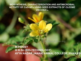
1. M.PRAISY
III B.Sc (BIO-TECHNOLOGY)
AYYA NADAR JANAKI AMMAL COLLEGE,SIVAKASI.
BIOSYNTHESIS, CHARACTERIZATION AND ANTIMICROBIAL
ACTIVITY OF CuO-NPs USING SEED EXTRACTS OF CLEOME
VISCOSA L.
2. Introduction
Objectives
Methodology
Preparation of seed extract
Green synthesis of CuO-NPs
Antibacterial Activity of CuO-NPs
Limitation
Expected outcome
3. The metal nanoparticles such as Cu, Ag, Pb and Zn are found to
have anti-bacterial activity.
Copper-based nanoparticles are an important class of materials
with applications as catalysts, conductive inks, and antimicrobial
agents.
Cleome viscosa is the Asian spiderflower.
It is a fast-growing herb of humid and warm habitats. It is
commonly found growing as a weed medicinal in disturbed sites,
and gardens.
The leaves are use as external application to wounds and
ulcers.
The dipyridodiazepinone derivatives of C. viscose was approved
as a therapeutic agent for the treatment of AIDS
The seed are anthelmintic and carminative and they contain
major fatty acids like palmitic acid,stearic acid,oleic acid and
linoleic acid.
4. To synthesis CuO-NPs using seed samples
of C. viscosa.
To characterize the CuO-NPs by UV-Vis
Spectrophotometer, FTIR, SEM and XRD
analysis.
To study the antimicrobial activity of CuO-
NPs against human pathogens.
5. 1
• Preparation of seed extract
2
• Green synthesis of CuO-NPs
3
• Antibacterial Activity of CuO-
NPs
6. Preparation of seed extract
25 g of seeds will be thoroughly washed, dried well and allowed to
boil for 5 min at 80°C with 100 mL of de-ionized water in a 250-mL
Erlenmeyer flask and then cooled down to room temperature.
The resulting solution will be filtered through a Whatman filter
paper of pore size 0.2 μm.
The filtrate will be stored at 4°C as a stock for the synthesis of
CuO-NPs.
7. Green synthesis of CuO-NPs
50 ml of 10 mM aqueous solution of copper nitrate (99.99 % purity, Hi-Media)
will be added to 5 mL C. viscose seed extract in a 100 mL Erlenmeyer flask
with constant stirring on a magnetic stirrer at 100–120 °C.
Colour change of the reaction mixture will be observed from deep blue to
colourless and then to brick red and dark red on vigorous stirring for 24 h.
Then the resultant solution will be centrifuged at 10,000 rpm for 10 min at room
temperature.
The mixture will be collected after discarding the supernatant.
The collected CuO-NPs will be allowed to dry in a watch glass.
The formation of black precipitate will be grinded for further characterization.
The morphological and structural characteristics will be confirmed by UV-Vis
Spectrophotometry, FTIR, SEM and XRD.
8. Antibacterial Activity of CuO-NPs
Green synthesized CuO-NPs will be tested for antibacterial activity
against human pathogens such as Pseudomonas aeruginosa,
Klebsilla pneumonia, Salmonella typhimurium and Serratia
marcescens by agar well diffusion method.
Minimum inhibitory concentration (MIC) of the CuONP against
each bacterial pathogen will be determined by micro-dilution broth
method. CFU value will be calculated for each dilution factor.
All bacterial dilutions will be standardized to match the McFarland
(turbidity) standard (1.5 X 10⁸ CFU/ml).
9. The synthesis of CuO-NPs will be depending upon the
concentration of Copper nitrate solution and seed extracts of C.
viscosa plants.
The surface plasmon bands of the UV–vis spectrum between 200 –
700 nm will indicates the existence of CuO-NPs nanoparticles.
The antimicrobial effect of the CuO-NPs is corresponding to
Human pathogenic bacteria in biomedical studies.
10. The Copper oxide nanoparticles will be synthesized in Copper nitrate solution
containing of seed sample extracts of C. viscose plants as bio-reducing agent.
The colloidal suspension will show the resonances at 200 to 700 nm by
Spectrophotometry reading and it will be indicating the formation of CuO-NPs.
FTIR spectra will record in solid phase using the KBr (potassium bromide)
pellet technique in the range of 4000–400 cm−1.
SEM analysis will show the actual size of copper oxide nanoparticles.
The crystalline nature will be produced for CuO-NPs of seed extract of C.
viscose plants by XRD based on the diffraction peaks at 2θ of 32.41, 35.61,
38.81, 48.91, 53.31, 58.21, 61.61 and 66.31, which will be assigned to (110),
(111), (200), (−202), (020), (202), (−113) and (022) planes respectively.
The green synthesized CuO-NPs will be reported as bactericidal properties
through studying antimicrobial activity against human pathogens of
Pseudomonas aeruginosa, Klebsilla pneumonia, Salmonella typhimurium and
Serratia marcescens by agar well diffusion method.
11. 1. Ponce AA. and Klabunde KJ (2005) Chemical and catalytic activity of copper
nanoparticles prepared via metal vapor synthesis. J. Mol. Catal. A. 225: 1–6.
2. Huang, Z., Cui, F., Kang, H., Chen, J., Zhang, X. and Xia, C
(2008) Highly dispersed silica-supported copper nanoparticles
prepared by precipitation−gel method: a simple but efficient
and stable catalyst for glycerol hydrogenolysis. Chem. Mater.
20: 5090–5099.
3. Mali RG (2010). Cleome viscosa (wild mustard): A review on ethnobotany,
phytochemistry, and pharmacology. Pharm. Biol. 48:105-112.
4. Panduraju T, Parvathi B, Rammohan M, Reddy CS (2011). Wound
healing properties of Cleome viscosa linn. Hygeia J. D. Med. 3:41-45.