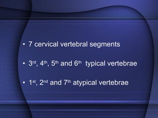
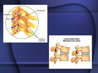
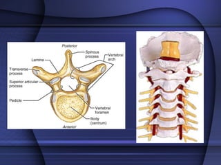
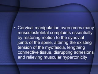
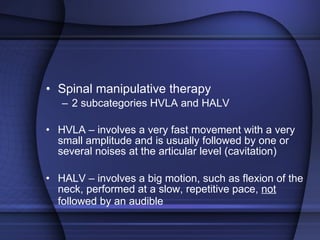
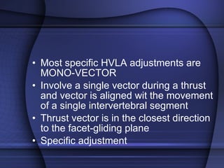
Occiput PRA625


Occipitofrontalis
Semispinalis Capitis
Suboccipitals
 
Cranial manipulation Developed mid 1930’s Controversy Cottom  Three Hypothesis Dysfunction dural tension CSF circulation Stress and pressure
Technique Saggital suture spread Cranial universal  Parietal lift
 
 
HW Read the article by Flynn et al  “ The audible pop is not necessary for successful spinal HV manipulation in individuals with low back pain”
 
 
Cervical Spine
Cervical Spine Most vulnerable site for injury Common area everyday complaints and articular dysfunction Tri-planar motion Restoration of joint mobility
7 cervical vertebral segments 3 rd , 4 th , 5 th  and 6 th   typical vertebrae 1 st , 2 nd  and 7 th  atypical vertebrae
 
Cervical Vertebrae Seven vertebrae (C 1 -C 7 ) are the smallest, lightest vertebrae C 3 -C 6  are distinguished with an oval body, short spinous processes, and large, triangular vertebral foramina Each transverse process contains a transverse foramen
 
 
Cervical Vertebrae Table 7.2
Cervical Vertebrae: The Atlas (C 1 ) The atlas  Has no body and no spinous process Consists of anterior and posterior arches, and two lateral masses The superior surface of lateral masses articulate with the occipital condyles
Cervical Vertebrae: The Atlas (C 1 ) Figure 7.16a, b
Cervical Vertebrae: The Axis (C 2 ) The axis has a body, spine, and vertebral arches as do other cervical vertebrae Figure 7.16c
Cervical Vertebrae: The Axis (C 2 ) Unique to the axis is the dens, or odontoid process, which projects superiorly from the body and is cradled in the anterior arch of the atlas Figure 7.16c
Cervical Vertebrae: The Axis (C 2 ) The dens is a pivot for the rotation of the atlas  Figure 7.16c
Biomechanics Cervical spine has numerous articulations Cervical spine – lordotic curve
 
 
Cervical spine motion Rotation contributes to the greatest disc degeneration
 
Cervical spine provides different motion characteristics Occipitoatlantal complex (C0-C1) Atlantoaxial complex  (C1-C2)
Greatest ROM occurs the middle of the cervical at  the level of C5-C6 during flexion and extension  Facet joints lie 45 degrees to the ventral and motion involves a coupled movement along both the X and Y axes Rash,PJ and Burke RK.  Kinesiology and Applied Anatomy . 1974 Bergman, DH and Peterson, TF. Chiropractic Technique. 2 nd  ed. 2002.
What is the coupled physiological effect of the cervical spine with left lateral flexion? (Hint: think facet, VB, Disc, SP)
Lateral bending produces concomitant PA rotation, with spinous movements to the contralateral side of lateral flexion Coupling movement actually decrease gradually from C2-C7 as the incline of the facet joints Bergman, DH and Peterson, TF. Chiropractic Technique. 2 nd  ed. 2002.
Biomechanics of Cervical Injury Mechanism  Structures    Affected Flexion  or axial compressive loading Posterior column elements Capsular ligaments Supraspinous Lig Interspinous Lig Erector spinae gp Trapezius muscle
Biomechanics of Cervical Injury Mechanism  Structures    Affected Lateral flexion Nerve root Dorsal sleeve Brachial plexus Scalene muscles
Biomechanics of Cervical Injury Mechanism  Structures    Affected Extension or axial rotation Anterior column elements ALL Outer annulus of disk Apophyseal joints SCM
Indications for manipulations Proper patient selection enhances the efficiency of CMT and minimizes the risk Complete H & P Radiographic studies A Pt with a sign of VBI or carotid artery disease should have a vascular examination and Doppler studies
H & P, neurological, orthopedic and radiography will generally indicate the appropriateness of CMT for the individual patients
Cervical manipulation overcomes many musculoskeletal complaints essentially by restoring motion to the synovial joints of the spine, altering the existing tension of the myofascia, lengthing connective tissue, disrupting adhesions and relieving muscular hypertonicity
Spinal manipulative therapy 2 subcategories HVLA and HALV HVLA – involves a very fast movement with a very small amplitude and is usually followed by one or several noises at the articular level (cavitation) HALV – involves a big motion, such as flexion of the neck, performed at a slow, repetitive pace,  not  followed by an audible
Most specific HVLA adjustments are MONO-VECTOR Involve a single vector during a thrust and vector is aligned wit the movement of a single intervertebral segment Thrust vector is in the closest direction to the facet-gliding plane Specific adjustment
Zygapophyseal joint locking theory Joint fixation is probably multi-factorial and may involve derangement of the posterior articular joints, intra-discal degrangements, muscle spasm, soft tissue fibrosis etc.
Mechanisms of Action in Manipulative Therapy HVLA causes changes in the joint capsule tension Excites the mechanoreceptors of the synovial joints, primarily the type I and type II Type I mechanoreceptors contribute to perception of position and kinesthetic awareness
Type II mechanoreceptors  inhibit pain and exert a phasic (relaxing) effect on motor neurons Endogenous opiate release that follows CMT combined with nociceptor suppression at the cord level, also inhibit pain
Upper cervical complex is critically important in manual therapy because of vascular insult Several investigators have reported moderate amounts of kinking of the vertebral arteries during moderate amounts  of axial rotation even in individuals with no prior vascular disease
Chiropractic profession is well aware of the cases of documented VBA infarct following manipulation by various practitioners of manual therapy A through practitioner should ALWAYS screen patients for neurovascular disturbances, especially VBAI, to ensure safety and efficacy of the cervical manipulation

Pra625 cervical spine

  • 1.
  • 2.
  • 3.
  • 4.
  • 5.
  • 6.
  • 7.
  • 8.
  • 9.
  • 10.
  • 11.
    Cranial manipulation Developedmid 1930’s Controversy Cottom Three Hypothesis Dysfunction dural tension CSF circulation Stress and pressure
  • 12.
    Technique Saggital suturespread Cranial universal Parietal lift
  • 13.
  • 14.
  • 15.
    HW Read thearticle by Flynn et al “ The audible pop is not necessary for successful spinal HV manipulation in individuals with low back pain”
  • 16.
  • 17.
  • 18.
  • 19.
    Cervical Spine Mostvulnerable site for injury Common area everyday complaints and articular dysfunction Tri-planar motion Restoration of joint mobility
  • 20.
    7 cervical vertebralsegments 3 rd , 4 th , 5 th and 6 th typical vertebrae 1 st , 2 nd and 7 th atypical vertebrae
  • 21.
  • 22.
    Cervical Vertebrae Sevenvertebrae (C 1 -C 7 ) are the smallest, lightest vertebrae C 3 -C 6 are distinguished with an oval body, short spinous processes, and large, triangular vertebral foramina Each transverse process contains a transverse foramen
  • 23.
  • 24.
  • 25.
  • 26.
    Cervical Vertebrae: TheAtlas (C 1 ) The atlas Has no body and no spinous process Consists of anterior and posterior arches, and two lateral masses The superior surface of lateral masses articulate with the occipital condyles
  • 27.
    Cervical Vertebrae: TheAtlas (C 1 ) Figure 7.16a, b
  • 28.
    Cervical Vertebrae: TheAxis (C 2 ) The axis has a body, spine, and vertebral arches as do other cervical vertebrae Figure 7.16c
  • 29.
    Cervical Vertebrae: TheAxis (C 2 ) Unique to the axis is the dens, or odontoid process, which projects superiorly from the body and is cradled in the anterior arch of the atlas Figure 7.16c
  • 30.
    Cervical Vertebrae: TheAxis (C 2 ) The dens is a pivot for the rotation of the atlas Figure 7.16c
  • 31.
    Biomechanics Cervical spinehas numerous articulations Cervical spine – lordotic curve
  • 32.
  • 33.
  • 34.
    Cervical spine motionRotation contributes to the greatest disc degeneration
  • 35.
  • 36.
    Cervical spine providesdifferent motion characteristics Occipitoatlantal complex (C0-C1) Atlantoaxial complex (C1-C2)
  • 37.
    Greatest ROM occursthe middle of the cervical at the level of C5-C6 during flexion and extension Facet joints lie 45 degrees to the ventral and motion involves a coupled movement along both the X and Y axes Rash,PJ and Burke RK. Kinesiology and Applied Anatomy . 1974 Bergman, DH and Peterson, TF. Chiropractic Technique. 2 nd ed. 2002.
  • 38.
    What is thecoupled physiological effect of the cervical spine with left lateral flexion? (Hint: think facet, VB, Disc, SP)
  • 39.
    Lateral bending producesconcomitant PA rotation, with spinous movements to the contralateral side of lateral flexion Coupling movement actually decrease gradually from C2-C7 as the incline of the facet joints Bergman, DH and Peterson, TF. Chiropractic Technique. 2 nd ed. 2002.
  • 40.
    Biomechanics of CervicalInjury Mechanism Structures Affected Flexion or axial compressive loading Posterior column elements Capsular ligaments Supraspinous Lig Interspinous Lig Erector spinae gp Trapezius muscle
  • 41.
    Biomechanics of CervicalInjury Mechanism Structures Affected Lateral flexion Nerve root Dorsal sleeve Brachial plexus Scalene muscles
  • 42.
    Biomechanics of CervicalInjury Mechanism Structures Affected Extension or axial rotation Anterior column elements ALL Outer annulus of disk Apophyseal joints SCM
  • 43.
    Indications for manipulationsProper patient selection enhances the efficiency of CMT and minimizes the risk Complete H & P Radiographic studies A Pt with a sign of VBI or carotid artery disease should have a vascular examination and Doppler studies
  • 44.
    H & P,neurological, orthopedic and radiography will generally indicate the appropriateness of CMT for the individual patients
  • 45.
    Cervical manipulation overcomesmany musculoskeletal complaints essentially by restoring motion to the synovial joints of the spine, altering the existing tension of the myofascia, lengthing connective tissue, disrupting adhesions and relieving muscular hypertonicity
  • 46.
    Spinal manipulative therapy2 subcategories HVLA and HALV HVLA – involves a very fast movement with a very small amplitude and is usually followed by one or several noises at the articular level (cavitation) HALV – involves a big motion, such as flexion of the neck, performed at a slow, repetitive pace, not followed by an audible
  • 47.
    Most specific HVLAadjustments are MONO-VECTOR Involve a single vector during a thrust and vector is aligned wit the movement of a single intervertebral segment Thrust vector is in the closest direction to the facet-gliding plane Specific adjustment
  • 48.
    Zygapophyseal joint lockingtheory Joint fixation is probably multi-factorial and may involve derangement of the posterior articular joints, intra-discal degrangements, muscle spasm, soft tissue fibrosis etc.
  • 49.
    Mechanisms of Actionin Manipulative Therapy HVLA causes changes in the joint capsule tension Excites the mechanoreceptors of the synovial joints, primarily the type I and type II Type I mechanoreceptors contribute to perception of position and kinesthetic awareness
  • 50.
    Type II mechanoreceptors inhibit pain and exert a phasic (relaxing) effect on motor neurons Endogenous opiate release that follows CMT combined with nociceptor suppression at the cord level, also inhibit pain
  • 51.
    Upper cervical complexis critically important in manual therapy because of vascular insult Several investigators have reported moderate amounts of kinking of the vertebral arteries during moderate amounts of axial rotation even in individuals with no prior vascular disease
  • 52.
    Chiropractic profession iswell aware of the cases of documented VBA infarct following manipulation by various practitioners of manual therapy A through practitioner should ALWAYS screen patients for neurovascular disturbances, especially VBAI, to ensure safety and efficacy of the cervical manipulation

Editor's Notes

  • #4 The occipital bone , a bone situated at the back and lower part of the cranium , is trapezoid in shape and curved on itself. It is pierced by a large oval aperture, the foramen magnum , through which the cranial cavity communicates with the vertebral canal .
  • #5 Localization of the occipital nerve. The nerve is positioned medial to the pulse of the occipital artery; approximately one third of the distance from the occipital protuberance to the mastoid, whereas the lesser occipital nerve is more lateral, approximately two thirds of this distance. The greater occipital nerve arises from the posterior primary ramus of the second cervical nerve root deep (Figure 18–8). It travels deep to the cervical paraspinousmusculature and to the cervical paraspinousmusculature and becomes superficial just inferior to the superior nuchal line and lateral to the occipital protuberance of the skull; at this point, the nerve is just lateral to the occipital artery. The lesser occipital nerve and greater auricular nerve are terminal branches of the superficial cervical plexus. Both arise from the posterior primary ramus of the second and third cervical nerve roots, travel through the cervical paraspinousmusculature, and become superficial over the inferior nuchal line of the skull, just superior and medial to the mastoid and just inferior to the tragus of the ear, respectively. The lateral section of the posterior scalp is supplied by the lesser occipital and great auricular nerves
  • #6 Cutaneous innervation of the head and neck. The trigeminal nerve, the fifth cranial nerve, supplies the majority of sensory innervation to the face (Figure 18–1). Preganglionic fibers exit the brainstem and travel anteriorly to synapse with second-order neurons within the trigeminal (gasserian) ganglion (Figure 18–2). The ganglion lies within the cranial vault at the base of the petrous portion of the temporal bone in a dural invagination containing cerebrospinal fluid known as Meckel’s cave. Postganglionic fibers exit the ganglion to form the ophthalmic (V1), maxillary (V2), and (V3) nerves
  • #7 The most common early presenting symptom is occipital pain referred to the vertex of the head and ipsilateral shoulder and arm pain, exacerbated by head movement. Presence of neurological signs are associated with lesions of the 9th, 10th, 11th, and 12th cranial nerves. Horner's syndrome is seen because of the proximity of the cervical sympathetic chain to the jugular foramen. Pain Type : Neuropathic Cause : Invasion of Bone By Tumor or Metastasis Effects : Neurological Signs Clinical Signs: Occipital Pain Referred to the Vertex of the head and Ipsilateral Shoulder and Arm Pain, Exacerbated by Head Movement, Horner's Syndrome , ( ptosis , miosis , anhidrosis )
  • #8 Trauma to the scalp fascia or the occipitalis at the back of the skull can transmit pain through the head and into the eye. Trauma may include a blow to the back of the head, strain from a tight ponytail or bun, or the weight of long, heavy hair. In one case I know of, a man struck the top of his head on the corner of a cabinet. Result: a slight puncture wound in the scalp, a brutal pain in the eye . Frontalis helps open the eyes, raises the eyebrows, and wrinkles the forehead into "worry lines."It is commonly used by biofeedback practitioners to monitor muscle tension. Trauma to frontalis (whether a blow to the forehead or habitual frowning) can cause severe frontal headache often diagnosed as "migraine." Frontalis is one of the muscles that definitively proved the muscle-migraine connection. Botox injections paralyzed the frontalis, eliminating "worry lines" but they also had the surprising side effect of halting chronic "migraines". Or maybe not so surprising, as frontalis entraps the supraorbital nerve. The related corrugator supercilii (at the top of the nose between the eyebrows) compresses branches of the supraorbital nerve along with the supratrochlear nerve and branches of the supraorbital nerve. Your frown may be giving you a migraine!
  • #9 (headpain and occipital neuralgia) Injured in whiplash and involved in "tension" and "cervicogenic" headache. Semispinalis capitis is commonly injured in auto accidents. You can injure it more slowly but just as effectively with a chronic head-forward position. When tight semispinalis entraps the greater occipital nerve which in turn causes numbness, tingling and/or burning pain extending over the back of the head to the top (vertex) of the head. It may be difficult to touch chin to chest, and sufferers may be unable to lay back of head on pillow. Relieve nerve pain with cold. Relieve muscle pain with moist heat. In either case, look for the origin of the pain which is rarely the spot where it hurts. Semispinalis Cervicis (even more head pain). Typically produces a vague band of pain from occiput along side of head to just behind orbit (similar to suboccipital pain pattern).
  • #10 The four pairs of suboccipital muscles cause deep aching pain running in a band from the back of the head to the orbit of the eye, possibly with balance problems and dizziness. One of these (the rectus capitis superior minor) attaches directly to the dura mater of the spinal cord. When traumatized it can produce odd visual and neurological symptoms to the point of seizures. Pain in the angle of the neck and along the vertebral border of the scapula may be so severe that patient cannot move the neck at all. Suboccipitals are often strained in persons who wear bifocals, children who watch TV lying with chin propped on hands, and anyone who holds the head in position with chin up and neck flexed backward.
  • #11 Small muscles below the occiput set off referred pain. One-sided headaches, which radiate from the occiput to the eye and forehead, can often be traced back to spasmodic contraction of one or more of four small (suboccipital) muscle pairs. These four small cervical vertebrae muscles are located at the deepest point inside the neck. They contain so-called trigger points which when activated by local spasmodic muscle contraction - usually in combination with the neck muscles located directly over them - set off the above-mentioned referred pain. The person experiencing the pain finds it difficult to define. Often it is termed a "ghost headache". These four small cervical vertebrae muscles are especially important to the movement of the two upper cervical vertebrae joints. The muscles execute a bending-stretching movement in the uppermost joint (between the atlas and head) as well as lateral tilting of the head to the left and right (10-degree bending and 25-degree stretching). The joint between the atlas and axis allows a 45-degree rotation to the left and right.
  • #20 The cervical spine has the precarious task of maintaining head posture while allowing for a great deal of mobility The c/s must balance the weight of the head atop a relatively thin and long lever, making it quite vulnerable to traumatic forces Vulnerability due to high level of mobility produced by its triplanar motion, this mobility provides those in manual therapy with a tremendous opportunity to correct postural and articular faults in the neck Corrective care that centers around early movements and aggressive restoration of joint mobility is the hallmark of treatment for mechanical disorders of the cervical spine The cervical facets allow movement in all directions, the c/s is therefore the most vulnerable portion of the vertebral column
  • #21 Typical cervical vertebrae posses same structural parts as all the other true vertebra, plus some distinct and unique physical features: SP’s are bifid which allow for better attachment of the ligaments and muscles , foramen transversarium, broad VB that are small, oval and wide transversely, superior margin of the VB is lipped for the uncinate processes, *** joints of Luschka are pseudojoints that have a synovial membrane but no joint capsule which serve as tracts that glide the motion of lateral flexion and coupled rotation, Sup and Inf articulating process and each transverse process from C6 upward has a foramen for the vertebral artery. ***Articular Facets are teardropped shape with Superior facing up and posteriorly and inferior facing down and anteriorly, placing the joint space at 45 degrees angle midway b/w coronal and transverse planes ***The disc-height to body height ratio is greatest 2:5 in the c/s therefore allowing the greatest possible ROM Atypical – C1 no VB and spinous process, C2 has a dens or odontoid process and C7 prominent SP, not bifid and vertebral artery does not enter the foramen transversarium at this point
  • #23 Typical vertebrae – bifid spinous process, foramen transversarium, broad VB, Sup and Inf articulating process and a foramen for the vertebral artery
  • #32 Intricate anatomic configurations of nerves, vessels, joint structures , ligamentous network tat provides vital support to mobile osseous structures The cervical spine forms a lordotic curve that develeps to secondary to the response of upright posture, the functions of the cervical curve and the A-P curves throughout the spine are to add resiliency to the spine in response to axial compression forces and to balance the center of gravity of the skull over the spine, the COG for the skull lies anterior to the FM Cervical lordosis is a convex curve anteriorly and is known as a compensatory or secondary curvature
  • #35 Flexion, extension, lateral bending and rotation Rotation pivotal role in degenerative process of the disk and it is the disk were cervical dysfunction and pain are prevalent
  • #37 OA C0-C1 provides extensive motion flexion/extension AA C1-C2 functions as a pivot point for PA rotation
  • #38 Middle region of the cervical vertebrae
  • #39 Primary Source Pg. 195 Fig 5-48, 5-49
  • #40 Bergman and Peterson pg 194-5 lateral flxn and rotation fig.5-48, 5-49
  • #46 Figure 6-16 draw on board
  • #53 ACC VBA lecture 2006 slide 35