
Recommended
More Related Content
Similar to Cervical_Spine.pptx
Similar to Cervical_Spine.pptx (20)
Recently uploaded
Recently uploaded (20)
Cervical_Spine.pptx
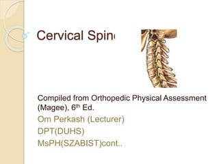
- 1. Cervical Spine Compiled from Orthopedic Physical Assessment (Magee), 6th Ed. Om Perkash (Lecturer) DPT(DUHS) MsPH(SZABIST)cont..
- 2. Learning Objective At the end of this lecture, the student will be able to do the subjective and objective examination of the cervical spine.
- 3. Scanning Examination Examination of the cervical spine involves determining whether the injury or pathology occurs in the cervical spine or in a portion of the upper limb.
- 6. Cervicobrachial area (C3 to C7) Pain in this area is commonly referred into the upper extremity.
- 7. Coupled movement Greatest flexion-extension of the facet joints occurs between C5-C6 Cervical Spine Nerve Supply Resting position Midway b/w flex & ext. Close packed position Full extension Capsular pattern Side flexion and rotation equally limited extension
- 8. For C3 to C7, the main ligaments are the anterior longitudinal ligament, posterior longitudinal ligament, the ligamentum flavum, and the supraspinal and interspinal ligaments. There are also ligaments b/w the transverse processes but in the cervical spine, they are rudimentary.
- 9. Uncinate joints or Joints of Luschka
- 10. Intervertebral disc make up approximately 25% of the height of the cervical spine. No disc b/w C0 to C1or b/w C1to C2. It is the discs rather than the vertebrae that give the cervical spine its lordotic shape.
- 11. Pateint History
- 12. Grading of patients suffering from Neck Pain : 1 to 10 Signs of headaches having a cervical origin Signs and symptoms of VBI OBSERVATION: Head and Neck posture
- 14. Kinematics of the craniocervical flexion
- 15. PASSIVE MOVEMENTS PPIVM CAPSULAR PATTERN RESISTED ISOMETRIC MOVEMENTS SCANNING EXAMINATION MYOTOMES OF THE UPPER LIMB SENSORY SCANNING EXAMINATION FUNCTIONAL ASSESSMENT SPECIAL TESTS
- 16. MANUAL THERAPY Cervical Spine Dr. Om Perkash (Lecturer) DPT(DUHS) MsPH(SZABIST)cont..
- 17. Lecture’s Objectives: Muscle palpation Palpation of spine and facet joint General rotation General lateral flexion Postero-anterior glide central Postero-anterior glide unilateral Transverse glide
- 20. Palpation: Sweating and temperature Soft tissue changes ◦ With three fingers ◦ With two fingers ◦ With one finger
- 21. We use these techniques to assess ◦ General muscular tightness ◦ Local areas of thickening adjacent to one/more spinous process In the largest part of the m/s bulk in mid- laminar trough area ◦ Soft thickening at post. Articular pillar ◦ Hard bony thickening ◦ Posterolateral margins of articular pillar ◦ Tight ligament nuchae
- 22. Palpation: Bony points Interspinous spaces PAIVMs ◦ Hypo mobility ◦ Hypermobility ◦ Crepitus ◦ Stiffness ◦ Muscle spasm ◦ End feel
- 23. Unilateral palpation Palpation with rotation
- 25. Longitudinal Movement: Uses: ◦ Gain patient’s confidence ◦ Useful guide ◦ Relieve pain ◦ Protective deformity (Wry neck) ◦ Pain stimulating migraine ◦ Cervical joint locking ◦ Shooting occipital pain
- 26. Postero-anterior Central Vertebral Uses: ◦ Pain ◦ Stiffness ◦ Increase ROM ◦ Muscular spasm ◦ Joint locking ◦ Different neck disorders ◦ Marked degenerative changes
- 27. Postero-anterior Unilateral Uses: ◦ Pain ◦ Stiffness ◦ Degenerative joint ◦ Muscular spasm ◦ Facet joint locking
- 28. Transverse Vertebral Pressure Uses: ◦ Degenerative changes ◦ Combine with PA unilateral ◦ Useful for gross pain ◦ Non- neurological ◦ Opening of facet joint ◦ Stretching of capsule
- 29. Rotation Uses: ◦ Pain ◦ Unilateral pain ◦ Increase ROM ◦ Muscular spasm ◦ Pain from scapula, tendon ◦ Shooting occipital pain ◦ Acute torticollis
- 30. Lateral Flexion Uses: ◦ Pain ◦ ROM ◦ Unilateral pain ◦ Muscular spasm ◦ Pain from neck, scapula or arm ◦ Torticollis
- 31. Reference Maitland’s Vertebral Manipulation (Seventh Edition)
- 33. Objective Assessment Postural Analysis Active ROM Passive ROM Accessory Movements Dermatomes Myotomes Reflexes Special Tests: Compression, Distraction, Spurling
- 34. Dermatomes
- 35. Myotomes Upper Limb C4: Shoulder elevation C5: Shoulder abduction C6: Elbow Flexion + wrist extension C7: Elbow Extension + wrist flexion C8: Fingers adduction T1: Fingers abduction
- 37. Objective Assessment Group Practice