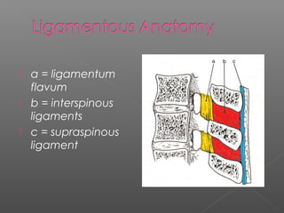
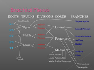
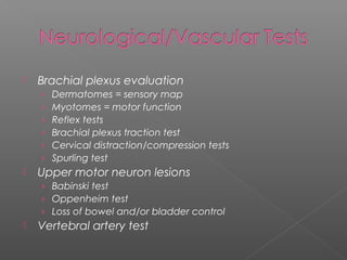
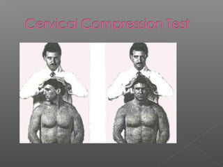
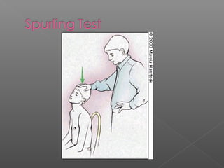
Cervical Spine Fractures
 C1 - C7
 Anterior longitudinal ligament
› Reinforces anterior discs, limits extension
 Posterior longitudinal ligament
› Reinforces posterior discs, limits flexion
 Ligamentum nuchae = supraspinous ligament
› Thicker than in thoracic/lumbar regions
› Limits flexion
 Interspinous/intertransverse ligaments
› Limit flexion and rotation/limits lateral flexion
 Ligamentum flavum
› Attach lamina of one vertebrae to another, reinforces
articular facets
› Limits flexion and rotation
 a = ligamentum
flavum
 b = interspinous
ligaments
 c = supraspinous
ligament
Dermatomes
Myotomes C5 – Abduction
C6 – Elbow Flexion/Wrist Extension
C7 – Elbow Extension/Wrist Flexion
C8 – Finger Flexion
T1 – Finger Abduction
C1-2 – Neck Flexion
C3 – Lateral Neck Flexion
C4 – Shoulder Elevation
C1 – top of head
C2 – Temporal
C3 – Side of
jaw/neck
C4 – top of
shoulders
ROOTS TRUNKS DIVISIONS CORDS BRANCHES
C5
C6
C7
C8
T1
Upper
Middle
Lower
Anterior
Posterior
Anterior
Posterior
Anterior
Posterior
Lateral
Posterior
Medial
Suprascapular
Lateral Pectoral
Musculotaneous
Axillary
Radial
Median
Ulnar
Long
Thoracic
Medial Pectoral
Medial Antebrachial
Medial Brachial Cutaneous
Thoracodorsal
Subscapular
Dorsal Scapular
 Muscles
Trapezius
Sternocleidomastoid
Scalenes
Splenius
Semispinalis, Spinalis, Longissimus
 Fairly uncommon in athletics(6-7%) - but greater
than 90% of all fatalities are cervical related.
 Cervical injuries are primarily technique related:
› Spearing
› Tackling or falling head first.
 Must have an emergency plan:
› All personnel know roles and equipment use.
› All unconscious athletes - suspect head/neck
› Always suspect the worse until proven otherwise
 Common MOIs
› Axial Loading
› Flexion Force
› Hyperextension Force
› Flexion-Rotation Force
› Hyperextension-Rotation
› Lateral Flexion
 Cervical Fracture or Dislocation
› Weakness or Paralysis
 Cervical Nerve Root Injury
› Herniated Disc
› Laceration
› Cord Shock (Central Cord Syndrome)
› Hemorrhage
› Contusion
› Cervical Stenosis
 Cervical Strain
› Active motion most painful
 Cervical Sprain (Whiplash)
› Passive and active motion painful
 Torticollis (WryNeck)
› Muscle spasm and facet irritation
 Brachial Plexus Stretch or Compression
 Contusions to Throat
 Localized pain
› Typically indicative of muscular strain,
ligamentous sprain, facet joint injury, fracture
and/or subluxation or dislocation
 Radiating pain
› Heightened risk of likely spinal cord, cervical
nerve root and/or brachial plexus injury
 Acute onset
› Generally associated with one specific
mechanism of injury/event
 Chronic or insiduous (unknown) onset
› Generally related to overuse injuries
(accumulative microtrauma) and/or postural
abnormalities and deficiencies
 Cervical spine curvature
 Position of head relative to shoulders
 Soft tissue symmetry
 Level of shoulders
 Normal cervical spine has lordotic curve
 Increased lordotic curve (forward head)
indicative of poor posture and muscular
weakness or imbalance
 Lessened lordotic curve indicative of
muscular spasm/guarding and/or nerve
root impingement
 Head should be seated symmetrically on
cervical spine
 Lateral flexion from unilateral spasm of
muscles – strain and/or spasm (guarding)
 Rotation from unilateral spasm of
sternomastoid muscle – strain and/or
spasm (guarding) or torticollis
 Inspect height of:
› Acromioclavicular (AC) joints
› Deltoids
› Clavicles
 Dominant extremity often appears
depressed relative to non-dominant
extremity
 Hyoid bone
› At level of C3 vertebrae, note movement with
swallowing
 Thyroid cartilage
› At level of C4/C5 vertebrae, also moves with
swallowing, protects larynx
› Aka – “Adam’s apple”
 Cricoid cartilage
› At level of C6/C7 vertebrae, point where
esophagus and trachea deviate, rings of
cartilage
 Sternomastoid
› Sternum (near SC joint) to mastoid process
 Scalenes
› Posterior/lateral to sternomastoid muscles
› Difficult to differentiate, palpate collectively
 Carotid artery
› Primary pulse point
 Lymph nodes
› Only discernable if enlarged due to illness
 Occiput
› Posterior aspect of skull, many ms. attachments
 Transverse processes
› Can only palpate C1 transverse processes
approx. one finger below mastoid processes
 Spinous processes
› Flex cervical spine, C7 and T1 are prominent
› Can palpate C5 and C6, maybe C3 and C4
 Trapezius
› Upper fibers from occiput and cervical spinous
processes to distal clavicle
 Range of motion testing
› Active
› Passive
› Resisted
 Ligamentous/capsular tests
 Neurological tests
› Brachial plexus evaluation
› Reflex tests
› Upper motor neuron lesions
 Best done in sitting or standing
 Flexion – touch chin to chest
 Extension – look straight above head
 Lateral flexion – approximately 45
degrees
 Rotation – nose over tip of shoulder
 Best done laying supine
 Flexion – firm end feel
 Extension – hard end feel (occiput on
cervical spinous processes)
 Lateral flexion – firm end feel (stabilize
opposite shoulder)
 Rotation – firm end feel
 Easiest to perform all in seated position –
stabilize proximally to avoid substitution
 Flexion – resistance to forehead
 Extension – resistance to occiput
 Lateral flexion – resistance to temporal and
parietal regions
 Rotation – resistance to temporal region or
side of face
 No specific named tests for cervical
spine
 End feels associated with passive ranges
of motion essentially become end points
for joint capsule and ligamentous stress
tests
 Brachial plexus evaluation
› Dermatomes = sensory map
› Myotomes = motor function
› Reflex tests
› Brachial plexus traction test
› Cervical distraction/compression tests
› Spurling test
 Upper motor neuron lesions
› Babinski test
› Oppenheim test
› Loss of bowel and/or bladder control
 Vertebral artery test
 All based upon anatomical position
 C5 – lateral arm
 C6 – lateral forearm, thumb, index finger
 C7 – posterior forearm, middle finger
 C8 – medial forearm, ring and little
fingers
 T1 – medial arm
 Minor differences will exist from one
resource to another
 C5 – shoulder abduction
 C6 – elbow flexion or wrist extension
 C7 – elbow extension or wrist flexion
 C8 – grip strength (shake hands)
 T1 – interossei (spread fingers)
 Dermatomes
 Reflexes
› Babinski
› Oppenheim
› Biceps
› Brachioradialis
› Triceps
 Myotomes
 C5 – biceps brachii reflex (anterior arm
near antecubital fossa)
 C6 – brachioradialis reflex (thumb side of
forearm)
 C7 – triceps brachii reflex (at insertion on
olecranon process)
 Mimics mechanism of injury
 Cervical spine laterally flexed and
opposite shoulder is depressed
 Positive if radiating/”burning” pain in
upper extremity
› If traction injury, symptoms noted on side of
depressed shoulder
› If compression injury, symptoms noted in
direction of lateral flexion
 Neurological findings!!
•Burning, achy pain
•Muscle weakness
•Point tenderness
•Mechanism of Injury
TESTS:
•Brachial Plexus
Traction Test
•Tinel’s Sign
•Spurling’s Test
•Cervical Distraction
 Treatment will depend on which of the
seven cervical vertebrae are damaged
and the kind of fracture sustained. A
minor compression fracture can be
treated with a cervical brace worn for 6
to 8 weeks until the bone heals. A more
complex or extensive fracture may
require traction, surgery, 2 to 3 months in
a rigid cast, or a combination of these
treatments.
C spine eval (ray) (1)

C spine eval (ray) (1)

  • 1.
  • 2.
  • 4.
     Anterior longitudinalligament › Reinforces anterior discs, limits extension  Posterior longitudinal ligament › Reinforces posterior discs, limits flexion  Ligamentum nuchae = supraspinous ligament › Thicker than in thoracic/lumbar regions › Limits flexion  Interspinous/intertransverse ligaments › Limit flexion and rotation/limits lateral flexion  Ligamentum flavum › Attach lamina of one vertebrae to another, reinforces articular facets › Limits flexion and rotation
  • 5.
     a =ligamentum flavum  b = interspinous ligaments  c = supraspinous ligament
  • 6.
    Dermatomes Myotomes C5 –Abduction C6 – Elbow Flexion/Wrist Extension C7 – Elbow Extension/Wrist Flexion C8 – Finger Flexion T1 – Finger Abduction C1-2 – Neck Flexion C3 – Lateral Neck Flexion C4 – Shoulder Elevation C1 – top of head C2 – Temporal C3 – Side of jaw/neck C4 – top of shoulders
  • 7.
    ROOTS TRUNKS DIVISIONSCORDS BRANCHES C5 C6 C7 C8 T1 Upper Middle Lower Anterior Posterior Anterior Posterior Anterior Posterior Lateral Posterior Medial Suprascapular Lateral Pectoral Musculotaneous Axillary Radial Median Ulnar Long Thoracic Medial Pectoral Medial Antebrachial Medial Brachial Cutaneous Thoracodorsal Subscapular Dorsal Scapular
  • 8.
  • 9.
     Fairly uncommonin athletics(6-7%) - but greater than 90% of all fatalities are cervical related.  Cervical injuries are primarily technique related: › Spearing › Tackling or falling head first.  Must have an emergency plan: › All personnel know roles and equipment use. › All unconscious athletes - suspect head/neck › Always suspect the worse until proven otherwise
  • 10.
     Common MOIs ›Axial Loading › Flexion Force › Hyperextension Force › Flexion-Rotation Force › Hyperextension-Rotation › Lateral Flexion
  • 11.
     Cervical Fractureor Dislocation › Weakness or Paralysis  Cervical Nerve Root Injury › Herniated Disc › Laceration › Cord Shock (Central Cord Syndrome) › Hemorrhage › Contusion › Cervical Stenosis
  • 13.
     Cervical Strain ›Active motion most painful  Cervical Sprain (Whiplash) › Passive and active motion painful  Torticollis (WryNeck) › Muscle spasm and facet irritation  Brachial Plexus Stretch or Compression  Contusions to Throat
  • 14.
     Localized pain ›Typically indicative of muscular strain, ligamentous sprain, facet joint injury, fracture and/or subluxation or dislocation  Radiating pain › Heightened risk of likely spinal cord, cervical nerve root and/or brachial plexus injury
  • 15.
     Acute onset ›Generally associated with one specific mechanism of injury/event  Chronic or insiduous (unknown) onset › Generally related to overuse injuries (accumulative microtrauma) and/or postural abnormalities and deficiencies
  • 16.
     Cervical spinecurvature  Position of head relative to shoulders  Soft tissue symmetry  Level of shoulders
  • 17.
     Normal cervicalspine has lordotic curve  Increased lordotic curve (forward head) indicative of poor posture and muscular weakness or imbalance  Lessened lordotic curve indicative of muscular spasm/guarding and/or nerve root impingement
  • 19.
     Head shouldbe seated symmetrically on cervical spine  Lateral flexion from unilateral spasm of muscles – strain and/or spasm (guarding)  Rotation from unilateral spasm of sternomastoid muscle – strain and/or spasm (guarding) or torticollis
  • 21.
     Inspect heightof: › Acromioclavicular (AC) joints › Deltoids › Clavicles  Dominant extremity often appears depressed relative to non-dominant extremity
  • 22.
     Hyoid bone ›At level of C3 vertebrae, note movement with swallowing  Thyroid cartilage › At level of C4/C5 vertebrae, also moves with swallowing, protects larynx › Aka – “Adam’s apple”  Cricoid cartilage › At level of C6/C7 vertebrae, point where esophagus and trachea deviate, rings of cartilage
  • 23.
     Sternomastoid › Sternum(near SC joint) to mastoid process  Scalenes › Posterior/lateral to sternomastoid muscles › Difficult to differentiate, palpate collectively  Carotid artery › Primary pulse point  Lymph nodes › Only discernable if enlarged due to illness
  • 24.
     Occiput › Posterioraspect of skull, many ms. attachments  Transverse processes › Can only palpate C1 transverse processes approx. one finger below mastoid processes  Spinous processes › Flex cervical spine, C7 and T1 are prominent › Can palpate C5 and C6, maybe C3 and C4  Trapezius › Upper fibers from occiput and cervical spinous processes to distal clavicle
  • 25.
     Range ofmotion testing › Active › Passive › Resisted  Ligamentous/capsular tests  Neurological tests › Brachial plexus evaluation › Reflex tests › Upper motor neuron lesions
  • 26.
     Best donein sitting or standing  Flexion – touch chin to chest  Extension – look straight above head  Lateral flexion – approximately 45 degrees  Rotation – nose over tip of shoulder
  • 27.
     Best donelaying supine  Flexion – firm end feel  Extension – hard end feel (occiput on cervical spinous processes)  Lateral flexion – firm end feel (stabilize opposite shoulder)  Rotation – firm end feel
  • 28.
     Easiest toperform all in seated position – stabilize proximally to avoid substitution  Flexion – resistance to forehead  Extension – resistance to occiput  Lateral flexion – resistance to temporal and parietal regions  Rotation – resistance to temporal region or side of face
  • 29.
     No specificnamed tests for cervical spine  End feels associated with passive ranges of motion essentially become end points for joint capsule and ligamentous stress tests
  • 30.
     Brachial plexusevaluation › Dermatomes = sensory map › Myotomes = motor function › Reflex tests › Brachial plexus traction test › Cervical distraction/compression tests › Spurling test  Upper motor neuron lesions › Babinski test › Oppenheim test › Loss of bowel and/or bladder control  Vertebral artery test
  • 31.
     All basedupon anatomical position  C5 – lateral arm  C6 – lateral forearm, thumb, index finger  C7 – posterior forearm, middle finger  C8 – medial forearm, ring and little fingers  T1 – medial arm
  • 32.
     Minor differenceswill exist from one resource to another  C5 – shoulder abduction  C6 – elbow flexion or wrist extension  C7 – elbow extension or wrist flexion  C8 – grip strength (shake hands)  T1 – interossei (spread fingers)
  • 33.
     Dermatomes  Reflexes ›Babinski › Oppenheim › Biceps › Brachioradialis › Triceps  Myotomes
  • 34.
     C5 –biceps brachii reflex (anterior arm near antecubital fossa)  C6 – brachioradialis reflex (thumb side of forearm)  C7 – triceps brachii reflex (at insertion on olecranon process)
  • 35.
     Mimics mechanismof injury  Cervical spine laterally flexed and opposite shoulder is depressed  Positive if radiating/”burning” pain in upper extremity › If traction injury, symptoms noted on side of depressed shoulder › If compression injury, symptoms noted in direction of lateral flexion
  • 39.
     Neurological findings!! •Burning,achy pain •Muscle weakness •Point tenderness •Mechanism of Injury TESTS: •Brachial Plexus Traction Test •Tinel’s Sign •Spurling’s Test •Cervical Distraction
  • 40.
     Treatment willdepend on which of the seven cervical vertebrae are damaged and the kind of fracture sustained. A minor compression fracture can be treated with a cervical brace worn for 6 to 8 weeks until the bone heals. A more complex or extensive fracture may require traction, surgery, 2 to 3 months in a rigid cast, or a combination of these treatments.