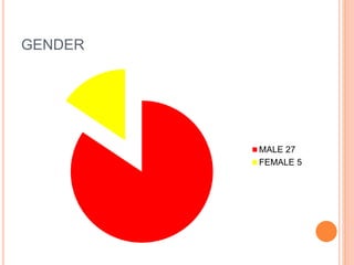
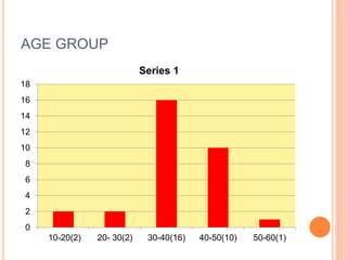
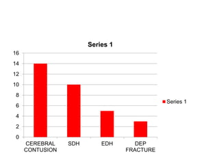
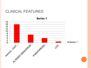
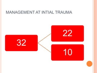
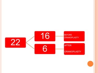
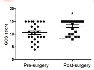
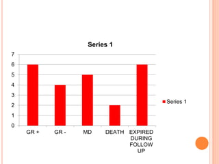
POST TRAUMATIC
HYDROCEPHALUS; A CASE
SERIES (INSTITUTIONAL EXPERIENCE)
DR . K .VAMSHI KRISHNA
M.ch (NEUROSURGERY)
NIZAMS INSTITUTE OF MEDICAL SCIENCES
HYDERABAD
INTRODUCTION
Post-traumatic hydrocephalus (PTH) is
a rare clinico- pathologic entity as a
result of sequelae of head injury.
Hydrocephalus following traumatic
brain injury (TBI) or post-traumatic
hydrocephalus (PTH) is not just a
ventricular enlargement but an active
and progressive disorder of
cerebrospinal fluid (CSF) accumulation
in the ventricular system, causing
compression of the brain parenchyma.
AIMS AND OBJECTIVES
To look at the Incidence, risk factors,
prognosis factors and Glasgow
outcome scale for post traumatic
hydrocephalus patients.
MATERIALS AND METHODS
 RETROSPECTIVE STUDY
 2004- 2016
 32 PATIENTS
 SURGICAL PROTOCOL
 FOLLOW UP
DEMOGRAPHIC PROFILE
 Data
 Mode of injury
 Time of injury
 Admission GCS
 CT findings
 Time of bone flap replacement
 Mode of management.
Outcome assessments were done
using Glasgow coma outcome (GCS)
scale.
Follow up data was obtained from
outpatient reviews and telephonic
interviews and death summaries.
GENDER
MALE 27
FEMALE 5
AGE GROUP
0
2
4
6
8
10
12
14
16
18
10-20(2) 20- 30(2) 30-40(16) 40-50(10) 50-60(1)
Series 1
0
2
4
6
8
10
12
14
16
CEREBRAL
CONTUSION
SDH EDH DEP
FRACTURE
Series 1
Series 1
CLINICAL FEATURES
0
2
4
6
8
10
12
14
16
18
Series 1
Series 1
MANAGEMENT AT INTIAL TRAUMA
32
22
10
22
16 BEFORE
CRANIOPLASTY
6
AFTER
CRANIOPLASTY
TIME PERIOD
0
2
4
6
8
10
12
14
3 MONTHS 3-6 MONTHS >6 MONTHS
Series 1
Series 1
32
• TOTAL
• PATIENTS
22
• DECOMPRESSIVE CRANIECTOMY
• 11 MONTHS
10
• NON DECOMPRESSION
• 17 MONTHS
TREATMENT MODALITIES
VP SHUNT 18
TP SHUNT 10
LP
 Of the 32 patients Preoperative mean GCS was
10.37 standard deviation (3.80).
 Post shunt 19 patients had improvement IN GCS.
 Post Treatment 6 patients had shunt related
complications.
*P<0.001
0
1
2
3
4
5
6
7
GR + GR - MD DEATH EXPIRED
DURING
FOLLOW
UP
Series 1
Series 1
DISCUSSION
The clinical entity of Post traumatic
hydrocephalus was first recognized in
1914 by Dandy and Blackfan who
described a case of hydrocephalus
developed in child after a severe fall.
The incidence of PTH in the World
literature is quite variable ranging from
0.7 to 29%
Variability in incidence is attributed to
complex Pathology of PTH and
discrepancy in diagnostic criteria.
Incidence in our study is 2.08% though
the hospital admission biases exist.
Patients who underwent
decompressive craniectomy had high
chance of developing hydrocephalus.
Various hypotheses have been
proposed
CELLULAR LEVEL
Schaller et al . In particular, reduction
cerebral metabolic rate of glucose
(CMRglc) documented the high
susceptibility of oxygen metabolism to
perfusion disturbance
The increase of glucose metabolism
not only correlates with the restitution
of CBF but is a good predictive value
for clinical outcome after cranioplasty.
Yoshida et al. observed decreased
activity of phosphocreatine (PCr)
before and a significant improvement
after cranioplasty
Phosphocreatinine plays a pivotal role
in cellular metabolism, and the
increase in its activity after
cranioplasty reflects profound changes
in mitochondria and neuronal
metabolism.
DC itself being risk factor for
development of hydrocephalus as the
trauma events lead to surgical debris
which may lead to mechanical
blockade.
Since Arachnoid granulations act as
pressure valves, and inflammation of
this lead to the circulation and
absorption disturbance of
cerebrospinal fluid
And when the skull is removed too
close to the midline, the external force
compressing the veins mainly during
the diastolic phase is reduced, causing
an increase in venous outflow and
extracellular fluid absorption and a
decrease in brain parenchyma volume,
which causes ventriculomegaly and
hydrocephalus.
In post decompressive craniectomy
there is loss of insulating factors
which alter temperature gradients and
reduction of the core temperature of
the brain leading to alteration of
cerebral blood flow ,which may
contribute to the reduction in
neurological function
In our study of the 32 patients 22
patients underwent decompressive
craniectomy and 16 patients
developed hydrocephalus before
cranioplasty.
Early cranioplasty will lead to
restoration of normal intracranial
pressure dynamics and resolution of
hydrocephalus
Our study has analysed that
advantages of placement of lumbar
drain before the cranioplasty in
patients where ventriculomegaly with
flap bulge is there but CT does not
show PVO .
CONCLUSION
 Clinical development of post traumatic
hydrocephalus is known to be multi factorial. This
study sought to determine risk factors and
prognostic correlation.
 Our retrospective analysis suggests that
Decompressive craniectomy and delay in bone
flap replacement increase the risk of development
of PTH, but due to small population size, statistical
significance was unable to be established.

Post traumatic hydrocephalus

  • 1.
    POST TRAUMATIC HYDROCEPHALUS; ACASE SERIES (INSTITUTIONAL EXPERIENCE) DR . K .VAMSHI KRISHNA M.ch (NEUROSURGERY) NIZAMS INSTITUTE OF MEDICAL SCIENCES HYDERABAD
  • 2.
    INTRODUCTION Post-traumatic hydrocephalus (PTH)is a rare clinico- pathologic entity as a result of sequelae of head injury.
  • 3.
    Hydrocephalus following traumatic braininjury (TBI) or post-traumatic hydrocephalus (PTH) is not just a ventricular enlargement but an active and progressive disorder of cerebrospinal fluid (CSF) accumulation in the ventricular system, causing compression of the brain parenchyma.
  • 4.
    AIMS AND OBJECTIVES Tolook at the Incidence, risk factors, prognosis factors and Glasgow outcome scale for post traumatic hydrocephalus patients.
  • 5.
    MATERIALS AND METHODS RETROSPECTIVE STUDY  2004- 2016  32 PATIENTS  SURGICAL PROTOCOL  FOLLOW UP
  • 6.
    DEMOGRAPHIC PROFILE  Data Mode of injury  Time of injury  Admission GCS  CT findings  Time of bone flap replacement  Mode of management.
  • 7.
    Outcome assessments weredone using Glasgow coma outcome (GCS) scale. Follow up data was obtained from outpatient reviews and telephonic interviews and death summaries.
  • 8.
  • 9.
    AGE GROUP 0 2 4 6 8 10 12 14 16 18 10-20(2) 20-30(2) 30-40(16) 40-50(10) 50-60(1) Series 1
  • 10.
  • 11.
  • 12.
    MANAGEMENT AT INTIALTRAUMA 32 22 10
  • 13.
  • 14.
    TIME PERIOD 0 2 4 6 8 10 12 14 3 MONTHS3-6 MONTHS >6 MONTHS Series 1 Series 1
  • 15.
    32 • TOTAL • PATIENTS 22 •DECOMPRESSIVE CRANIECTOMY • 11 MONTHS 10 • NON DECOMPRESSION • 17 MONTHS
  • 16.
  • 17.
     Of the32 patients Preoperative mean GCS was 10.37 standard deviation (3.80).  Post shunt 19 patients had improvement IN GCS.  Post Treatment 6 patients had shunt related complications.
  • 18.
  • 19.
    0 1 2 3 4 5 6 7 GR + GR- MD DEATH EXPIRED DURING FOLLOW UP Series 1 Series 1
  • 20.
    DISCUSSION The clinical entityof Post traumatic hydrocephalus was first recognized in 1914 by Dandy and Blackfan who described a case of hydrocephalus developed in child after a severe fall.
  • 21.
    The incidence ofPTH in the World literature is quite variable ranging from 0.7 to 29% Variability in incidence is attributed to complex Pathology of PTH and discrepancy in diagnostic criteria. Incidence in our study is 2.08% though the hospital admission biases exist.
  • 22.
    Patients who underwent decompressivecraniectomy had high chance of developing hydrocephalus. Various hypotheses have been proposed
  • 23.
    CELLULAR LEVEL Schaller etal . In particular, reduction cerebral metabolic rate of glucose (CMRglc) documented the high susceptibility of oxygen metabolism to perfusion disturbance The increase of glucose metabolism not only correlates with the restitution of CBF but is a good predictive value for clinical outcome after cranioplasty.
  • 24.
    Yoshida et al.observed decreased activity of phosphocreatine (PCr) before and a significant improvement after cranioplasty Phosphocreatinine plays a pivotal role in cellular metabolism, and the increase in its activity after cranioplasty reflects profound changes in mitochondria and neuronal metabolism.
  • 25.
    DC itself beingrisk factor for development of hydrocephalus as the trauma events lead to surgical debris which may lead to mechanical blockade.
  • 26.
    Since Arachnoid granulationsact as pressure valves, and inflammation of this lead to the circulation and absorption disturbance of cerebrospinal fluid
  • 27.
    And when theskull is removed too close to the midline, the external force compressing the veins mainly during the diastolic phase is reduced, causing an increase in venous outflow and extracellular fluid absorption and a decrease in brain parenchyma volume, which causes ventriculomegaly and hydrocephalus.
  • 28.
    In post decompressivecraniectomy there is loss of insulating factors which alter temperature gradients and reduction of the core temperature of the brain leading to alteration of cerebral blood flow ,which may contribute to the reduction in neurological function
  • 29.
    In our studyof the 32 patients 22 patients underwent decompressive craniectomy and 16 patients developed hydrocephalus before cranioplasty. Early cranioplasty will lead to restoration of normal intracranial pressure dynamics and resolution of hydrocephalus
  • 30.
    Our study hasanalysed that advantages of placement of lumbar drain before the cranioplasty in patients where ventriculomegaly with flap bulge is there but CT does not show PVO .
  • 31.
    CONCLUSION  Clinical developmentof post traumatic hydrocephalus is known to be multi factorial. This study sought to determine risk factors and prognostic correlation.  Our retrospective analysis suggests that Decompressive craniectomy and delay in bone flap replacement increase the risk of development of PTH, but due to small population size, statistical significance was unable to be established.