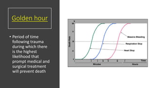
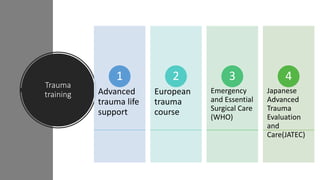
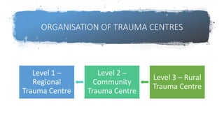
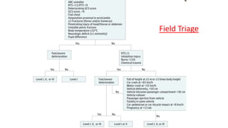
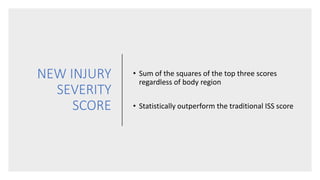
Resuscitation Principles and recent advances
Polytrauma
Moderator : Dr Samarth Mittal
Co-Moderator: Dr Siva G Presenter: Dr Namith Rangaswamy
• ‘Significant injuries of three or more points in
two or more different anatomic AIS regions in
conjunction with one or more additional
variables from the five physiologic parameters’
• Hypotension (SBP <90 mm Hg)
• Level of consciousness (GCS < 8)
• Acidosis (base excess ≤ -6.0)
• Coagulopathy (INR≥1.4/ PT≥ 40 seconds)
• Age (>70 years)
EVOLUTION OF DEFINITION
• Solely the Berlin definition resulted in a patient number
reflecting clinical reality
INJURY SEVERITY SCORE
• Anatomic scoring
• Patient data is reduced to
number
• Degree of critical illness
• 6 body regions
• Baker SP et al, "The Injury Severity Score: a
method for describing patients with multiple
injuries and evaluating emergency care", J
Trauma 14:187-196;1974
ABBREVIATED INJURY SCALE
• Introduced in 1969 - updated against survival
• Ranked on a scale of 1 to 6
• Over 9 anatomic regions
Copes WS, Sacco WJ, Champion HR, Bain LW, "Progress in Characterising Anatomic Injury", In Proceedings of the 33rd Annual Meeting of the
Association for the Advancement of Automotive Medicine, Baltimore, MA, USA 205-218
Injury Severity Score
• Each injury is assigned an AIS and is allocated to one of six body
regions
• 3 most severely injured body regions have their score squared and
added together to produce the ISS score
• Values from 0 to 75
Epidemiology
• 9people die every minute from injuries or violence
• 5.8million people of all ages and economic groups die every year from
unintentional injuries and violence
• 20-50million significant injuries
• No 1 cause of death in < 40 years
* Data from ATLS Course Manual
INDIA
•4,13,457 deaths in 2015
•32.8 deaths per hour
•53 cases of road accidents took place every one hour during 2015,
wherein 17 persons were killed
*NCRBI, Annual Report 2015
Mechanism of
Injury
Non-penetrating blunt
Penetrating
Blast
Thermal
Chemical
Others - crush & barotrauma
Trimodal
distribution of
death in trauma
• Immediate death (50%): 0 to 1
hour
• Early death (30%): 1 to 3 hours
• Late death ( 20%): 1 to 6 weeks
Golden hour
• Period of time
following trauma
during which there
is the highest
likelihood that
prompt medical and
surgical treatment
will prevent death
Trauma
training Advanced
trauma life
support
1
European
trauma
course
2
Emergency
and Essential
Surgical Care
(WHO)
3
Japanese
Advanced
Trauma
Evaluation
and
Care(JATEC)
4
ORGANISATION OF TRAUMA CENTRES
Level 1 –
Regional
Trauma Centre
Level 2 –
Community
Trauma Centre
Level 3 – Rural
Trauma Centre
Prehospital Phase
• Field triage
• Emphasize airway maintenance
• Control of external bleeding and
shock
• Immobilization of the patient
• Immediate transport to the closest
appropriate facility
• Page the hospital for preparation
• Oestern HJ, Garg B, Kotwal P. Trauma care in India and Germany. Clin Orthop Relat Res. 2013;471(9):2869–2877.
Field Triage
Centralised
Accident and
Trauma
Services
(CATS)
• Instituted in 1991
• Under Aegis of AIIMS New Delhi
• Proposed fleet: 141 BLS and 9 ALS vehicles
• Average response time: 10 min
It is better to ‘’scoop and run’’ than ‘’stay and play’’
Air Ambulance
• Far from Trauma Centers
• Pitfalls : Under triage, High cost
and Crash
• 27% of US residents transported
within Golden hour
• $5000-$6000 more than ground
transport
• Expectation: 17% reduction in
mortality
Trauma Team
Hospital Triage
CASE
Pt: 500183877
60year old male
Alleged history of fall from wooden ladder from
about 20 feet
No loss of consciousness
No ENT bleed
Severe pain in abdomen and pelvis
CASE
Awake
Spontaneous respiration
Radial Pulse 106 bpm
Reid et al
Rapid Primary
Survey
Simultaneous
resuscitation
Secondary
survey
Reassessment
of ABCD
Transfer to
Definitive Care
Primary
survey with
simultaneous
resuscitation
Airway maintenance with
restriction of cervical spine motion
Breathing and ventilation
Circulation with hemorrhage
control
Disability(assessment of neurologic
status)
Exposure/Environmental control
Adjuncts to
Primary
survey
ECG – dysrhythmias
Pulse oximetry
Ventilatory rate, Capnography, arterial
blood gases and TEG
Urinary and gastric catheters
X-Ray
eFAST, FAST and DPL
Airway with C-spine control
• SPEECH
• Hoarseness
• Noisy
• Gurgle
• Stridor
• LOOK
• Obvious Airway
injury
• Agitation
• Rib retraction
• Deformity
• Foreign body
• FEEL
• Blunt/Penetrating
injury
• Crepitus
• Tracheal deviation
• Hematoma
say “hello”
C-Spine stabilization and safe removal of helmet
Observation
• Threatened
• GCS<8
• Skull base #
• Oral bleed
• Obstructed
• Injury to Airway
• Massive Max-Facial
fracture
• Neck Hematoma
Sequence
Chin lift
Jaw thrust
Finger sweep
Suction
Reassess
Airway Decision Scheme
Preoxygenate
Able to oxygenate? Definite airway
Assess airway anatomy
and ease of intubation
Consider Adjunct
Intubation +/- drug
assisted
Consider awake
intubation
Call for assistance
Definite airway│surgical
airway
easy
No
yes
difficult
unsuccessful
• Oropharyngeal airway
• Laryngeal mask airway
• Laryngeal tube airway
• Multilumen esophageal
airway
Definitive
airway
Inability to maintain a patent airway by
other means
Inability to maintain adequate
oxygenation
Obtundation
GCS <8
Orophalyngeal Intubation
Surgical Airway
• Needle Cricothyroidotomy
• Surgical Cricothyroidotomy
 Edema of the glottis
 Fracture of the larynx
 Severe oropharyngeal hemorrhage
CASE
Airway patent
Cervical spine stabilized with collar
Breathing and
ventilation
• Look
• Listen
• Feel
AIM : to hunt and treat life threatening
thoracic condition
•LISTEN
• Chest bilaterally
•FEEL
• Dullness
• Hyper-resonance
• Crepitus
• Deformity
• CCT
•LOOK
• Rate and Depth
• Engorged veins
• Tracheal deviation
• Chest movements
• Accessory muscles
• Signs of injury
Management
Administer OXYGEN
(10L/min)
Watch
Oxygen saturation
Arterial blood gases
Sr. Lactate
CXR
Ventilate
Life threatening conditions
• Tension Pneumothorax
• Respiratory distress
• Tracheal deviation
• Distended neck veins
• Diminished breath sounds
• Immediate needle thoracostomy in 2nd intercostal
space through mid clavicular line
Open
pneumothorax
Flial chest
• Paradoxical respiration
• Distress
• Local pressure
• Endotracheal intubation
• Mechanical ventilation
Massive Hemothorax
• Rapid accumulation of more than 1500 mL of
blood or one-third or more of the patient’s
blood volume in the chest cavity
• Volume replacement + chest decompression
• Accumulation of
fluid in the
pericardial sac
• Becks triad
• FAST/eFAST is a
rapid and accurate
• Emergency
thoracotomy or
sternotomy
• Subxiphoid
pericardiocentesis
CASE
BREATHING – Spontaneous ;RR-20/Min , SPO2-100%;
Chest – Decreased Air Entry Lt Side;
CCT- Positive
Pnemo Scan- Negative
Left sided ICD was inserted in 5th ICS under LA ,
gush of air and minimal blood with good column
movemnt
CASE
CXR pre and post ICD
Circulation
• The first step in managing shock in
trauma patients is to recognize its
presence
Assessment
• Pulses (carotid and radial)
• Skin colour
• Temperature
• Blood pressure
• Capillary refill time
• Any injured patient who is cool to the touch and is
tachycardic should be considered to be in shock until
proven otherwise
• Causes
• Hypovolemic
• Cardiogenic
• Neurogenic
• Septic
• 90% of all trauma
patients could not be
classified according to
the ATLS1
classification of
hypovolemic shock.
• Raised critical
appraisal
Hypovolemic Shock
Hemorrhage is the most common cause of shock in trauma patients
Hypovolemic
shock
• Assess blood loss
• External or obvious
• Internal or Covert
• Chest
• Abdomen
• Limbs
• Pelvis
‘’Floor and four more’’
The basic management principle is to stop the bleeding and replace the
volume loss.
Controlling obvious
hemorrhage
Obtaining adequate
intravenous access
Assessing tissue
perfusion
The priority is to stop
the bleeding, not to
calculate the volume
of fluid lost
Fluid
replacement
Large bore cannula
Fluid warmer
Rapid infusion pump
Intraosseous access
Isotonic crystalloid solution 1 liter or
20ml/kg (in children)
‘’Permissive hypotension’’
Age Urine output
Adult >0.5ml/hour
Children >1ml/hour
<2years >2ml/hour
Not responding
to initial fluid
replacement
Look for
ongoing blood
loss
Begin blood
and blood
products
Non
hemorrhagic
cause
Surgical consult
Blood
replacement
• Ratio 1:1:1
• Restore the oxygen-carrying capacity
• Complete crossmatching - 1 hour
Massive
Transfusion
Protocol
Designed to interrupt the lethal triad
Activated after transfusion of 4-10 units
Predefined ratio
Type O pRBCs
AB plasma
Patil V, Shetmahajan M. Massive transfusion and massive transfusion protocol. Indian J Anaesth 2014;58:590-5
Pitfalls
• Not standardized: trigger is
undefined
• Lactic acidosis
• DIC
• SIRS
• Circulatory overload
• Dilutional coagulopathy
• Citrate toxicity
• Hypokalemia
• Hypothermia
Rapid
Responsders
Quickly respond to the initial
fluid bolus
Hemodynamically normal
Slow the fluids to maintenance
rates
Transient responders
• to the initial fluid bolusRespond
• if fluid/blood is slowedDeteriorate
• ongoing blood loss or inadequate resuscitationIndicate
• MTPInitiate
• rapid surgical interventionRequire
Non responders
Failure to
respond to
crystalloid and
blood
administration
1
Initiate MTP
2
Look for non
hemorrhagic
shock
3
Rush to OR
4
Autotransfusion
• Sterile collection
• Anticoagulation
• Cell salvage system
• Washed / preset hematocrit
• Transfusion of shed blood
Control of hemorrhage
Wound packingPelvic binder
splinting
CASE
Circulation - Pulse-106/min, BP-102/56 mmHg,
IVF prewarmed Ringers lactate 1000ml bolus stat
with maintainance @ 100ml/hr
2 units of O-negative blood transfused
eFAST- Negative
Neurologic
Evaluation(Disability)
• GCS
• Motor score correlates with outcome
• AVPU scale
• Pupillary reactions
• Consult a neurosurgeon once a brain injury is
recognized
Exposure and
Environmental Control
• Completely undress the patient
• Cover the patient with warm blankets
• External warming device
• Warm intravenous fluid
CASE
D) Disability - E4V5M6,
Pupils B/L NSRL;
E) Exposure - HYPOTHERMIA PREVENTED,
X-Ray
• CXR – AP
• Pelvis with B/L Hips
eFAST
• Trained ER personnel
• 5 quadrants
• Fluid/Air
• Sensitivity – 64-96%
• Specificity – 96-99%
• DPL has decreased from
9% to 1%
• Management has
changed in 32.8%
• FAST has excellent
diagnostic accuracy
• High negative
predictive value
99%
Rapid Ultrasound for Shock
and Hypotension
•Patients with
undifferentiated shock
•RUSH protocol
• The Pump
• The Tank
• The Pipes
Seif, Dina et al. “Bedside ultrasound in resuscitation and the rapid ultrasound in shock protocol.” Critical care research and practice vol. 2012 (2012): 503254.
CASE
PCT – Positive
Open book pelvic fracture
Single point 6th to 11th rib #
with sx emphysema
Secondary
Survey
Head to toe examination
Detailed history
Continued monitoring
Fingers and tubes in every orifice
Adjunct: X-Rays, Trauma CT
Pan CT led to 16% increase in detection and diagnosis of traumatic
intra-thoracic injuries
CASE
Secondary survery
Clinically right upperlimb # - wrist
deformity
Foleys catheterization done – Hematuria
seen
Log roll: No spinal tenderness/ crepitus/
step deformity
CASE
Fracture Distal both bone forearm – right
CASE
CECT Torso with Cystogram
Confirmed CXR findings
Left spr & ipr #,Zone 2 sacral # left side, left L1
to L5 transverse process #, L2 to L4 spinous
process #.
Extraperitoneal bladder rupture with
extravasation of contrast in the anteroinferior
aspect of bladder, left lumbar artery muscular
branch contrast extravasation.
Hybrid OR.
Future of
Trauma
care?
• Operating Rooms with integrated Imaging
equipment
• Integration of IVR to surgical treatment
• Shorten the time-to-surgery
• highest mortality was from
falls >6 m.
• The ISS was 29 in survivors,
36 in non-survivors, and 54 in
the pelvic death subgroup.
• Type C fracture was a
predictor of mortality
• Pelvic death subgroup
received a mean of 10.7 units
of blood
• No improvement
in shock after
implementation
of MTP in pelvic
fracture patients
World Society Of Emergency Surgery
WSES Grading
• Results in a shorter time
to intervention
• Lower mortality
compared to AE
Preperitoneal packing
• Successful outcome
• Justified surgery
• Efficient
hemorrhage control
• Can be definitive
fixation in some
cases
Resuscitative Endovascular Balloon Occlusion
of Aorta
• SBP increased from
60(35-75)mmHg to
115(91-128)mmHg
• Non invasive
• Balloon inflation in
zone 3
• Initial response to
resuscitation with
ANGIO is 73%.
• Also identifies arterial
injury
Angio Embolization
Spine / SCI
• Level
• Severity of neurological deficit
• Spinal cord syndrome
• Always consider spine injury to be
unstable unless proved
Canadian C-
spine rule
National Emergency X-Radiography Utilization
Study (NEXUS) Criteria
• No posterior midline cervical-spine tenderness
• No evidence of intoxication
• A normal level of alertness
• No focal neurologic deficit
• No painful distracting injuries
NO NO X-RAY
Meets ALL low-risk criteria?
Immobilize
and Log roll
Open fracture
• Tourniquet if
bleeding is ongoing
• Wound wash
• Antibiotics
• Assess vascular and
Neurological status
• Splinting
Blunt Trauma Abdomen
• Direct blow/ Shearing injuries
• Spleen (40% to 55%), liver (35% to 45%),
and small bowel
• FAST or DPL
• Laparotomy – surgical judgement to
determine timing of surgery
Damage control orthopedics
• Limited early surgical intervention
• Limit ongoing hemorrhage and soft-tissue
injury , reduce contamination
• External fixation of long bone/pelvis
fractures
• Debridement of open fractures
• Prevent second hit – and development of
lethal triad
Whom?
• Polytrauma + ISS of >20 points and additional thoracic trauma
• Polytrauma with abdominal/pelvic trauma and hemorrhagic shock
• ISS of ≥40 points in the absence of additional thoracic injury
• Radiographic findings of bilateral lung contusion
• Initial mean pulmonary arterial pressure of >24 mm Hg
• >6 mm Hg in pulmonary arterial pressure during intramedullary
nailing
CASE
Admitted under Trauma Surgery for
observation
Continuous vitals monitoring
Continuous urinary bladder irrigation
Pelvic binder with Supracondylar femoral
skeletal traction with 8 kg weight
CASE
Surgery on day 16 Discharge on day 21
• Nearly 4.4 lakh
deaths due to
preventable medical
error
• Patient safety –
priority
• Learn from errors
Q1. A 5-year-old boy is struck by an automobile and brought to the
emergency department. He is lethargic, but withdraws purposefully
from painful stimuli. His blood pressure is 90 mm Hg systolic, heart rate
is 140 beats per minute, and his respiratory rate is 36 breaths per
minute. The preferred route of venous access in this patient is
Percutaneous femoral vein cannulation
Cutdown on the saphenous vein at the ankle
Intraosseous catheter placement in the proximal tibia
Percutaneous peripheral veins in the upper extremities
Central venous access via the subclavian or internal jugular vein
Q2. A 32 year old man’s right leg is trapped beneath his overturned car
for nearly 2 hours before he is extricated. On arrival in the ER, his right
lower extremity is cool, mottled, insensate and motionless. Pulses cannot
be palpated below femoral vessels. During initial management of this
patient, which of the following is most likely to improve the chances for
limb salvage?
Applying skeletal traction
Anticoagulant therapy
Thrombolytic therapy
Right lower limb fasciotomy
Immediately transfer patient to Trauma Centre
THANK YOU
Next week: ‘’Perilunate Dislocation’’ – Dr. Devansh Goyal
EXTRA SLIDES
Massive Blood Transfusion
• Replacement of one entire blood volume within 24 h
• Transfusion of >10 units of packed red blood cells (PRBCs) in 24 h
• Transfusion of >20 units of PRBCs in 24 h
• Transfusion of >4 units of PRBCs in 1 h when on-going need is
foreseeable
• Replacement of 50% of total blood volume (TBV) within 3 h
NEW INJURY
SEVERITY
SCORE
• Sum of the squares of the top three scores
regardless of body region
• Statistically outperform the traditional ISS score
EXPONENTIAL
INJURY
SEVERITY
SCORE
• Kuo SCH, Kuo PJ, Chen YC, Chien PC, Hsieh HY, et al.
(2017) Comparison of the new Exponential Injury Severity Score
with the Injury Severity Score and the New Injury Severity Score
in trauma patients: A cross-sectional study. PLOS ONE 12(11):
e0187871.
REVISED TRAUMA SCORE
• Physiologic scoring
• range 0 to 7.8408
Champion HR et al, "A Revision of the Trauma Score", J Trauma 29:623-629,1989
Outcome = Anatomic injury + Physiologic injury
• How are the numerical identifiers structured ?
ex: 851814.3
• 8 = Body Region: Lower Extremity
• 5 =Type of Anatomic Structure: Skeletal
• 18 =Specific Anatomic Structure: Femur
• 14= Level of injury: Shaft
• .3 = AIS: Severity score
INTERNATIONAL CLASSIFICATION OF DISEASES
INJURY SEVERITY SCORE (ICISS)
• Utilizes the ICD-9 codes assigned to each patient
• Measured survival risk ratios are assigned to all ICD-9 trauma codes
• Simple product of all such ratios
• ICISS = (SRR)injury1 x (SRR)injury2 x (SRR)injury3 X (SRR)injury4…
• Can be calculated from existing hospital information without the need
for a dedicated trauma registrar
• Even non-clinical hospital coders are able to accurately interpret and
document the injuries sustained
Pape et al, 2009

Polytrauma in orthopaedics

  • 1.
    Resuscitation Principles andrecent advances Polytrauma Moderator : Dr Samarth Mittal Co-Moderator: Dr Siva G Presenter: Dr Namith Rangaswamy
  • 2.
    • ‘Significant injuriesof three or more points in two or more different anatomic AIS regions in conjunction with one or more additional variables from the five physiologic parameters’ • Hypotension (SBP <90 mm Hg) • Level of consciousness (GCS < 8) • Acidosis (base excess ≤ -6.0) • Coagulopathy (INR≥1.4/ PT≥ 40 seconds) • Age (>70 years)
  • 3.
  • 4.
    • Solely theBerlin definition resulted in a patient number reflecting clinical reality
  • 5.
    INJURY SEVERITY SCORE •Anatomic scoring • Patient data is reduced to number • Degree of critical illness • 6 body regions • Baker SP et al, "The Injury Severity Score: a method for describing patients with multiple injuries and evaluating emergency care", J Trauma 14:187-196;1974
  • 6.
    ABBREVIATED INJURY SCALE •Introduced in 1969 - updated against survival • Ranked on a scale of 1 to 6 • Over 9 anatomic regions Copes WS, Sacco WJ, Champion HR, Bain LW, "Progress in Characterising Anatomic Injury", In Proceedings of the 33rd Annual Meeting of the Association for the Advancement of Automotive Medicine, Baltimore, MA, USA 205-218
  • 7.
    Injury Severity Score •Each injury is assigned an AIS and is allocated to one of six body regions • 3 most severely injured body regions have their score squared and added together to produce the ISS score • Values from 0 to 75
  • 8.
    Epidemiology • 9people dieevery minute from injuries or violence • 5.8million people of all ages and economic groups die every year from unintentional injuries and violence • 20-50million significant injuries • No 1 cause of death in < 40 years * Data from ATLS Course Manual
  • 9.
    INDIA •4,13,457 deaths in2015 •32.8 deaths per hour •53 cases of road accidents took place every one hour during 2015, wherein 17 persons were killed *NCRBI, Annual Report 2015
  • 10.
  • 11.
    Trimodal distribution of death intrauma • Immediate death (50%): 0 to 1 hour • Early death (30%): 1 to 3 hours • Late death ( 20%): 1 to 6 weeks
  • 12.
    Golden hour • Periodof time following trauma during which there is the highest likelihood that prompt medical and surgical treatment will prevent death
  • 13.
    Trauma training Advanced trauma life support 1 European trauma course 2 Emergency andEssential Surgical Care (WHO) 3 Japanese Advanced Trauma Evaluation and Care(JATEC) 4
  • 14.
    ORGANISATION OF TRAUMACENTRES Level 1 – Regional Trauma Centre Level 2 – Community Trauma Centre Level 3 – Rural Trauma Centre
  • 15.
    Prehospital Phase • Fieldtriage • Emphasize airway maintenance • Control of external bleeding and shock • Immobilization of the patient • Immediate transport to the closest appropriate facility • Page the hospital for preparation • Oestern HJ, Garg B, Kotwal P. Trauma care in India and Germany. Clin Orthop Relat Res. 2013;471(9):2869–2877.
  • 16.
  • 17.
    Centralised Accident and Trauma Services (CATS) • Institutedin 1991 • Under Aegis of AIIMS New Delhi • Proposed fleet: 141 BLS and 9 ALS vehicles • Average response time: 10 min
  • 18.
    It is betterto ‘’scoop and run’’ than ‘’stay and play’’
  • 19.
    Air Ambulance • Farfrom Trauma Centers • Pitfalls : Under triage, High cost and Crash • 27% of US residents transported within Golden hour • $5000-$6000 more than ground transport • Expectation: 17% reduction in mortality
  • 20.
  • 21.
  • 23.
    CASE Pt: 500183877 60year oldmale Alleged history of fall from wooden ladder from about 20 feet No loss of consciousness No ENT bleed Severe pain in abdomen and pelvis
  • 24.
  • 26.
  • 27.
  • 28.
    Primary survey with simultaneous resuscitation Airway maintenancewith restriction of cervical spine motion Breathing and ventilation Circulation with hemorrhage control Disability(assessment of neurologic status) Exposure/Environmental control
  • 29.
    Adjuncts to Primary survey ECG –dysrhythmias Pulse oximetry Ventilatory rate, Capnography, arterial blood gases and TEG Urinary and gastric catheters X-Ray eFAST, FAST and DPL
  • 30.
    Airway with C-spinecontrol • SPEECH • Hoarseness • Noisy • Gurgle • Stridor • LOOK • Obvious Airway injury • Agitation • Rib retraction • Deformity • Foreign body • FEEL • Blunt/Penetrating injury • Crepitus • Tracheal deviation • Hematoma say “hello”
  • 31.
    C-Spine stabilization andsafe removal of helmet
  • 32.
    Observation • Threatened • GCS<8 •Skull base # • Oral bleed • Obstructed • Injury to Airway • Massive Max-Facial fracture • Neck Hematoma
  • 33.
  • 34.
    Airway Decision Scheme Preoxygenate Ableto oxygenate? Definite airway Assess airway anatomy and ease of intubation Consider Adjunct Intubation +/- drug assisted Consider awake intubation Call for assistance Definite airway│surgical airway easy No yes difficult unsuccessful
  • 35.
    • Oropharyngeal airway •Laryngeal mask airway • Laryngeal tube airway • Multilumen esophageal airway
  • 36.
    Definitive airway Inability to maintaina patent airway by other means Inability to maintain adequate oxygenation Obtundation GCS <8 Orophalyngeal Intubation
  • 37.
    Surgical Airway • NeedleCricothyroidotomy • Surgical Cricothyroidotomy  Edema of the glottis  Fracture of the larynx  Severe oropharyngeal hemorrhage
  • 38.
    CASE Airway patent Cervical spinestabilized with collar
  • 39.
    Breathing and ventilation • Look •Listen • Feel AIM : to hunt and treat life threatening thoracic condition
  • 40.
    •LISTEN • Chest bilaterally •FEEL •Dullness • Hyper-resonance • Crepitus • Deformity • CCT •LOOK • Rate and Depth • Engorged veins • Tracheal deviation • Chest movements • Accessory muscles • Signs of injury
  • 41.
  • 42.
    Life threatening conditions •Tension Pneumothorax • Respiratory distress • Tracheal deviation • Distended neck veins • Diminished breath sounds • Immediate needle thoracostomy in 2nd intercostal space through mid clavicular line
  • 43.
  • 44.
    Flial chest • Paradoxicalrespiration • Distress • Local pressure • Endotracheal intubation • Mechanical ventilation
  • 45.
    Massive Hemothorax • Rapidaccumulation of more than 1500 mL of blood or one-third or more of the patient’s blood volume in the chest cavity • Volume replacement + chest decompression
  • 46.
    • Accumulation of fluidin the pericardial sac • Becks triad • FAST/eFAST is a rapid and accurate • Emergency thoracotomy or sternotomy • Subxiphoid pericardiocentesis
  • 47.
    CASE BREATHING – Spontaneous;RR-20/Min , SPO2-100%; Chest – Decreased Air Entry Lt Side; CCT- Positive Pnemo Scan- Negative Left sided ICD was inserted in 5th ICS under LA , gush of air and minimal blood with good column movemnt
  • 48.
  • 49.
    Circulation • The firststep in managing shock in trauma patients is to recognize its presence
  • 50.
    Assessment • Pulses (carotidand radial) • Skin colour • Temperature • Blood pressure • Capillary refill time
  • 51.
    • Any injuredpatient who is cool to the touch and is tachycardic should be considered to be in shock until proven otherwise • Causes • Hypovolemic • Cardiogenic • Neurogenic • Septic
  • 52.
    • 90% ofall trauma patients could not be classified according to the ATLS1 classification of hypovolemic shock. • Raised critical appraisal
  • 53.
    Hypovolemic Shock Hemorrhage isthe most common cause of shock in trauma patients
  • 54.
    Hypovolemic shock • Assess bloodloss • External or obvious • Internal or Covert • Chest • Abdomen • Limbs • Pelvis ‘’Floor and four more’’
  • 55.
    The basic managementprinciple is to stop the bleeding and replace the volume loss. Controlling obvious hemorrhage Obtaining adequate intravenous access Assessing tissue perfusion The priority is to stop the bleeding, not to calculate the volume of fluid lost
  • 56.
    Fluid replacement Large bore cannula Fluidwarmer Rapid infusion pump Intraosseous access Isotonic crystalloid solution 1 liter or 20ml/kg (in children) ‘’Permissive hypotension’’
  • 57.
    Age Urine output Adult>0.5ml/hour Children >1ml/hour <2years >2ml/hour
  • 58.
    Not responding to initialfluid replacement Look for ongoing blood loss Begin blood and blood products Non hemorrhagic cause Surgical consult
  • 59.
    Blood replacement • Ratio 1:1:1 •Restore the oxygen-carrying capacity • Complete crossmatching - 1 hour
  • 60.
    Massive Transfusion Protocol Designed to interruptthe lethal triad Activated after transfusion of 4-10 units Predefined ratio Type O pRBCs AB plasma Patil V, Shetmahajan M. Massive transfusion and massive transfusion protocol. Indian J Anaesth 2014;58:590-5
  • 61.
    Pitfalls • Not standardized:trigger is undefined • Lactic acidosis • DIC • SIRS • Circulatory overload • Dilutional coagulopathy • Citrate toxicity • Hypokalemia • Hypothermia
  • 62.
    Rapid Responsders Quickly respond tothe initial fluid bolus Hemodynamically normal Slow the fluids to maintenance rates
  • 63.
    Transient responders • tothe initial fluid bolusRespond • if fluid/blood is slowedDeteriorate • ongoing blood loss or inadequate resuscitationIndicate • MTPInitiate • rapid surgical interventionRequire
  • 64.
    Non responders Failure to respondto crystalloid and blood administration 1 Initiate MTP 2 Look for non hemorrhagic shock 3 Rush to OR 4
  • 65.
    Autotransfusion • Sterile collection •Anticoagulation • Cell salvage system • Washed / preset hematocrit • Transfusion of shed blood
  • 66.
    Control of hemorrhage WoundpackingPelvic binder splinting
  • 67.
    CASE Circulation - Pulse-106/min,BP-102/56 mmHg, IVF prewarmed Ringers lactate 1000ml bolus stat with maintainance @ 100ml/hr 2 units of O-negative blood transfused eFAST- Negative
  • 68.
    Neurologic Evaluation(Disability) • GCS • Motorscore correlates with outcome • AVPU scale • Pupillary reactions • Consult a neurosurgeon once a brain injury is recognized
  • 69.
    Exposure and Environmental Control •Completely undress the patient • Cover the patient with warm blankets • External warming device • Warm intravenous fluid
  • 70.
    CASE D) Disability -E4V5M6, Pupils B/L NSRL; E) Exposure - HYPOTHERMIA PREVENTED,
  • 71.
    X-Ray • CXR –AP • Pelvis with B/L Hips
  • 72.
    eFAST • Trained ERpersonnel • 5 quadrants • Fluid/Air
  • 73.
    • Sensitivity –64-96% • Specificity – 96-99% • DPL has decreased from 9% to 1% • Management has changed in 32.8%
  • 74.
    • FAST hasexcellent diagnostic accuracy • High negative predictive value 99%
  • 75.
    Rapid Ultrasound forShock and Hypotension •Patients with undifferentiated shock •RUSH protocol • The Pump • The Tank • The Pipes Seif, Dina et al. “Bedside ultrasound in resuscitation and the rapid ultrasound in shock protocol.” Critical care research and practice vol. 2012 (2012): 503254.
  • 76.
    CASE PCT – Positive Openbook pelvic fracture Single point 6th to 11th rib # with sx emphysema
  • 77.
    Secondary Survey Head to toeexamination Detailed history Continued monitoring Fingers and tubes in every orifice Adjunct: X-Rays, Trauma CT
  • 78.
    Pan CT ledto 16% increase in detection and diagnosis of traumatic intra-thoracic injuries
  • 79.
    CASE Secondary survery Clinically rightupperlimb # - wrist deformity Foleys catheterization done – Hematuria seen Log roll: No spinal tenderness/ crepitus/ step deformity
  • 80.
    CASE Fracture Distal bothbone forearm – right
  • 81.
    CASE CECT Torso withCystogram Confirmed CXR findings Left spr & ipr #,Zone 2 sacral # left side, left L1 to L5 transverse process #, L2 to L4 spinous process #. Extraperitoneal bladder rupture with extravasation of contrast in the anteroinferior aspect of bladder, left lumbar artery muscular branch contrast extravasation.
  • 84.
    Hybrid OR. Future of Trauma care? •Operating Rooms with integrated Imaging equipment • Integration of IVR to surgical treatment • Shorten the time-to-surgery
  • 85.
    • highest mortalitywas from falls >6 m. • The ISS was 29 in survivors, 36 in non-survivors, and 54 in the pelvic death subgroup. • Type C fracture was a predictor of mortality • Pelvic death subgroup received a mean of 10.7 units of blood
  • 86.
    • No improvement inshock after implementation of MTP in pelvic fracture patients
  • 87.
    World Society OfEmergency Surgery
  • 88.
  • 90.
    • Results ina shorter time to intervention • Lower mortality compared to AE Preperitoneal packing
  • 91.
    • Successful outcome •Justified surgery • Efficient hemorrhage control • Can be definitive fixation in some cases
  • 92.
    Resuscitative Endovascular BalloonOcclusion of Aorta • SBP increased from 60(35-75)mmHg to 115(91-128)mmHg • Non invasive • Balloon inflation in zone 3
  • 93.
    • Initial responseto resuscitation with ANGIO is 73%. • Also identifies arterial injury Angio Embolization
  • 94.
    Spine / SCI •Level • Severity of neurological deficit • Spinal cord syndrome • Always consider spine injury to be unstable unless proved
  • 95.
  • 96.
    National Emergency X-RadiographyUtilization Study (NEXUS) Criteria • No posterior midline cervical-spine tenderness • No evidence of intoxication • A normal level of alertness • No focal neurologic deficit • No painful distracting injuries NO NO X-RAY Meets ALL low-risk criteria?
  • 97.
  • 98.
    Open fracture • Tourniquetif bleeding is ongoing • Wound wash • Antibiotics • Assess vascular and Neurological status • Splinting
  • 100.
    Blunt Trauma Abdomen •Direct blow/ Shearing injuries • Spleen (40% to 55%), liver (35% to 45%), and small bowel • FAST or DPL • Laparotomy – surgical judgement to determine timing of surgery
  • 102.
    Damage control orthopedics •Limited early surgical intervention • Limit ongoing hemorrhage and soft-tissue injury , reduce contamination • External fixation of long bone/pelvis fractures • Debridement of open fractures • Prevent second hit – and development of lethal triad
  • 103.
    Whom? • Polytrauma +ISS of >20 points and additional thoracic trauma • Polytrauma with abdominal/pelvic trauma and hemorrhagic shock • ISS of ≥40 points in the absence of additional thoracic injury • Radiographic findings of bilateral lung contusion • Initial mean pulmonary arterial pressure of >24 mm Hg • >6 mm Hg in pulmonary arterial pressure during intramedullary nailing
  • 105.
    CASE Admitted under TraumaSurgery for observation Continuous vitals monitoring Continuous urinary bladder irrigation Pelvic binder with Supracondylar femoral skeletal traction with 8 kg weight
  • 106.
    CASE Surgery on day16 Discharge on day 21
  • 107.
    • Nearly 4.4lakh deaths due to preventable medical error • Patient safety – priority • Learn from errors
  • 108.
    Q1. A 5-year-oldboy is struck by an automobile and brought to the emergency department. He is lethargic, but withdraws purposefully from painful stimuli. His blood pressure is 90 mm Hg systolic, heart rate is 140 beats per minute, and his respiratory rate is 36 breaths per minute. The preferred route of venous access in this patient is Percutaneous femoral vein cannulation Cutdown on the saphenous vein at the ankle Intraosseous catheter placement in the proximal tibia Percutaneous peripheral veins in the upper extremities Central venous access via the subclavian or internal jugular vein
  • 109.
    Q2. A 32year old man’s right leg is trapped beneath his overturned car for nearly 2 hours before he is extricated. On arrival in the ER, his right lower extremity is cool, mottled, insensate and motionless. Pulses cannot be palpated below femoral vessels. During initial management of this patient, which of the following is most likely to improve the chances for limb salvage? Applying skeletal traction Anticoagulant therapy Thrombolytic therapy Right lower limb fasciotomy Immediately transfer patient to Trauma Centre
  • 111.
    THANK YOU Next week:‘’Perilunate Dislocation’’ – Dr. Devansh Goyal
  • 114.
  • 118.
    Massive Blood Transfusion •Replacement of one entire blood volume within 24 h • Transfusion of >10 units of packed red blood cells (PRBCs) in 24 h • Transfusion of >20 units of PRBCs in 24 h • Transfusion of >4 units of PRBCs in 1 h when on-going need is foreseeable • Replacement of 50% of total blood volume (TBV) within 3 h
  • 119.
    NEW INJURY SEVERITY SCORE • Sumof the squares of the top three scores regardless of body region • Statistically outperform the traditional ISS score
  • 120.
    EXPONENTIAL INJURY SEVERITY SCORE • Kuo SCH,Kuo PJ, Chen YC, Chien PC, Hsieh HY, et al. (2017) Comparison of the new Exponential Injury Severity Score with the Injury Severity Score and the New Injury Severity Score in trauma patients: A cross-sectional study. PLOS ONE 12(11): e0187871.
  • 122.
    REVISED TRAUMA SCORE •Physiologic scoring • range 0 to 7.8408 Champion HR et al, "A Revision of the Trauma Score", J Trauma 29:623-629,1989
  • 123.
    Outcome = Anatomicinjury + Physiologic injury
  • 125.
    • How arethe numerical identifiers structured ? ex: 851814.3 • 8 = Body Region: Lower Extremity • 5 =Type of Anatomic Structure: Skeletal • 18 =Specific Anatomic Structure: Femur • 14= Level of injury: Shaft • .3 = AIS: Severity score
  • 127.
    INTERNATIONAL CLASSIFICATION OFDISEASES INJURY SEVERITY SCORE (ICISS) • Utilizes the ICD-9 codes assigned to each patient • Measured survival risk ratios are assigned to all ICD-9 trauma codes • Simple product of all such ratios • ICISS = (SRR)injury1 x (SRR)injury2 x (SRR)injury3 X (SRR)injury4… • Can be calculated from existing hospital information without the need for a dedicated trauma registrar • Even non-clinical hospital coders are able to accurately interpret and document the injuries sustained
  • 130.

Editor's Notes

  • #3 number of definitions of polytrauma have been reported in the literature generally referring to trauma patients whose injuries involve multiple body regions and in whom the combination of injuries would cause a life-threatening condition To objectify New definition proposed in 2014 in Berlin with consensus of various international trauma associations
  • #5 Various Definitions put to test by Frenzel, they found that
  • #6 To understand the definition better it is necessary to know ISS One of very few scoring system according to consider anatomic region Which is a process by which complex and variable patient data is reduced to a single number And indicate degree of critical illness
  • #7  Injuries are ranked on a scale of 1 to 6, with 1 being minor, 5 severe, and 6 a nonsurvivable injury The AIS is monitored by a scaling committee of the Association for the Advancement of Automotive Medicine
  • #8 If an injury is assigned an AIS of 6 (unsurvivable injury), the ISS score is automatically assigned to 75 drawback of the ISS is that it only considers one injury in each body region The ISS score is virtually the only anatomical scoring system in use and correlates linearly with mortality, morbidity, hospital stay and other measures of severity Its weaknesses are that any error in AIS scoring increases the ISS error
  • #9 Global stats, ATLS MANUAL
  • #10 In India, NCRBI, ANUUAL REPORT 2015
  • #12  Death in trauma can be divided into three time periods The first peak occurs within seconds to minutes of injury During this immediate period, deaths generally result from apnea due to severe brain or high spinal cord injury or rupture of the heart, aorta, or other large blood vessels. Very few of these patients can be saved because of the severity of their injuries. Only prevention can significantly reduce this peak of trauma-related deaths second peak occurs within minutes to several hours Deaths that occur during this period are usually due to subdural and epidural hematomas, hemopneumothorax, ruptured spleen, lacerations of the liver, pelvic fractures, and/or multiple other injuries associated with significant blood loss third peak, which occurs several days to weeks after the initial injury often due to sepsis and multiple organ system dysfunctions
  • #13 Generally considered to be 1st hour Causes of death during that period is stoppage of heart, disturbed airway followed by hemorrhage. Needs care in that sequence
  • #14 ATLS – American College of surgeon ETC – European Resuscitation council JATEC- Japanese Association for the surgery in trauma
  • #18 Only 56% of the ambulances have one or more paramedics To cut short such crisis, CATS was started in New Delhi
  • #19 Smith published a study in Injury Journal comparing two strategies of Prehospital care and found it is always better to scoop and run than stay and play
  • #20 2014 Apr 25, annals of emergency medicine Delgado in his study found that
  • #27 ZPS is intended to interrogate and manage AND optimize non-clinical processes before and during a resuscitation
  • #31 Look Inspect feel
  • #32 One person always stabilizes spine while the other expands the helmet to remove it
  • #37 Indicated when there is Inability to maintain a patent airway by other means, with impending or potential airway compromise (e.g., following inhalation injury, facial fractures, or retropharyngeal hematoma Inability to maintain adequate oxygenation by facemask oxygen supplementation, or the presence of apnea Obtundation or combativeness resulting from cerebral hypoperfusion (Glasgow Coma Scale [GCS] score of 8 or less
  • #42 Principle of management is to
  • #44 Promptly close the defect with a sterile occlusive dressing that is large enough to overlap the wound’s edges. Tape it securely on three sides to provide a flutter-valve effect.
  • #47 FAST is 90–95% accurate in identifying the presence of pericardial fluid for the experienced operator Emergency thoracotomy or sternotomy Subxiphoid pericardiocentesis
  • #50 The second step in managing shock is to identify the probable cause of shock and adjust treatment accordingly
  • #51 Look for
  • #53 Mutschler published a study in INJURY where he criticized the earlier classification given by ATLS of Hypovolaemic shock.
  • #54 Blood loss of <15, 15-30, 31-40 and over 40 percent for class I,II,III and IV respectively Mutschler added Base deficit and GCS in this newer classification
  • #55 Richard Buckley General Principles of Fracture Care  Canadian Orthopaedic Association, Orthopaedic Trauma Association
  • #57 Earlier recommendation for to use upto 2L of crystalloid, However newer studies and so ATLS currently recommends 1 liter of crystalloid Permissive hypotension – organ perfusion and tissue oxygenation with the avoidance of rebleeding by accepting a lower-than-normal blood pressure Also called as “controlled resuscitation,” “balanced resuscitation,” “hypotensive resuscitation
  • #59  • Obtain surgical consultation for definitive hemorrhage control. table
  • #60 PRBC, plasma and platelets given in ratio 1:1:1 Restore the oxygen-carrying capacity of the intravascular volume complete crossmatching process requires approximately 1 hour type O pRBCs (Rh negative preferred in female) AB plasma
  • #61 MTP describes the process of management of blood transfusion requirements in major bleeding episodes, assisting the interactions of the treating clinicians and the blood bank and ensuring judicious use of blood and blood components Main aim is to interrupt the lethal triad Generally this is activated after transfusion of 4-10 units. MTPs have a predefined ratio of RBCs, FFP/cryoprecipitate and platelets units (random donor platelets) in each pack Once the patient is in the protocol, the blood bank ensures rapid and timely delivery of all blood components together to facilitate resuscitation
  • #66 Sterile collection of blood from body cavities and processing it through anticoagulants and cell saver system where the packed cells are prepared to preset hematocrit.
  • #69  The GCS is a quick, simple, and objective method of determining the level of consciousness Prevention of secondary brain injury by maintaining adequate oxygenation and perfusion are the main goals of initial manage ment
  • #70 After completing the assessment, cover the patient with warm blankets or an external warming device to prevent him or her from developing hypothermia in the trauma receiving area
  • #76 bedside ultrasound in patients with undifferentiated shock allows for rapid evaluation of reversible causes of shock Pump: pericardial effusion, ventricular sizes, contractility Tank: reserve- venecava, leakiness-fast/thoracic, compromise – pneumothorax Pipe: leak-aneurysm,rupture /obstruction-dvt
  • #79 Trauma CT protocol – Vertex to coccyx, preferred than selective CT
  • #85 They save the time needed to transfer patients, who are medically unstable, from the operating room to the angiography suite All though evidence are limited to case reports this could be future of integrated trauma care One such case report was published international journal of em medicine by Japanese medical experts where they saved almost 1 hour of acute care time with such integration
  • #86 Coming to importance of pelvic fractures, study by palmcrantz showed that
  • #87 And hence need for definite control of bleeding
  • #88 World society of emergency surgery has very recently given guidelines and classification of pelvic trauma severity
  • #91 Study by clay cothren burlew showed that preperitoneal packing lowers mortality compared to AE and is time saving
  • #92 Prospective study
  • #94 Study comparing Exfix with angio and determined that initial response to resuscitation with angio was 73% .
  • #95 Determine the level and severity of neurological deficit Identify cord syndromes
  • #96 Presence of high risk or absence of low risk of if patient is voluntarily able to rotate neck 45degree
  • #99  judicious use of a tourniquet can be lifesaving and/or limb-saving in the presence of ongoing hemorrhage
  • #102 Instructional course material was put together by American academy of Orthopaedic Surgeons and published in JBJS
  • #103 Definite care not later than 2nd week
  • #104 Hannover criteria Louisville criteria Ph/temp/coagulo/operative time>90/geriartric patient/>10units transfused
  • #108 Medical errors – higher in developing countries
  • #121 RECENTLY INTRODUCED, 2004  The EISS was computed as the simple change in AIS values by raising each AIS severity score (1–6) by 3 taking a power of AIS minus 2, and then summing the three most severe scores (i.e., highest AIS values), regardless of body regions. If 
  • #128  utilizes the ICD-9 codes assigned to each patient Measured survival risk ratios are assigned to all ICD-9 trauma codes The simple product of all such ratios for an individual patient's injuries have been found to predict outcome more accurately than ISS ICISS = (SRR)injury1 x (SRR)injury2 x (SRR)injury3 X (SRR)injury4… ICISS is promoted as being able to be calculated from existing hospital information without the need for a dedicated trauma registrar. This assumes, however, that the non-clinical hospital coders are able to accurately interpret and document the injuries sustained.