Download to read offline
![International Research Journal of Engineering and Technology (IRJET) e-ISSN: 2395-0056
Volume: 09 Issue: 04 | Apr 2022 www.irjet.net p-ISSN: 2395-0072
© 2022, IRJET | Impact Factor value: 7.529 | ISO 9001:2008 Certified Journal | Page 1462
Pneumonia Detection Using X-Ray
Keval Shah 1, Veer Patel1, Indraneel Sarmalkar1, Suchetadevi Gaikwad2
1Dept. of Information Technology, Atharva College of Engineering, Maharashtra, India
2Assistant Professor, Dept. of Information Technology, Atharva Collegeof Engineering, Maharashtra, India
400061
-----------------------------------------------------------------------***---------------------------------------------------------------------
Abstract -Pneumonia is an infection that can lead to a
fatal outcome, it is caused because of various bacteria, fungi,
and viruses due to this inflammation in air sacs is caused. To
diagnose pneumonia, a chest X-ray is used by the medical
practitioners as the best imaging modality and it can be
managed effectively with medicines and proper treatment.
The visual analysis of a patient’s X-ray chest radiogram by a
knowledgeable doctor takes about five to fifteen minutes.
once cases are targeted, this may doubtless place
tremendous pressure on the doctor’s clinical diagnosis. We
used TensorFlow and Keras as our main libraries and
performed in the Jupyter notebook. CNN (ConvNet)
Algorithm is used for the detection as it is better than others,
as we studied in different papers. This CNN Algorithm
contains Conv2D and Maxpooling2D layers which are used
to reduce the error by fitting a function appropriately on the
given training set and avoiding the overfitting of images. We
downloaded the dataset from Kaggle which has around
5,800 chest X-Ray images.
Key Words:CNN Algorithm, Pneumonia Detection,
Tensor Flow, X-Ray, Diagnose Pneumonia, Bacteria,
Viruses
1. INTRODUCTION
Pneumonia is an infection in one or each lung. Bacteria,
viruses, and fungi cause it. The infection causes
inflammation within the air sacs in your lungs, creating it
troublesome to breathe. To diagnose pneumonia, a chest
X-ray is used by the medical practitioners as the best
imaging modality and it can be managed effectively with
medicines and proper treatment. The current system to
detect pneumonia is not very précised, so we propose a
system that will give more accuracy and the type of
pneumonia.
2. LITERATURE REVIEW
In [1], Tatiana Gabruseva, Dmytro Poplavskiy, Alexandr
Kalinin, IEEE 2020:” Deep Learning For Automatic
Pneumonia Detection.” The main aim of this proposed
research paper is to provide a simple and effective
algorithm for the localization of lung opacities region.
In [2], V. Sirish Kaushik, Anand Nayyar, Gaurav Kataria,
and Rachna Jain, Research Gate, 2020: “Pneumonia
Detection Using Convolution Neural Networks(CNNs).”
This paper consists of the usage of the CNN algorithm. CNN
classifier model 3 with three convolutional layers are
92.31%, 98%, and 94%, respectively, which are quite high
compared to other models that were trained.
In [3], Gaurav Labhane, Rutuja Pansare, Saumil
Maheshwari, Anupam Shukla, Ritu Tiwari, IEEE, 2020:
“Detection Of Pediatric Pneumonia From Chest X-Ray
Images Using CNN And Transfer Learning.” The models
proposed here for the detection of pediatric pneumonia on
the frontal chest x-ray images using CNN and Transfer
learning approaches have significant results.
In [4], Zebinjiang, IEEE(ICBAIE), 2020: “Chest X-Ray
Pneumonia Detection Based On Convolution Neural
Network.” A number of convolutional neural networks are
used to deal with chest x-ray pneumonia detection tasks.
In [5], Sheikh Rafiul, Islam, Santi P. Maity, Ajoy Kumar, Ray
and Mrinal Mandal, IEEE(CCECE), 2019: “Automatic
Detection Of Pneumonia On Compressed Sensing Images
Using Deep Learning.” Here is an automatic computerized
system for detecting pneumonia using a CS-based DL
model. The study shows that the use of DL in the CS
framework reduces the required observations for
detecting pneumonia.
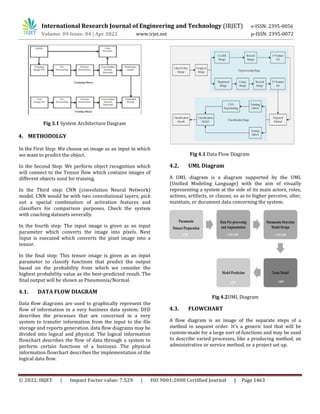
3. SYSTEM CONFIGURATION
The Purpose of Pneumonia detection system is for
research purposes which would we helpful in medical
sector. It contains models which describes that a particular
patient’s x-ray is Pneumonia positive or not. It also
suggests some basics remedies for patients to be followed
if detected pneumonia positive.Developing a user-friendly
web-based system for people. The dataset used in this
project is imported from Kaggle. Dataset consist of 5216
chest X-rays with 3875 images having Pneumonia and
1341 images shown to be normal.](https://image.slidesharecdn.com/irjet-v9i4256-220927113128-55523195/85/Pneumonia-Detection-Using-X-Ray-1-320.jpg)
![International Research Journal of Engineering and Technology (IRJET) e-ISSN: 2395-0056
Volume: 09 Issue: 04 | Apr 2022 www.irjet.net p-ISSN: 2395-0072
© 2022, IRJET | Impact Factor value: 7.529 | ISO 9001:2008 Certified Journal | Page 1464
Fig 4.3 Flow Chart
5. RESULT
Here we successfully got the prediction for the pneumonia,
when inserted pneumonia infected patient’s chest X-ray.
From our System, we tend to get an accuracy of eighty-six.
86% (~90%).
6. FUTURE SCOPE
In the future, we will additionally classify and observe its
varieties that are infectious agents or microorganisms by
adding a lot of datasets of respiratory disease varieties.
Further, we are able to conjointly offer necessary steps
and remedies to be taken if the patient is detected as
pneumonia infected.
7. CONCLUSION
The main aim of the planned resolution is to implement
the model having the best accuracy and bottom loss error
for respiratory illness Detection. The rule can sight
whether or not the given x-ray of a collection of lungs has
pneumonia or not. On applying this CNN model with a pair
of layers of Conv2D and MaxPooling2D.
8. REFERENCES
[1] Gaurav Labhane, Rutuja Pansare, Saumil Maheshwari,
Anupam Shukla,Ritu Tiwari” Detection of Pediatric
Pneumonia from Chest X-Ray Images using CNN and
Transfer Learning” IEEE 2020
[2] Tatiana Gabruseva, Dmytro Poplavskiy, Alexandr
Kalinin” Deep Learning for Automatic Pneumonia
Detection” IEEE 2020
[3] V. Sirish Kaushik, Anand Nayyar,Gaurav Kataria and
Rachna Jain”Pneumonia Detection Using Convolutional
Neural Networks (CNNs)” ResearchGate 2020
[4] Sheikh Rafiul Islam, Santi P. Maity, Ajoy Kumar Ray and
Mrinal Mandal “AutomaticDetection of Pneumonia on
Compressed Sensing Images using Deep Learning”
IEEE(CCECE) 2019
[5] Zebinjiang “Chest Xray pneumonia detection based on
convolutional neural network”(ICBAIE) 2019](https://image.slidesharecdn.com/irjet-v9i4256-220927113128-55523195/85/Pneumonia-Detection-Using-X-Ray-3-320.jpg)

1) The document discusses a study that used a convolutional neural network (CNN) model to detect pneumonia from chest x-rays. 2) The researchers trained the CNN model using a dataset of over 5,800 chest x-ray images from Kaggle to classify images as showing pneumonia or being normal. 3) The model achieved an accuracy of 86% in detecting pneumonia, demonstrating the potential for deep learning approaches to help diagnose pneumonia from medical images more quickly and accurately than human experts.