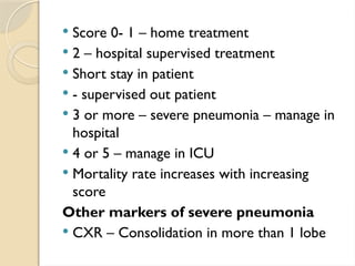
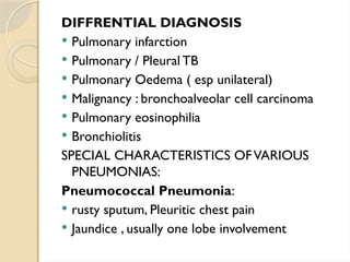
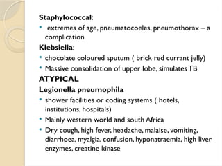
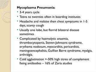
Professor Chibueze Haggai Njoku
Department of Internal Medicine
Faculty of Clinical Sciences
College of Medical Sciences
University of Calabar
PNEUMONIA
 DEFINITION:
Inflammation of the lung parenchyma, the
portion distal to the bronchioles and
comprising the respiratory bronchioles,
alveolar duct, alveolar sac and alveoli
Normal Lung Resolving Pneumonia
EPIDEMIOLOGY
 Estimated worldwide incidence of CAP is 1.5-14 cases per 1000-
affected by geography, season and population characteristics.
 Animal incidence in US – 24.8 cases /10,000 with higher rates as
age increases (2021 August)
 Affects approximately 450 people a year in all parts of the world
 Resulted in 1.4 million deaths in 2010 (7% of the worlds yearly
total )
 3.0 million deaths in 2016 ( the 4th
leading cause of death
worldwide )
 Rates are greatest in children less than five and adults > 75 years
 Five times more frequent in the developing world compared to
developed world.
 Estimated 4 million cases and 200,000 deaths in adults per year
in sub Saharan African (2013 study) published in thorax
ASSOCIATED FACTORS
 Rains and may peak in early dry season
 Clustering – mining, military
 Habits – smoking increasing risk in US X4
 Decreased immunity – DM, CKD,
Nephrotic Syndrome, Cirrhosis,
Alcoholism
 Commoner among the blacks
IN AFRICA
 2nd
commonest cause of adult admission after
malaria
 Commoner in young adults <40years
 Loss of active work in 60% of survivors for up
to 3 weeks
 Mortality highest in the elderly
 Predisposing Factors – URT, Smoking,Alcohol,
Steroid Therapy, old age,Air pollution,
HIV/AIDS Pre-existing Lung disease, recent
influenza, pregnancy, SCD, bronchiectasis
(cystic fibrosis) , Ca Lung , IV drug users
CLASSIFICATION
By anatomy:
 Lobar – immunocompetent
 Broncho – low immunity
By infection:
 Bacterial
 Fungal
 Viral
By Epidemiology:
 Community acquired
 Hospital acquired - nosocomial
 Through inhalation – Aspiration
Lobar Pneumonia
Bronchopneumonia
AETIOLOGY
 Unknown in 10-50% of cases
 Empirical treatment – assumption that
organisms in an environment are likely
the same
CONSTRAINTTO DIAGNOSIS
 Standard blood cultures may not be
totally revealing and insensitive
 Accessible diagnostic tool like sputum
culture – non specific
COMMON ORGANISMS
BACTERIAL
Gram Positive:
 Strep pneumonia 30-60% (Bacteremia 20-
30%)
 Haemophilus influenza 3-10%
 Staph aureus (including NRSA) 3-5%
Gram Negative:
 Klebsiella pneumoniae, Acinobacter,
Pseudomonas aeroginosa 3-10%
 Haemophilus influenza 3-12%
 Moraxella cataralis 3-5%
ATYPICAL
 Legionella, Mycoplasma pneumonia, Chlamydia
pneumonia (2-10%)
FUNGAL
 Histoplasma, Blastomyces, Coccidiodes (variable
depending on location)
VIRAL
 Influenza A – most common
 Parainfluenza
 Respiratory Syncitial virus (commonest in children)
2-15%
 Aspiration Pneumonia 6-10%
 Mycobacterium tuberculosis variable
 Unkown 30-60%
OTHERS
 Salmonella spp,Adenovirus, Cytomegalovirus,
Measles, Herpes simplex,Varicella, CoronaVirus
(Urban SARS- associated corona virus
The unknown may include : Pneumocytes jiroveci,
Escherichia coli anerobic bacteria, Chlamydia
psittaci, legionella pneumophila, Coxiella burnetti,
Hstoplasma capsilatum,Actinomyces isrealli,
Norcardia asteroidis,Aspergillus fumigatus ,
Paragonimus and other migrating parasites
 Empirical use of penicillins is permitted because
70% of pneumonias may be due to Streptococcus
pneumonia
Pathology – 4 stages
Congestion
 pneumococcal cell wall components stimulate
release of protein rich serous fluids within the
alveoli
 Activate pro-coagulant cascade leading to fibrin
deposition
Red hepatization
 Cell wall components bind to epithelial and
endothelial cells
 Those separate from each other, barrier function
disrupted
 Red blood cells infiltrate the lesion
Red Hepatization Grey Hepatization
Gray hepatization
 Expression of adhesion molecules or
integrin on endothelial cells
 Recruitment of large numbers of
leucocytes – grossly appears gray
 Amplification of inflammation by
complement cascade
Resolution
 Healing of the inflammatory process
without scarring
 Fibrosis and abscess formation may result.
Consolidation
Solidification of the lungs due to filling of the alveoli
with inflammatory exudates
PATHOPHYSIOLOGY OF HYPOXIA
 i) consolidation makes the lungs stiff
 ii) voluntary splinting to reduce pain
 fibrin deposition in small pulmonary vessels leads to
poor perfusion and shunting
CLINICAL FEATURES
 sudden illness
 fever – up to 39.5o
C or higher
 Rigors, shivering, vomiting, poor appetite, headaches
PULMONARY SYMPTOMS:
breathlessness, cough – initially dry but
later mucopurulent with pleuritic chest
psin.Tachypnoea /Tachycardia . rusty
sputum ( Strep pneumonia)
INVESTIGATION
 Sputum – gram stain of direct smear . Z-
N- Staining, m/c/s
 May induce sputum or do bronchial
lavage.Tracheal aspirate
 Blood – cultures
 Serology – mycoplasm, chlamydia, legionella, viral
infections
 PCR- mycoplasma in throath swabs, legionella, chlamydia
 Cold agglutinins
 Examination of pleural aspirate if para-pneumonic effusion
 Urine antigen test – legionella, pneumococcus
 CXR – homogenous opacity (affected lobe or segment)
within 12-18 hours of illness with treatment assess changes
7-10 days by repeat. After cure, clears within 6 weeks
 Reveals complications e.g effusion, empyema, abscess
 Diffuse alveolar or interstitial infiltrates
 FBC- leukocytosis
 Procalcitonin level (70.5microgram /l)
 Pulse oximetry
 PSI – pneumonia severity index
 CURB.65 (BTS – 1987)
ASSESSMENT OF SEVERITY
 1 – C – Confusion
 1 – U- Urea> 7mmol/l
 1 – R – Respiratory Rate > 30/min
 1 – B – BP systolic <90mmHg or Diastolic
<60mmHg
 1 – 65 – Age >65
 Score 0- 1 – home treatment
 2 – hospital supervised treatment
 Short stay in patient
 - supervised out patient
 3 or more – severe pneumonia – manage in
hospital
 4 or 5 – manage in ICU
 Mortality rate increases with increasing
score
Other markers of severe pneumonia
 CXR – Consolidation in more than 1 lobe
 PaO2 - < 8kpa
 Low Albumin <35g/l
 WBC < 4X10⁹
/L or > 20 X
109
/L
 Blood culture – positive
DIFFRENTIAL DIAGNOSIS
 Pulmonary infarction
 Pulmonary / Pleural TB
 Pulmonary Oedema ( esp unilateral)
 Malignancy : bronchoalveolar cell carcinoma
 Pulmonary eosinophilia
 Bronchiolitis
SPECIAL CHARACTERISTICS OFVARIOUS
PNEUMONIAS:
Pneumococcal Pneumonia:
 rusty sputum, Pleuritic chest pain
 Jaundice , usually one lobe involvement
Staphylococcal:
 extremes of age, pneumatocoeles, pneumothorax – a
complication
Klebsiella:
 chocolate coloured sputum ( brick red currant jelly)
 Massive consolidation of upper lobe, simulates TB
ATYPICAL
Legionella pneumophila
 shower facilities or coding systems ( hotels,
institutions, hospitals)
 Mainly western world and south Africa
 Dry cough, high fever, headache, malaise, vomiting,
diarrhoea, myalgia, confusion, hyponatraemia, high liver
enzymes, creatine kinase
Mycoplasma Pneumonia:
 3-4 years cycle
 Teens to twenties often in boarding institutes
 Headache and malaise then chest symptoms in 1-5
days, scanty cough
 Usually one lobe, but florrid bilateral disease
sometimes
 Complicated by haemolytic anaemia,
thrombocytopenia, Steven-Johnsons syndrome,
erythema nodosum, myocarditis, pericarditis,
meningoencephalitis, Guillian Barre syndrome, myalgia,
arthralgia.
 Cold agglutination +-50% high titres of complement
fixing antibodies – 16% of Zaria studies
Chlamydia psittaci:
 Zoonosis- exposure to birds – parrots, budgerigers,
pigeons, cockatoos, . reported in S.Africa
 Protracted illness, high or low grade fever
 Headache, meningism, photophobia, rose spots,
hepatosplenomegaly at times
 Rising titres of complement fixing antibodies
Chlamydia pneumonia:
 Droplet infection, crowded places, runny stuffy nose,
fatigue, low grade fever, hoarseness of voice,
headache, slowly worsening cough that can last for
months, headache, type specific
microimmunoflorescense test, PCR.
Rickettsia – Coxiella burnetti (Q fever):
 Contact with goats, sheep, cattle and domestic animals –
cats, dogs, rabbits
 Workers in dairy farm, abattoirs and hide factories
 Amniotic fluid and placenta carry higher risk also urine,
faeces and milk of the pets
 Q-query – until true cause discovered 1930s
 M>F Acute and Chronic forms
 Headache, fever, malaise, multiple lesion on CXR
 Acute – Severe headache, high fever, hepatitis, myalgia,
conjunctivitis
 Chronic – endocarditis, hepatomegaly
 Increasing titre of complement fixing antibodies PCR
 Vaccine-once in life. Effect last 5 years only adults > 15
years
Hospital acquired pneumonia (nosocomial):
 Develops > 3 days after admission to hospital
Predisposing Factors
 Coma, malnutrition, severe metabolic acidosis,
alcoholism, burns, COPD, pre-existing neurological
disease, elderly, sedation, steroid therapy,
instrumentation – intubation – pharynx, larynx and for
ventilation
Aetiology
 Gram negative bacilli, Staph aureus,Anaerobic bacteria,
H. influenza, S. pneumonia,Viruses, fungi,TB
Aspiration Pneumonia
Aspiration of pharyngeal contents in patients with
depressed consciousness dysphagia.
 Effects – due to combination of chemicals and
infections of periodontal disease
 Multiple aetiology – bacteroides, strept,
fusobacteria
TREATMENT
 Oxygen
 Fluid balance
 Drugs
 CAP – Amoxycillin, Macrolides,
Fluoroquinolones
 Severe – Co-Amoxyclav, Cephalosporins, IV-
macrolides
ATYPICAL
 Legionaire – Clarithromycin + Rifampicin
 Chlamydia – Tetracycline or Macrolides
 Pneumoystis jiroveci – high dose cotrimoxazole
 Mycoplasma – Tetracycline / IV erythromycin
 Coxiella – Tetracycline, doxycycline 18 months
course for chronic forms
 Actinomyces – penicillins
 Staph – flucloxacillin
 Klebsiella – Gentamycin, Ceftazidine,
Ciprofloxacin
 Viral – Acyclovir, Vidarabine
HOSPITALACQUIRED – Cefuroxime
Metronidazole
Complications:
 Parapneumonic effusion
 Empyema
 Retension of sputum causing lung collapse
 DVT leading to Pulmonary Embolism
 Suppurative pneumonia – lung Absess
 ARD with renal failure and multiple organ failure
 Ectopic abscess formation (esp Staph aureus)
 Hepatitis, pericarditis, myocarditis , meningo
encephalitis
 Fibrosis
CAUSES OF SLOW RESOLUTION OF
PNEUMONIA
 Bronchial Obstruction – Neoplasm, Foreign
body especially peanuts
 Inappropriate Chemotherapy esp for staph,
Klebsiella , TB, Myocosis
 Decreased Immunity – cachexia,
agranulocytosis, immunoglobulin defects,
LVF
 Pharyngeal Pouch – with spilling
 Formation of abscess, emphysema, effusion
 Other causes of pulmonary fibrosis

Pneumonia presentation Prof Njoku Power Point.pptx

  • 1.
    Professor Chibueze HaggaiNjoku Department of Internal Medicine Faculty of Clinical Sciences College of Medical Sciences University of Calabar PNEUMONIA
  • 2.
     DEFINITION: Inflammation ofthe lung parenchyma, the portion distal to the bronchioles and comprising the respiratory bronchioles, alveolar duct, alveolar sac and alveoli Normal Lung Resolving Pneumonia
  • 3.
    EPIDEMIOLOGY  Estimated worldwideincidence of CAP is 1.5-14 cases per 1000- affected by geography, season and population characteristics.  Animal incidence in US – 24.8 cases /10,000 with higher rates as age increases (2021 August)  Affects approximately 450 people a year in all parts of the world  Resulted in 1.4 million deaths in 2010 (7% of the worlds yearly total )  3.0 million deaths in 2016 ( the 4th leading cause of death worldwide )  Rates are greatest in children less than five and adults > 75 years  Five times more frequent in the developing world compared to developed world.  Estimated 4 million cases and 200,000 deaths in adults per year in sub Saharan African (2013 study) published in thorax
  • 4.
    ASSOCIATED FACTORS  Rainsand may peak in early dry season  Clustering – mining, military  Habits – smoking increasing risk in US X4  Decreased immunity – DM, CKD, Nephrotic Syndrome, Cirrhosis, Alcoholism  Commoner among the blacks
  • 5.
    IN AFRICA  2nd commonestcause of adult admission after malaria  Commoner in young adults <40years  Loss of active work in 60% of survivors for up to 3 weeks  Mortality highest in the elderly  Predisposing Factors – URT, Smoking,Alcohol, Steroid Therapy, old age,Air pollution, HIV/AIDS Pre-existing Lung disease, recent influenza, pregnancy, SCD, bronchiectasis (cystic fibrosis) , Ca Lung , IV drug users
  • 6.
    CLASSIFICATION By anatomy:  Lobar– immunocompetent  Broncho – low immunity By infection:  Bacterial  Fungal  Viral By Epidemiology:  Community acquired  Hospital acquired - nosocomial  Through inhalation – Aspiration Lobar Pneumonia Bronchopneumonia
  • 7.
    AETIOLOGY  Unknown in10-50% of cases  Empirical treatment – assumption that organisms in an environment are likely the same CONSTRAINTTO DIAGNOSIS  Standard blood cultures may not be totally revealing and insensitive  Accessible diagnostic tool like sputum culture – non specific
  • 8.
    COMMON ORGANISMS BACTERIAL Gram Positive: Strep pneumonia 30-60% (Bacteremia 20- 30%)  Haemophilus influenza 3-10%  Staph aureus (including NRSA) 3-5% Gram Negative:  Klebsiella pneumoniae, Acinobacter, Pseudomonas aeroginosa 3-10%  Haemophilus influenza 3-12%  Moraxella cataralis 3-5%
  • 9.
    ATYPICAL  Legionella, Mycoplasmapneumonia, Chlamydia pneumonia (2-10%) FUNGAL  Histoplasma, Blastomyces, Coccidiodes (variable depending on location) VIRAL  Influenza A – most common  Parainfluenza  Respiratory Syncitial virus (commonest in children) 2-15%  Aspiration Pneumonia 6-10%  Mycobacterium tuberculosis variable  Unkown 30-60%
  • 10.
    OTHERS  Salmonella spp,Adenovirus,Cytomegalovirus, Measles, Herpes simplex,Varicella, CoronaVirus (Urban SARS- associated corona virus The unknown may include : Pneumocytes jiroveci, Escherichia coli anerobic bacteria, Chlamydia psittaci, legionella pneumophila, Coxiella burnetti, Hstoplasma capsilatum,Actinomyces isrealli, Norcardia asteroidis,Aspergillus fumigatus , Paragonimus and other migrating parasites  Empirical use of penicillins is permitted because 70% of pneumonias may be due to Streptococcus pneumonia
  • 11.
    Pathology – 4stages Congestion  pneumococcal cell wall components stimulate release of protein rich serous fluids within the alveoli  Activate pro-coagulant cascade leading to fibrin deposition Red hepatization  Cell wall components bind to epithelial and endothelial cells  Those separate from each other, barrier function disrupted  Red blood cells infiltrate the lesion
  • 12.
  • 13.
    Gray hepatization  Expressionof adhesion molecules or integrin on endothelial cells  Recruitment of large numbers of leucocytes – grossly appears gray  Amplification of inflammation by complement cascade Resolution  Healing of the inflammatory process without scarring  Fibrosis and abscess formation may result.
  • 14.
    Consolidation Solidification of thelungs due to filling of the alveoli with inflammatory exudates PATHOPHYSIOLOGY OF HYPOXIA  i) consolidation makes the lungs stiff  ii) voluntary splinting to reduce pain  fibrin deposition in small pulmonary vessels leads to poor perfusion and shunting CLINICAL FEATURES  sudden illness  fever – up to 39.5o C or higher  Rigors, shivering, vomiting, poor appetite, headaches
  • 15.
    PULMONARY SYMPTOMS: breathlessness, cough– initially dry but later mucopurulent with pleuritic chest psin.Tachypnoea /Tachycardia . rusty sputum ( Strep pneumonia) INVESTIGATION  Sputum – gram stain of direct smear . Z- N- Staining, m/c/s  May induce sputum or do bronchial lavage.Tracheal aspirate
  • 16.
     Blood –cultures  Serology – mycoplasm, chlamydia, legionella, viral infections  PCR- mycoplasma in throath swabs, legionella, chlamydia  Cold agglutinins  Examination of pleural aspirate if para-pneumonic effusion  Urine antigen test – legionella, pneumococcus  CXR – homogenous opacity (affected lobe or segment) within 12-18 hours of illness with treatment assess changes 7-10 days by repeat. After cure, clears within 6 weeks  Reveals complications e.g effusion, empyema, abscess  Diffuse alveolar or interstitial infiltrates
  • 17.
     FBC- leukocytosis Procalcitonin level (70.5microgram /l)  Pulse oximetry  PSI – pneumonia severity index  CURB.65 (BTS – 1987) ASSESSMENT OF SEVERITY  1 – C – Confusion  1 – U- Urea> 7mmol/l  1 – R – Respiratory Rate > 30/min  1 – B – BP systolic <90mmHg or Diastolic <60mmHg  1 – 65 – Age >65
  • 18.
     Score 0-1 – home treatment  2 – hospital supervised treatment  Short stay in patient  - supervised out patient  3 or more – severe pneumonia – manage in hospital  4 or 5 – manage in ICU  Mortality rate increases with increasing score Other markers of severe pneumonia  CXR – Consolidation in more than 1 lobe
  • 19.
     PaO2 -< 8kpa  Low Albumin <35g/l  WBC < 4X10⁹ /L or > 20 X 109 /L  Blood culture – positive
  • 20.
    DIFFRENTIAL DIAGNOSIS  Pulmonaryinfarction  Pulmonary / Pleural TB  Pulmonary Oedema ( esp unilateral)  Malignancy : bronchoalveolar cell carcinoma  Pulmonary eosinophilia  Bronchiolitis SPECIAL CHARACTERISTICS OFVARIOUS PNEUMONIAS: Pneumococcal Pneumonia:  rusty sputum, Pleuritic chest pain  Jaundice , usually one lobe involvement
  • 21.
    Staphylococcal:  extremes ofage, pneumatocoeles, pneumothorax – a complication Klebsiella:  chocolate coloured sputum ( brick red currant jelly)  Massive consolidation of upper lobe, simulates TB ATYPICAL Legionella pneumophila  shower facilities or coding systems ( hotels, institutions, hospitals)  Mainly western world and south Africa  Dry cough, high fever, headache, malaise, vomiting, diarrhoea, myalgia, confusion, hyponatraemia, high liver enzymes, creatine kinase
  • 22.
    Mycoplasma Pneumonia:  3-4years cycle  Teens to twenties often in boarding institutes  Headache and malaise then chest symptoms in 1-5 days, scanty cough  Usually one lobe, but florrid bilateral disease sometimes  Complicated by haemolytic anaemia, thrombocytopenia, Steven-Johnsons syndrome, erythema nodosum, myocarditis, pericarditis, meningoencephalitis, Guillian Barre syndrome, myalgia, arthralgia.  Cold agglutination +-50% high titres of complement fixing antibodies – 16% of Zaria studies
  • 23.
    Chlamydia psittaci:  Zoonosis-exposure to birds – parrots, budgerigers, pigeons, cockatoos, . reported in S.Africa  Protracted illness, high or low grade fever  Headache, meningism, photophobia, rose spots, hepatosplenomegaly at times  Rising titres of complement fixing antibodies Chlamydia pneumonia:  Droplet infection, crowded places, runny stuffy nose, fatigue, low grade fever, hoarseness of voice, headache, slowly worsening cough that can last for months, headache, type specific microimmunoflorescense test, PCR.
  • 24.
    Rickettsia – Coxiellaburnetti (Q fever):  Contact with goats, sheep, cattle and domestic animals – cats, dogs, rabbits  Workers in dairy farm, abattoirs and hide factories  Amniotic fluid and placenta carry higher risk also urine, faeces and milk of the pets  Q-query – until true cause discovered 1930s  M>F Acute and Chronic forms  Headache, fever, malaise, multiple lesion on CXR  Acute – Severe headache, high fever, hepatitis, myalgia, conjunctivitis  Chronic – endocarditis, hepatomegaly  Increasing titre of complement fixing antibodies PCR  Vaccine-once in life. Effect last 5 years only adults > 15 years
  • 25.
    Hospital acquired pneumonia(nosocomial):  Develops > 3 days after admission to hospital Predisposing Factors  Coma, malnutrition, severe metabolic acidosis, alcoholism, burns, COPD, pre-existing neurological disease, elderly, sedation, steroid therapy, instrumentation – intubation – pharynx, larynx and for ventilation Aetiology  Gram negative bacilli, Staph aureus,Anaerobic bacteria, H. influenza, S. pneumonia,Viruses, fungi,TB Aspiration Pneumonia Aspiration of pharyngeal contents in patients with depressed consciousness dysphagia.
  • 26.
     Effects –due to combination of chemicals and infections of periodontal disease  Multiple aetiology – bacteroides, strept, fusobacteria TREATMENT  Oxygen  Fluid balance  Drugs  CAP – Amoxycillin, Macrolides, Fluoroquinolones  Severe – Co-Amoxyclav, Cephalosporins, IV- macrolides
  • 27.
    ATYPICAL  Legionaire –Clarithromycin + Rifampicin  Chlamydia – Tetracycline or Macrolides  Pneumoystis jiroveci – high dose cotrimoxazole  Mycoplasma – Tetracycline / IV erythromycin  Coxiella – Tetracycline, doxycycline 18 months course for chronic forms  Actinomyces – penicillins  Staph – flucloxacillin  Klebsiella – Gentamycin, Ceftazidine, Ciprofloxacin  Viral – Acyclovir, Vidarabine
  • 28.
    HOSPITALACQUIRED – Cefuroxime Metronidazole Complications: Parapneumonic effusion  Empyema  Retension of sputum causing lung collapse  DVT leading to Pulmonary Embolism  Suppurative pneumonia – lung Absess  ARD with renal failure and multiple organ failure  Ectopic abscess formation (esp Staph aureus)  Hepatitis, pericarditis, myocarditis , meningo encephalitis  Fibrosis
  • 29.
    CAUSES OF SLOWRESOLUTION OF PNEUMONIA  Bronchial Obstruction – Neoplasm, Foreign body especially peanuts  Inappropriate Chemotherapy esp for staph, Klebsiella , TB, Myocosis  Decreased Immunity – cachexia, agranulocytosis, immunoglobulin defects, LVF  Pharyngeal Pouch – with spilling  Formation of abscess, emphysema, effusion  Other causes of pulmonary fibrosis