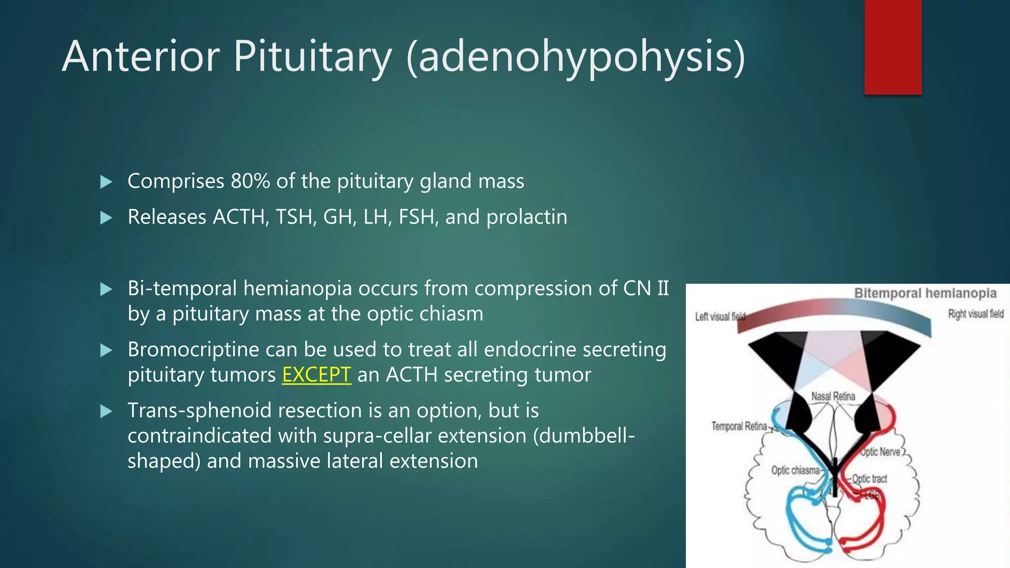
Pituitary and
Adrenals
ABSITE STUDY
Pituitary Gland
 Anterior has no direct
blood supply. There is a
portal system with blood
passing through the
posterior pituitary first
 Located in the sella turcica
Hypothalamus
 Releases TRH, CRH, GnRH and dopamine into median eminence
 Hypothalamus  Posterior Pituitary  Anterior Pituitary
 Dopamine inhibits prolactin secretion
Posterior Pituitary (neurohypohysis)
 Axons from the supraoptic and paraventricular nuclei of the hypothalamus to the
posterior pituitary
 Secretes 2 hormones
 ADH
 Release is mostly controlled by the supraoptic nuclei
 Regulated by osmolar receptors in the hypothalamus released in response to high osmolarity
 Causes increased water absorption in the renal collecting ducts
 Oxytocin
 Release is mostly controlled by the supraoptic nuclei
 Mainly functions in uterine contraction and lactation
*Posterior pituitary does NOT contain cell bodies
Anterior Pituitary (adenohypohysis)
 Comprises 80% of the pituitary gland mass
 Releases ACTH, TSH, GH, LH, FSH, and prolactin
 Bi-temporal hemianopia occurs from compression of CN II
by a pituitary mass at the optic chiasm
 Bromocriptine can be used to treat all endocrine secreting
pituitary tumors EXCEPT an ACTH secreting tumor
 Trans-sphenoid resection is an option, but is
contraindicated with supra-cellar extension (dumbbell-
shaped) and massive lateral extension
Prolactinoma
 Most common pituitary tumor (most are micro-adenomas)
 Symptoms: amenorrhea, galactorrhea, infertility, poor libido, visual problems
 Dx: Elevated prolactin (>150), MRI and visual field testing
 Tx: Most will not require surgery
 Asymptomatic AND micro (≤1cm)= follow with MRI
 Symptomatoc OR macro (>1cm)=
 Bromocryptine or cabergoline (dopamine agonist)- 85% success (safe with pregnancy)
 If failed medical Tx, hemorrhage, significant visual loss, young female wanting pregnancy, or
CSF leak then trans-sphenoid surgery- 85% success (15% recurrence)
Acromegaly
 Excessive GH, which stimulates secretion of IGF-1
 Most are macro-adenomas
 Symptoms: jaw enlargement, macroglossia, headaches, HTN, amenorrhea,
DM, giganticism and visual problems
 Can have cardiac issues (valvular or cardiomyopathy)
 Dx: Elevated IGF-1 (random GH is NOT useful), MRI
 Tx: Trans-sphenoid resection is first choice if no invasion to surrounding
tissue
 Octreotide and bromocriptine can shrink tumor and improve symptoms
 Pegvisomant (GH receptor antagonist)
Pituitary Potpourri
 Sheehan’s Syndrome
 Pituitary insufficiency in mother after delivery
 Caused by pituitary ischemia 2/2 hypovolemia/shock following childbirth
 Symptoms: Most common initial sign in difficulty with lactation; can also have amenorrhea, adrenal
insufficiency or hypothyroid
 Posterior pituitary is usually unaffected 2/2 direct blood supply
 Tx- exogenous replacement of deficiency
 Craniopharyngioma
 Most commonly occur in children 5-10yo and are benign
 Calcified cyst near anterior pituitary, remants of Rathke’s pouch (diverticulum arising from the embryonic
buccal cavity, from which the anterior pituitary gland develops)
 Symptoms: headaches, growth failure, visual problems, endocrine abnormalities, hydrocephalus
 Tx: Surgery if symptomatic (DI is a common post-op complication)
* Keep metastatic disease in mind (women with hx of breast CA and men with hx of lung CA)
Adrenal Anatomy
 Weighs 4-6 grams
 Arterial Supply – Inferior Phrenic, Unnamed Aorta, Renal
A. Branches
 Anterior and posterior surface are avascular
 Venous Drainage
 L Adrenal
 Drains from the lower pole into the L Renal V
 R Adrenal
 Drains the anterior surface of the gland into the posteriolateral
IVC; short
 Innervation
 Cortex= none
 Medulla= splanchnic
Adrenal Anatomy
 Cortex
 Originates from the mesoderm. From the adrenocortical ridge near the gonads during the 5th week
of gestation. Can have adrenal nests in the ovaries, testes or kidneys
 3 zones
 Glomerulosa (salt)– Aldosterone
 Fasciculata (sugar)– Cortisol
 Reticularis (sex)– Testosterone/estrogen
 Medulla
 Originates from the ectoderm from the neural crest
 Epinephrine (20%) – Requires PMNT – medulla or zuckerkandl (paraganglia near aortic bifrication)
 NorEpi (80%)
Physiology
 Renin-angiotensin-aldosterone pathway
 Decrease Na and volume stimulate renin secretion from the JXA
 Renin converts angiotensinogen to angiotensin I
 Angiotensin I is converted to angiotensin II by ACE in the lungs
 Angiotensin II then causes vasoconstriction and aldosterone secretion
 Aldosterone causes sodium retention and potassium excretion in the renal tubules
 Cortisol pathway
 Hypothalamus releases CRH
 CRH stimulates ACTH from the anterior pituitary
 ACTH stimulates cortisol synthesis and release from adrenal
 Cortisol acts in negative feedback to inhibit CRH/ACTH and also has effects on hepatic gluconeogenesis,
glycogen synthesis, protein catabolism, lipolysis
 Also causes hyperglycemia, impaired wound healing, collagen and bone loss
Primary Hyperaldosteronism
Conn’s Syndrome
 HTN, Hypokalemia, Polyuria, Polydipsia, Muscle Weakness
 Adenoma (60%), Bilateral Hyperplasia (40%)
 Elevated Aldosterone, Low Renin (Ratio >20:1)
 Elevated urine aldosterone following a salt loading test
 CT – adenoma vs hyperplasia
 If microadenoma (<1cm) or no mass then adrenal venous sampling to distinguish unilateral
adenoma vs B/L hyperplasia
 If macroadenoma (>1cm)- resect
 Adenoma- Resect (preop with spironolactone to normalize potassium)
 18-hydroxycorticosterone elevated (>100)with adenomas and low in hyperplasia
 Hyperplasia – Medical Tx – Spironolactone, Ca2+Blockers, Potassium
Hyperaldosteronism
 Secondary
 Secondary to renovascular HTN
 Elevated aldosterone AND renin
 Tertiary
 From diuretic use
Hypercortisolism
Cushing’s Syndrome
 Cushing’s Syndrome – Adrenal Hyperplasia, adenoma, carcinoma or ectopic ACTH or
coticotropin releasing factor (small cell carcinoma)
 Cushing’s Disease – ACTH Hypersecretion Pituitary Adenoma (60-70%)
 Weight gain, HTN, DM, Centrpetal obesity, hirsutism, striae, osteopenia
 24H Urinary/Cortisol (not serum cortisol)
 Low Dose Dexamethasone – 1mg
 Low Plasma ACTH – adrenal adenoma, hyperplasia, carcinoma
 High Plasma ACTH – Pituitary adenoma or Ectopic
 High Dose Dex – 2mg Q6h for 48H
 Etopic – Will not be suppressed
 Pituitary adenoma – Will be suppressed
Hypercortisolism Work-up
Hypercortisolism
 Source
 If adrenal suspected  CT abdomen (adrenal protocol)
 If pituitary suspected  MRI sella turcica
 If ectopic suspected  CT chest (suspected small cell carcinoma)
 Adrenalectomy
 Unilateral for adenoma or carcinoma
Need perioperative steroid coverage
 Bilateral for hyperplasia or in some cases of secondary
hypercortisolism without identification of primary tumor
Will require steroid replacement
*Exogenous steroid use is the MCC of cushingoid appearance
Congenital Adrenal Hyperplasia
Deficiency Aldosterone Testosterone
21-Hydroxylase Normal ↑
17-α-Hydroxylase ↑ Normal
11-α-Hydroxylase ↑ ↑
 Aldosterone
 HTN and hypokalemia
 Tertiary
 Virilization
Adrenal Insufficiency
 Primary – Most common is autoimmune
 Infection – Histoplasmosis, TB (MC in developing world), Meningococcus (Waterhouse Friderichsen
syndrome)
 Secondary-Iatrogenic; Exogenous Corticosteriods, most common
 Labs
 Elevated ACTH, Hyperkalemmia, Hyponatremia, Hypochloremia, Hypoglycemia, Acidosis
 Cosyntropin Stim Test – 250micrograms IV
 Cortisol level at 30-60 min if <18 is suggestive of insufficiency
 Hydrocortisone will interfere with this; can use dexamethasone
 Tx – Glucocorticoids and hydration
 Addisonian crisis
 Symptoms: hypotension, tachycardia, refractory shock
 Treat immediately. Don’t wait for labs
Evaluating Adrenal Mass
 Functioning or Non Functioning
 Majority are non functioning, if functioning resect regardless of size
 Blood/Urine levels – VMA, Catecholamines, Metanephrines
 Benign or Malignant
 Vast Majority are benign
 Carcinomas are rare – 50% stage IV at diagnosis
 Primary or Secondary
 Metastatic lesions from lung, breast, renal cell, melanoma, prostate, colon
Adrenal Incidentalomas
 Adrenal Mass discovered during imaging for unrelated reason
 DDx – Cortical adenoma, carcinoma, pheo, ganglioneuroma, cyast,
hemorrhage, fibrosis
 Evaluation
 Plasma Cortisol, Estradiol, testosterone, androstenedione
 24H urinary cortisol
 Low Dose Dexamethasone cortisol suppression test – should suppress pituitary
ACTH yielding decreased levels. No depression means cushing’s
 Renin/Aldosterone ratio
 Urinary catecholamines, metanephrines, VMA
Adrenal Incidentaloma
 Benign
 Round, smooth, homogenous
 Increased fat content
 Malignant
Irregular, focal hemorrhage/necrosis
 Percutaneous Bx
 Contraindicated if surgery indicated
 Perform if multiple masses present with primary unknown
 Not appropriate for hormonally active large tumor or if pheo is suspected
Indication for Surgery
 Hyperfunctioning – Conn, Cushing, Pheo
 Malignancy – based on CT
 Size
 Non functioning <cm 4 – Do not operate if benign imaging characteristics (smooth,
homogenous, <10 HU, >60% washout on 15min delay phase)
 CT at 3, 6, 12 months, resect if increase in size
 Non functioning 4-6cm – Individualize,
 <40yo resect, elderly observe
 Non Functioning - >6cm – Operate due to increased risk of malignancy unless clearly
benign cyst or myolipoma on imaging
 Begin metastatic workup
Adrenal Cortical Carcinoma
 Rare, aggressive, 60% hyperfunctioning
 Women, 40-50yo, Large (90%>6cm)
 Left sided more common
 Can present with liver mets
 Tx – Open Adrenalectomy (concern for tumor spillage), resection en bloc
 5 yr survival 20%
 No benefit to chemo/rads
 If unresectable, consider mitotane (adrenolytic agent that binds mitochondrial
proteins  adrenal atrophy and tumor death)
Pheochromocytoma
 Tumor of adrenal medulla or extra adrenal adrenergic tissue (neuroectodermal origin)
 Rule of 10s – 10% familial (MEN II A/B, von Hippel Lindau, Neurofibromatosis), extraadrenal
(secrete NE), multiple, bilateral, pediatric, malignant
 Sx- paroxysmal HTN, tachycardia, HTN, palpitations, flushing, sweating
 Dx – Initial plasma catecholamines/metaneprhines
 24h Urinary catecholamines (dopamine, epi, NE) and metabolites (VMA, metanephrines)
 CT abdomen
 MRI – bright T2
 Pheo Scan – MIBG I131 – concentrates in adrenergic vesicles – identify extra adrenal
 Preop – Alpha blockade first – Phenoxybenzamine (7-10d preop), then beta blockade 48 H
prior, hydrate
 If beta blockade first yields unopposed alpha yields HTN crisis
 Tx: Laparoscopic adrenalectomy
MEN
 MEN I
 Pituitary – Prolactinoma
 Initial Tx with bromocriptine; rare surgical excision
 Parathyroid – 4 gland hyperplasia (MC manifestation of MEN I)
 Pancreas – Neuroendocrine (MC non-functional)
 MEN IIA – Autosomal Dominant
 Medullary Thyroid Cancer (100%)
 Pheochromacytoma
 Hyperparathyroid
 MEN IIB – Autosomal Dominant
 Medullary Thyroid Carcinoma (100%)
 Pheochromacytoma
 Mucosal Neuroma/Marfanoid Habitus
Genes:
MEN I-
MEN II-
MTC-
MEN I
RET
RET, NTRK1

Pituitary and Adrenals ABSITE review

  • 1.
  • 2.
    Pituitary Gland  Anteriorhas no direct blood supply. There is a portal system with blood passing through the posterior pituitary first  Located in the sella turcica
  • 3.
    Hypothalamus  Releases TRH,CRH, GnRH and dopamine into median eminence  Hypothalamus  Posterior Pituitary  Anterior Pituitary  Dopamine inhibits prolactin secretion
  • 4.
    Posterior Pituitary (neurohypohysis) Axons from the supraoptic and paraventricular nuclei of the hypothalamus to the posterior pituitary  Secretes 2 hormones  ADH  Release is mostly controlled by the supraoptic nuclei  Regulated by osmolar receptors in the hypothalamus released in response to high osmolarity  Causes increased water absorption in the renal collecting ducts  Oxytocin  Release is mostly controlled by the supraoptic nuclei  Mainly functions in uterine contraction and lactation *Posterior pituitary does NOT contain cell bodies
  • 5.
    Anterior Pituitary (adenohypohysis) Comprises 80% of the pituitary gland mass  Releases ACTH, TSH, GH, LH, FSH, and prolactin  Bi-temporal hemianopia occurs from compression of CN II by a pituitary mass at the optic chiasm  Bromocriptine can be used to treat all endocrine secreting pituitary tumors EXCEPT an ACTH secreting tumor  Trans-sphenoid resection is an option, but is contraindicated with supra-cellar extension (dumbbell- shaped) and massive lateral extension
  • 6.
    Prolactinoma  Most commonpituitary tumor (most are micro-adenomas)  Symptoms: amenorrhea, galactorrhea, infertility, poor libido, visual problems  Dx: Elevated prolactin (>150), MRI and visual field testing  Tx: Most will not require surgery  Asymptomatic AND micro (≤1cm)= follow with MRI  Symptomatoc OR macro (>1cm)=  Bromocryptine or cabergoline (dopamine agonist)- 85% success (safe with pregnancy)  If failed medical Tx, hemorrhage, significant visual loss, young female wanting pregnancy, or CSF leak then trans-sphenoid surgery- 85% success (15% recurrence)
  • 7.
    Acromegaly  Excessive GH,which stimulates secretion of IGF-1  Most are macro-adenomas  Symptoms: jaw enlargement, macroglossia, headaches, HTN, amenorrhea, DM, giganticism and visual problems  Can have cardiac issues (valvular or cardiomyopathy)  Dx: Elevated IGF-1 (random GH is NOT useful), MRI  Tx: Trans-sphenoid resection is first choice if no invasion to surrounding tissue  Octreotide and bromocriptine can shrink tumor and improve symptoms  Pegvisomant (GH receptor antagonist)
  • 8.
    Pituitary Potpourri  Sheehan’sSyndrome  Pituitary insufficiency in mother after delivery  Caused by pituitary ischemia 2/2 hypovolemia/shock following childbirth  Symptoms: Most common initial sign in difficulty with lactation; can also have amenorrhea, adrenal insufficiency or hypothyroid  Posterior pituitary is usually unaffected 2/2 direct blood supply  Tx- exogenous replacement of deficiency  Craniopharyngioma  Most commonly occur in children 5-10yo and are benign  Calcified cyst near anterior pituitary, remants of Rathke’s pouch (diverticulum arising from the embryonic buccal cavity, from which the anterior pituitary gland develops)  Symptoms: headaches, growth failure, visual problems, endocrine abnormalities, hydrocephalus  Tx: Surgery if symptomatic (DI is a common post-op complication) * Keep metastatic disease in mind (women with hx of breast CA and men with hx of lung CA)
  • 9.
    Adrenal Anatomy  Weighs4-6 grams  Arterial Supply – Inferior Phrenic, Unnamed Aorta, Renal A. Branches  Anterior and posterior surface are avascular  Venous Drainage  L Adrenal  Drains from the lower pole into the L Renal V  R Adrenal  Drains the anterior surface of the gland into the posteriolateral IVC; short  Innervation  Cortex= none  Medulla= splanchnic
  • 10.
    Adrenal Anatomy  Cortex Originates from the mesoderm. From the adrenocortical ridge near the gonads during the 5th week of gestation. Can have adrenal nests in the ovaries, testes or kidneys  3 zones  Glomerulosa (salt)– Aldosterone  Fasciculata (sugar)– Cortisol  Reticularis (sex)– Testosterone/estrogen  Medulla  Originates from the ectoderm from the neural crest  Epinephrine (20%) – Requires PMNT – medulla or zuckerkandl (paraganglia near aortic bifrication)  NorEpi (80%)
  • 11.
    Physiology  Renin-angiotensin-aldosterone pathway Decrease Na and volume stimulate renin secretion from the JXA  Renin converts angiotensinogen to angiotensin I  Angiotensin I is converted to angiotensin II by ACE in the lungs  Angiotensin II then causes vasoconstriction and aldosterone secretion  Aldosterone causes sodium retention and potassium excretion in the renal tubules  Cortisol pathway  Hypothalamus releases CRH  CRH stimulates ACTH from the anterior pituitary  ACTH stimulates cortisol synthesis and release from adrenal  Cortisol acts in negative feedback to inhibit CRH/ACTH and also has effects on hepatic gluconeogenesis, glycogen synthesis, protein catabolism, lipolysis  Also causes hyperglycemia, impaired wound healing, collagen and bone loss
  • 12.
    Primary Hyperaldosteronism Conn’s Syndrome HTN, Hypokalemia, Polyuria, Polydipsia, Muscle Weakness  Adenoma (60%), Bilateral Hyperplasia (40%)  Elevated Aldosterone, Low Renin (Ratio >20:1)  Elevated urine aldosterone following a salt loading test  CT – adenoma vs hyperplasia  If microadenoma (<1cm) or no mass then adrenal venous sampling to distinguish unilateral adenoma vs B/L hyperplasia  If macroadenoma (>1cm)- resect  Adenoma- Resect (preop with spironolactone to normalize potassium)  18-hydroxycorticosterone elevated (>100)with adenomas and low in hyperplasia  Hyperplasia – Medical Tx – Spironolactone, Ca2+Blockers, Potassium
  • 13.
    Hyperaldosteronism  Secondary  Secondaryto renovascular HTN  Elevated aldosterone AND renin  Tertiary  From diuretic use
  • 14.
    Hypercortisolism Cushing’s Syndrome  Cushing’sSyndrome – Adrenal Hyperplasia, adenoma, carcinoma or ectopic ACTH or coticotropin releasing factor (small cell carcinoma)  Cushing’s Disease – ACTH Hypersecretion Pituitary Adenoma (60-70%)  Weight gain, HTN, DM, Centrpetal obesity, hirsutism, striae, osteopenia  24H Urinary/Cortisol (not serum cortisol)  Low Dose Dexamethasone – 1mg  Low Plasma ACTH – adrenal adenoma, hyperplasia, carcinoma  High Plasma ACTH – Pituitary adenoma or Ectopic  High Dose Dex – 2mg Q6h for 48H  Etopic – Will not be suppressed  Pituitary adenoma – Will be suppressed
  • 15.
  • 16.
    Hypercortisolism  Source  Ifadrenal suspected  CT abdomen (adrenal protocol)  If pituitary suspected  MRI sella turcica  If ectopic suspected  CT chest (suspected small cell carcinoma)  Adrenalectomy  Unilateral for adenoma or carcinoma Need perioperative steroid coverage  Bilateral for hyperplasia or in some cases of secondary hypercortisolism without identification of primary tumor Will require steroid replacement *Exogenous steroid use is the MCC of cushingoid appearance
  • 17.
    Congenital Adrenal Hyperplasia DeficiencyAldosterone Testosterone 21-Hydroxylase Normal ↑ 17-α-Hydroxylase ↑ Normal 11-α-Hydroxylase ↑ ↑  Aldosterone  HTN and hypokalemia  Tertiary  Virilization
  • 18.
    Adrenal Insufficiency  Primary– Most common is autoimmune  Infection – Histoplasmosis, TB (MC in developing world), Meningococcus (Waterhouse Friderichsen syndrome)  Secondary-Iatrogenic; Exogenous Corticosteriods, most common  Labs  Elevated ACTH, Hyperkalemmia, Hyponatremia, Hypochloremia, Hypoglycemia, Acidosis  Cosyntropin Stim Test – 250micrograms IV  Cortisol level at 30-60 min if <18 is suggestive of insufficiency  Hydrocortisone will interfere with this; can use dexamethasone  Tx – Glucocorticoids and hydration  Addisonian crisis  Symptoms: hypotension, tachycardia, refractory shock  Treat immediately. Don’t wait for labs
  • 19.
    Evaluating Adrenal Mass Functioning or Non Functioning  Majority are non functioning, if functioning resect regardless of size  Blood/Urine levels – VMA, Catecholamines, Metanephrines  Benign or Malignant  Vast Majority are benign  Carcinomas are rare – 50% stage IV at diagnosis  Primary or Secondary  Metastatic lesions from lung, breast, renal cell, melanoma, prostate, colon
  • 20.
    Adrenal Incidentalomas  AdrenalMass discovered during imaging for unrelated reason  DDx – Cortical adenoma, carcinoma, pheo, ganglioneuroma, cyast, hemorrhage, fibrosis  Evaluation  Plasma Cortisol, Estradiol, testosterone, androstenedione  24H urinary cortisol  Low Dose Dexamethasone cortisol suppression test – should suppress pituitary ACTH yielding decreased levels. No depression means cushing’s  Renin/Aldosterone ratio  Urinary catecholamines, metanephrines, VMA
  • 21.
    Adrenal Incidentaloma  Benign Round, smooth, homogenous  Increased fat content  Malignant Irregular, focal hemorrhage/necrosis  Percutaneous Bx  Contraindicated if surgery indicated  Perform if multiple masses present with primary unknown  Not appropriate for hormonally active large tumor or if pheo is suspected
  • 22.
    Indication for Surgery Hyperfunctioning – Conn, Cushing, Pheo  Malignancy – based on CT  Size  Non functioning <cm 4 – Do not operate if benign imaging characteristics (smooth, homogenous, <10 HU, >60% washout on 15min delay phase)  CT at 3, 6, 12 months, resect if increase in size  Non functioning 4-6cm – Individualize,  <40yo resect, elderly observe  Non Functioning - >6cm – Operate due to increased risk of malignancy unless clearly benign cyst or myolipoma on imaging  Begin metastatic workup
  • 23.
    Adrenal Cortical Carcinoma Rare, aggressive, 60% hyperfunctioning  Women, 40-50yo, Large (90%>6cm)  Left sided more common  Can present with liver mets  Tx – Open Adrenalectomy (concern for tumor spillage), resection en bloc  5 yr survival 20%  No benefit to chemo/rads  If unresectable, consider mitotane (adrenolytic agent that binds mitochondrial proteins  adrenal atrophy and tumor death)
  • 24.
    Pheochromocytoma  Tumor ofadrenal medulla or extra adrenal adrenergic tissue (neuroectodermal origin)  Rule of 10s – 10% familial (MEN II A/B, von Hippel Lindau, Neurofibromatosis), extraadrenal (secrete NE), multiple, bilateral, pediatric, malignant  Sx- paroxysmal HTN, tachycardia, HTN, palpitations, flushing, sweating  Dx – Initial plasma catecholamines/metaneprhines  24h Urinary catecholamines (dopamine, epi, NE) and metabolites (VMA, metanephrines)  CT abdomen  MRI – bright T2  Pheo Scan – MIBG I131 – concentrates in adrenergic vesicles – identify extra adrenal  Preop – Alpha blockade first – Phenoxybenzamine (7-10d preop), then beta blockade 48 H prior, hydrate  If beta blockade first yields unopposed alpha yields HTN crisis  Tx: Laparoscopic adrenalectomy
  • 25.
    MEN  MEN I Pituitary – Prolactinoma  Initial Tx with bromocriptine; rare surgical excision  Parathyroid – 4 gland hyperplasia (MC manifestation of MEN I)  Pancreas – Neuroendocrine (MC non-functional)  MEN IIA – Autosomal Dominant  Medullary Thyroid Cancer (100%)  Pheochromacytoma  Hyperparathyroid  MEN IIB – Autosomal Dominant  Medullary Thyroid Carcinoma (100%)  Pheochromacytoma  Mucosal Neuroma/Marfanoid Habitus Genes: MEN I- MEN II- MTC- MEN I RET RET, NTRK1