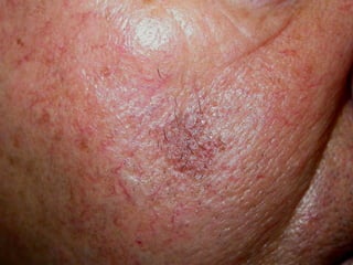
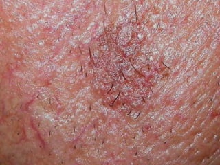
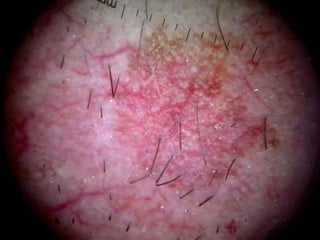
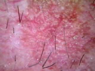
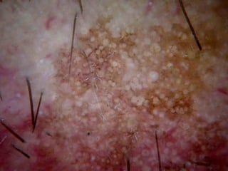
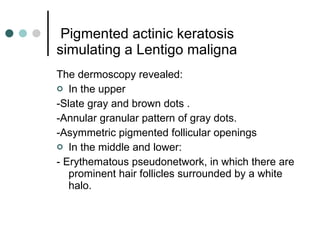
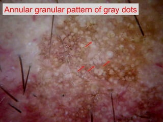
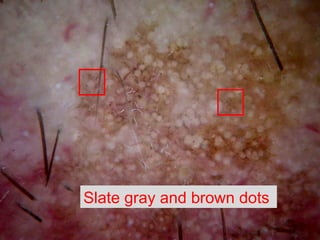
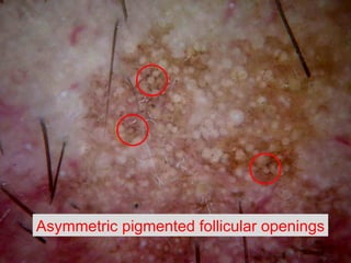
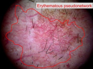
Pigmented actinic keratosis simulating a Lentigo maligna . F. Peral Rubio, M.D. Department of Dermatology  Hospital Universitario Virgen Macarena,  Seville, Spain
Pigmented actinic keratosis simulating a Lentigo maligna . A 56-years-old men. The patient was referred   to us for the assessment of a  pigmented lesion on the cheek  7 years  previously.


  Pigmented actinic keratosis simulating a Lentigo maligna The dermoscopy revealed: In the upper -Slate gray and brown dots . -Annular granular pattern  of gray dots. - Asymmetric pigmented follicular openings In the middle and lower: - E rythematous pseudonetwork, in which there are prominent hair follicles surrounded by a white halo.
Annular granular pattern  of gray dots
Slate gray and brown dots
Asymmetric pigmented follicular openings
E rythematous pseudonetwork
Pigmented actinic keratosis simulating a Lentigo maligna A biopsy was performed on the pigmented area and pathology revealed a  pigmented actinic keratosis.
Pigmented actinic keratosis simulating a Lentigo maligna Dermoscopy of  no pigmented actinic keratosis: Typical feature is a composite  pattern named “strawberry ”  characterized by: A  erythematous pseudonetwork on facial skin, in which there are prominent hair follicles surrounded by a white halo.  If the folllicle has a yellow central plaque they may look like a target.
Pigmented actinic keratosis simulating a Lentigo maligna Dermoscopy of  pigmented actinic keratosis : Facial pigmented actinic keratosis  is a differential diagnosis of lentigo maligna because it can have the same clinical and dermoscopic aspects: Gray or brown dots and globules. Asymmetric pigmented follicles. Annular-granular structures. Possible clues for pigmented actinic keratosis are scales and a superficial broken-up network usually not present in lentigo maligna.
Pigmented actinic keratosis simulating a Lentigo maligna Conclusions The dermatoscopical picture of pigmented actinic keratosis in this patient was practically indistinguishable from the early stage of Lentigo Maligna.  Pigmented actinic keratosis has a striking similarity to lentigo maligna in clinical and dermatoscopic features, thus representing a diagnostic challange.
  Pigmented actinic keratosis simulating a Lentigo maligna . F. Peral Rubio, M.D. Department of Dermatology  Hospital Universitario Virgen Macarena,  Seville, Spain

Pigmented actinic keratosis

  • 1.
    Pigmentedactinic keratosis simulating a Lentigo maligna . F. Peral Rubio, M.D. Department of Dermatology Hospital Universitario Virgen Macarena, Seville, Spain
  • 2.
    Pigmented actinic keratosissimulating a Lentigo maligna . A 56-years-old men. The patient was referred to us for the assessment of a pigmented lesion on the cheek 7 years previously.
  • 3.
  • 4.
  • 5.
  • 6.
  • 7.
  • 8.
    Pigmentedactinic keratosis simulating a Lentigo maligna The dermoscopy revealed: In the upper -Slate gray and brown dots . -Annular granular pattern of gray dots. - Asymmetric pigmented follicular openings In the middle and lower: - E rythematous pseudonetwork, in which there are prominent hair follicles surrounded by a white halo.
  • 9.
  • 10.
    Slate gray andbrown dots
  • 11.
  • 12.
  • 13.
    Pigmented actinic keratosissimulating a Lentigo maligna A biopsy was performed on the pigmented area and pathology revealed a pigmented actinic keratosis.
  • 14.
    Pigmented actinic keratosissimulating a Lentigo maligna Dermoscopy of no pigmented actinic keratosis: Typical feature is a composite pattern named “strawberry ” characterized by: A erythematous pseudonetwork on facial skin, in which there are prominent hair follicles surrounded by a white halo. If the folllicle has a yellow central plaque they may look like a target.
  • 15.
    Pigmented actinic keratosissimulating a Lentigo maligna Dermoscopy of pigmented actinic keratosis : Facial pigmented actinic keratosis is a differential diagnosis of lentigo maligna because it can have the same clinical and dermoscopic aspects: Gray or brown dots and globules. Asymmetric pigmented follicles. Annular-granular structures. Possible clues for pigmented actinic keratosis are scales and a superficial broken-up network usually not present in lentigo maligna.
  • 16.
    Pigmented actinic keratosissimulating a Lentigo maligna Conclusions The dermatoscopical picture of pigmented actinic keratosis in this patient was practically indistinguishable from the early stage of Lentigo Maligna. Pigmented actinic keratosis has a striking similarity to lentigo maligna in clinical and dermatoscopic features, thus representing a diagnostic challange.
  • 17.
    Pigmentedactinic keratosis simulating a Lentigo maligna . F. Peral Rubio, M.D. Department of Dermatology Hospital Universitario Virgen Macarena, Seville, Spain