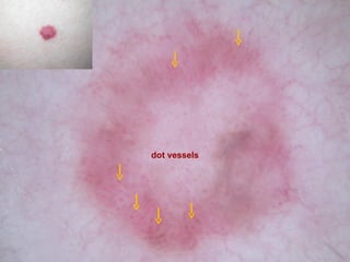
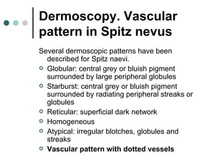
Dermoscopy. Vascular
             pattern in Spitz nevus


F. Peral Rubio, M.D.
Department of Dermatology
Hospital Universitario Virgen Macarena
Seville, Spain
Dermoscopy. Vascular
    pattern in Spitz nevus

 A 35-years-old women.
 The patient was referred to us for the
  assessment of a lesion on the right
  leg 3 years
Dermoscopy. Vascular
pattern in Spitz nevus

Dermoscopy revealed:
 A Vascular pattern with peripheral dot
  vessels
dot vessels
Dermoscopy. Vascular
pattern in Spitz nevus

   Pathology revealed a Spizt nevus.
Dermoscopy. Vascular
pattern in Spitz nevus

 Spitz nevus present as evolving
  lesions and tend to be pink or black.
 They may be clinically,
  dermoscopically and histologically
  difficult to distinguish from melanoma.
Dermoscopy. Vascular
pattern in Spitz nevus
Several dermoscopic patterns have been
  described for Spitz naevi.
 Globular: central grey or bluish pigment
  surrounded by large peripheral globules
 Starburst: central grey or bluish pigment
  surrounded by radiating peripheral streaks or
  globules
 Reticular: superficial dark network
 Homogeneous
 Atypical: irregular blotches, globules and
  streaks
 Vascular pattern with dotted vessels
Dermoscopy. Vascular
             pattern in Spitz nevus


F. Peral Rubio, M.D.
Department of Dermatology
Hospital Universitario Virgen Macarena
Seville, Spain

Spitz nevus

  • 1.
    Dermoscopy. Vascular pattern in Spitz nevus F. Peral Rubio, M.D. Department of Dermatology Hospital Universitario Virgen Macarena Seville, Spain
  • 2.
    Dermoscopy. Vascular pattern in Spitz nevus  A 35-years-old women.  The patient was referred to us for the assessment of a lesion on the right leg 3 years
  • 3.
    Dermoscopy. Vascular pattern inSpitz nevus Dermoscopy revealed:  A Vascular pattern with peripheral dot vessels
  • 6.
  • 7.
    Dermoscopy. Vascular pattern inSpitz nevus  Pathology revealed a Spizt nevus.
  • 8.
    Dermoscopy. Vascular pattern inSpitz nevus  Spitz nevus present as evolving lesions and tend to be pink or black.  They may be clinically, dermoscopically and histologically difficult to distinguish from melanoma.
  • 9.
    Dermoscopy. Vascular pattern inSpitz nevus Several dermoscopic patterns have been described for Spitz naevi.  Globular: central grey or bluish pigment surrounded by large peripheral globules  Starburst: central grey or bluish pigment surrounded by radiating peripheral streaks or globules  Reticular: superficial dark network  Homogeneous  Atypical: irregular blotches, globules and streaks  Vascular pattern with dotted vessels
  • 10.
    Dermoscopy. Vascular pattern in Spitz nevus F. Peral Rubio, M.D. Department of Dermatology Hospital Universitario Virgen Macarena Seville, Spain